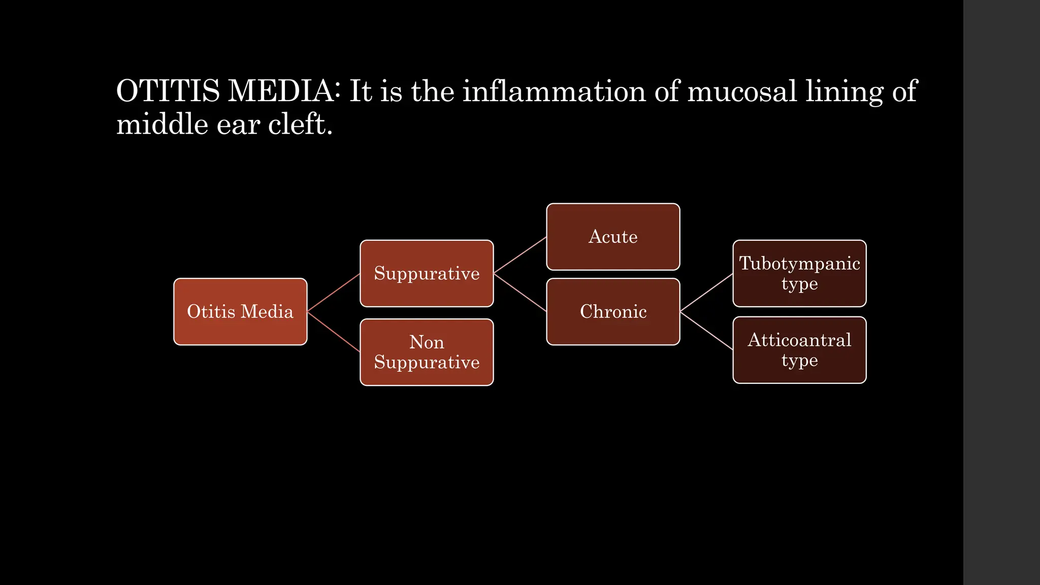
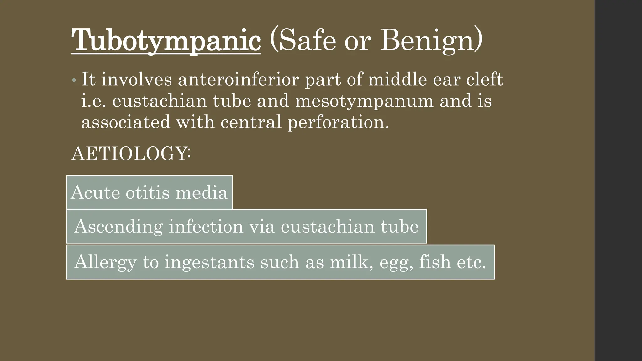
Chronic Suppurative
Otitis Media (CSOM)
Azan Rid
Roll No. : 100
Batch 15
OTITIS MEDIA: It is the inflammation of mucosal lining of
middle ear cleft.
Otitis Media
Suppurative
Acute
Chronic
Tubotympanic
type
Atticoantral
type
Non
Suppurative
• Chronic suppurative otitis media is an infection of
middle ear cleft characterized by otorrhea (ear
discharge) from a permanently perforated
tympanic membrane for more than 3 months.
• The perforation can occur from an acute otitis
media or a chronic middle ear effusion.
• Otorrhea can be the result of secretions entering
the middle ear from the eustachian tube or from
water exposure of the middle ear mucosa.
• EPIDEMIOLOGY: CSOM has higher incidence in
developing countries. It affects both sexes and all
age groups. CSOM is the most important cause of
hearing impairment mostly in rural population.
Types of CSOM
• Tubotympanic
• Atticoantral
Tubotympanic (Safe or Benign)
• It involves anteroinferior part of middle ear cleft
i.e. eustachian tube and mesotympanum and is
associated with central perforation.
AETIOLOGY:
Acute otitis media
Ascending infection via eustachian tube
Allergy to ingestants such as milk, egg, fish etc.
Pathology
• Perforation of Pars Tensa
• Middle Ear Mucosa
• Polyp
• Ossicular chain
• Tympanosclerosis
• Fibrosis and Adhesions
Bacteriology
• Aerobes:
Pseudomonas aeruginosa
Proteus
Escherichia coli
Staphylococcus aureus
• Anaerobes:
Bacteroides fragilis
Anaerobic streptococci
Commonest bacterial isolate in CSOM is
Pseudomonas aeruginosa.
Clinical Features
• Ear discharge
• Hearing loss
• Perforation
• Middle Ear Mucosa
Assessment
• Examination under microscope: Provides useful
information regarding presence of granulations,
in-growth of squamous epithelium from the edges
of perforations, status of ossicular chain,
tympanosclerosis and adhesions.
• Audiogram: Degree of hearing and its type.
• Culture & Sensitivity of ear discharge: To select
proper antibiotic ear drops.
• Mastoid X-Rays/ CT Scan Temporal bone:
Mastoid is usually sclerotic but may be
pneumatized with clouding of air cells.
Treatment
Aural toilet
• Remove all discharge and debris by dry
mopping, suction clearance or irrigation.
Ear Drops
• Antibiotic ear drops containing neomycin,
polymyxin, gentamicin + steroids.
Systemic
Antiobiotics
• Useful for acute.
Precautions
• Keep water out of ear.
Treatment of
contributory
cause
•Such as infected tonsils, adenoids,
maxillary antra and nasal allergy.
Surgical
•Removal of aural polyp/
granulations.
Reconstructive
Surgery
•Myringoplasty with or without
ossicular reconstruction.
Atticoantral Type (Unsafe or Dangerous
type)
• It involves posterosuperior part of middle ear
cleft (attic, antrum, posterior tympanum and
mastoid) and is associated with cholesteatoma.
• This area of infection is lined by flat epithelium
that responds to infection by granulations and
exudations of scanty pus.
• Aetiology:
1. It is seen in sclerotic mastoid.
2. Cholesteatoma
Pathology
• Cholesteatoma
• Osteitis and granulation tissue
• Ossicular necrosis
• Cholesterol granuloma
Bacteriology: Same as in tubotympanic.
Symptoms:
• Ear discharge
• Hearing loss
• Bleeding
Signs
• Perforation: Either attic or posterosuperior
marginal type.
• Retraction pockets: Invagination of tympanic
membrane in the attic or posterosuperior area of
pars tensa.
• Stages of retraction pockets
Stage I: TM retracted but does not contact the incus.
Stage II: TM retracted deep and contacts the incus.
Stage III ( Middle ear atelectasis): TM lies on promontory and
ossicles and middle ear space is obliterated.
Stage IV (Adhesive otitis media): Mucosal lining of middle ear is
absent and TM is adherent to promontory. Retraction pockets are
formed which may collect keratin plugs and form cholesteatoma.
Cholesteatoma
• Presence of keratinized
stratified squamous epithelium
in the middle ear cleft. It is a
skin in wrong place.
It is the main pathological
finding in atticoantral type of
CSOM.
Long process of incus is
commonly destroyed by
cholesteatoma.
Pearly white flakes of
cholesteatoma can be sucked
from the retraction pockets.
Assessment
• Examination under microscope: It reveals presence
of cholesteatoma, its site and extent, evidence of
bone destruction, granuloma, conditions of ossicles
and pockets of discharge.
• Tuning fork test and audiogram: Confirms the degree
and type of hearing loss.
• X-Ray mastoids/ CT Scan Temporal bone: They
indicate extent of bone destruction and degree of
mastoid pneumatization.
• Culture and sensitivity of ear discharge: Helps to
select proper antibiotic for local and systemic use.
Features Indicating Complications in
CSOM
• Pain
• Vertigo
• Persistent Headache
• Facial weakness
• A listless child refusing to take feeds
• Fever, Nausea and Vomiting
• Irritability and neck rigidity
• Diplopia
• Ataxia
• Abscess round the ear
Treatment
• Surgical
• Reconstructive Surgery
• Conservative treatment
Surgical
• Canal Wall Down
Procedure: Removal of
all or part of the
posterior bony canal
wall.
• Canal Wall Up
Procedure: The posterior
bony canal wall is not
removed.
• Reconstructive Surgery:
Hearing can be restored by
myringoplasty or tympanoplasty.
• Conservative treatment: Has
role in management of
cholesteatoma which is small
easily accessible to suction
clearance under operating
microscope.
 Polyp and granulations can also
be surgically removed by cup
forceps or cauterized by chemical
agents.
 Aural toilet and dry ear
precautions.

Chronic Suppurative Otitis Media (CSOM)

  • 1.
    Chronic Suppurative Otitis Media(CSOM) Azan Rid Roll No. : 100 Batch 15
  • 2.
    OTITIS MEDIA: Itis the inflammation of mucosal lining of middle ear cleft. Otitis Media Suppurative Acute Chronic Tubotympanic type Atticoantral type Non Suppurative
  • 3.
    • Chronic suppurativeotitis media is an infection of middle ear cleft characterized by otorrhea (ear discharge) from a permanently perforated tympanic membrane for more than 3 months. • The perforation can occur from an acute otitis media or a chronic middle ear effusion. • Otorrhea can be the result of secretions entering the middle ear from the eustachian tube or from water exposure of the middle ear mucosa. • EPIDEMIOLOGY: CSOM has higher incidence in developing countries. It affects both sexes and all age groups. CSOM is the most important cause of hearing impairment mostly in rural population.
  • 6.
    Types of CSOM •Tubotympanic • Atticoantral
  • 7.
    Tubotympanic (Safe orBenign) • It involves anteroinferior part of middle ear cleft i.e. eustachian tube and mesotympanum and is associated with central perforation. AETIOLOGY: Acute otitis media Ascending infection via eustachian tube Allergy to ingestants such as milk, egg, fish etc.
  • 9.
    Pathology • Perforation ofPars Tensa • Middle Ear Mucosa • Polyp • Ossicular chain • Tympanosclerosis • Fibrosis and Adhesions
  • 10.
    Bacteriology • Aerobes: Pseudomonas aeruginosa Proteus Escherichiacoli Staphylococcus aureus • Anaerobes: Bacteroides fragilis Anaerobic streptococci Commonest bacterial isolate in CSOM is Pseudomonas aeruginosa.
  • 11.
    Clinical Features • Eardischarge • Hearing loss • Perforation • Middle Ear Mucosa
  • 12.
    Assessment • Examination undermicroscope: Provides useful information regarding presence of granulations, in-growth of squamous epithelium from the edges of perforations, status of ossicular chain, tympanosclerosis and adhesions. • Audiogram: Degree of hearing and its type. • Culture & Sensitivity of ear discharge: To select proper antibiotic ear drops. • Mastoid X-Rays/ CT Scan Temporal bone: Mastoid is usually sclerotic but may be pneumatized with clouding of air cells.
  • 13.
    Treatment Aural toilet • Removeall discharge and debris by dry mopping, suction clearance or irrigation. Ear Drops • Antibiotic ear drops containing neomycin, polymyxin, gentamicin + steroids. Systemic Antiobiotics • Useful for acute. Precautions • Keep water out of ear.
  • 14.
    Treatment of contributory cause •Such asinfected tonsils, adenoids, maxillary antra and nasal allergy. Surgical •Removal of aural polyp/ granulations. Reconstructive Surgery •Myringoplasty with or without ossicular reconstruction.
  • 15.
    Atticoantral Type (Unsafeor Dangerous type) • It involves posterosuperior part of middle ear cleft (attic, antrum, posterior tympanum and mastoid) and is associated with cholesteatoma. • This area of infection is lined by flat epithelium that responds to infection by granulations and exudations of scanty pus. • Aetiology: 1. It is seen in sclerotic mastoid. 2. Cholesteatoma
  • 17.
    Pathology • Cholesteatoma • Osteitisand granulation tissue • Ossicular necrosis • Cholesterol granuloma Bacteriology: Same as in tubotympanic. Symptoms: • Ear discharge • Hearing loss • Bleeding
  • 18.
    Signs • Perforation: Eitherattic or posterosuperior marginal type.
  • 19.
    • Retraction pockets:Invagination of tympanic membrane in the attic or posterosuperior area of pars tensa. • Stages of retraction pockets Stage I: TM retracted but does not contact the incus. Stage II: TM retracted deep and contacts the incus. Stage III ( Middle ear atelectasis): TM lies on promontory and ossicles and middle ear space is obliterated. Stage IV (Adhesive otitis media): Mucosal lining of middle ear is absent and TM is adherent to promontory. Retraction pockets are formed which may collect keratin plugs and form cholesteatoma.
  • 20.
    Cholesteatoma • Presence ofkeratinized stratified squamous epithelium in the middle ear cleft. It is a skin in wrong place. It is the main pathological finding in atticoantral type of CSOM. Long process of incus is commonly destroyed by cholesteatoma. Pearly white flakes of cholesteatoma can be sucked from the retraction pockets.
  • 21.
    Assessment • Examination undermicroscope: It reveals presence of cholesteatoma, its site and extent, evidence of bone destruction, granuloma, conditions of ossicles and pockets of discharge. • Tuning fork test and audiogram: Confirms the degree and type of hearing loss. • X-Ray mastoids/ CT Scan Temporal bone: They indicate extent of bone destruction and degree of mastoid pneumatization. • Culture and sensitivity of ear discharge: Helps to select proper antibiotic for local and systemic use.
  • 22.
    Features Indicating Complicationsin CSOM • Pain • Vertigo • Persistent Headache • Facial weakness • A listless child refusing to take feeds • Fever, Nausea and Vomiting • Irritability and neck rigidity • Diplopia • Ataxia • Abscess round the ear
  • 23.
    Treatment • Surgical • ReconstructiveSurgery • Conservative treatment
  • 24.
    Surgical • Canal WallDown Procedure: Removal of all or part of the posterior bony canal wall. • Canal Wall Up Procedure: The posterior bony canal wall is not removed.
  • 25.
    • Reconstructive Surgery: Hearingcan be restored by myringoplasty or tympanoplasty. • Conservative treatment: Has role in management of cholesteatoma which is small easily accessible to suction clearance under operating microscope.  Polyp and granulations can also be surgically removed by cup forceps or cauterized by chemical agents.  Aural toilet and dry ear precautions.