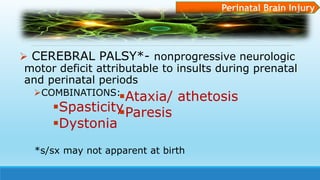
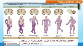
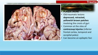
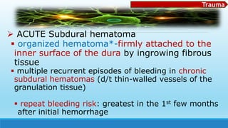
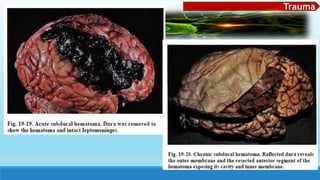
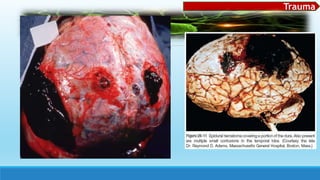
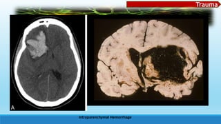
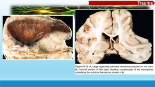
This document discusses cellular reactions and pathologies in the central nervous system. It begins by outlining the typical reactions of neurons, astrocytes, microglia, and other glial cells to injury. It then describes various inclusion bodies, degenerative changes, and proliferative reactions that may occur. The rest of the document covers topics like cerebral edema, hydrocephalus, herniation, malformations and developmental disorders of the brain. It includes detailed descriptions and diagrams of various neural tube defects, forebrain anomalies, and other congenital central nervous system abnormalities.
Pathology of Central nervous system /certified fixed orthodontic courses by I...Indian dental academy
The Indian Dental Academy is the Leader in continuing dental education , training dentists in all aspects of dentistry and offering a wide range of dental certified courses in different formats.
Indian dental academy provides dental crown & Bridge,rotary endodontics,fixed orthodontics,
Dental implants courses.for details pls visit www.indiandentalacademy.com ,or call
0091-9248678078
Introduction to the histology and pathology of the nervous system. Brief overview of most common brain cancers and histological changes of neurons and glial cells
- Coagulative Necrosis / summarized
- Description of the GROSS appearance of Coagulative necrosis.
- Description of the MICROSCOPIC appearance of Coagulative necrosis.
IT INCLUDES ANATOMY, PHYSIOLOGY AND PATHOLOGY OF LIVER .
THE SOURCES ARE:-
THE MEDICAL TEXT BOOK OF ROBBIN'S PATHOLOGY
AND OTHERS
IMAGES SOURCE :- ATLAS BOOKS AND INTERNET
This lesson is about writing agony letter , it appears in the productive tasks of the New Slimming of the syllabus.
Teachers can use the letters or Reading and Writing as brainstorming without referring to the Second Conditional ( since it is omitted according to the New Slimming )
Central nervous Pathology by dr sadaf hussainDureSameen19
The central nervous system (CNS) comprises the brain and spinal cord and is the most complex organ system in the human body. The CNS differs from other organ systems in the variety of functions that it provides and in the localization of these functions to specialized areas of the CNS. The localization of specialized functions means that a relatively small, focal lesion in the CNS can produce a profound deficit, for example, loss of speech. This localization also results in the various populations of neurons within the CNS having unique capabilities and also unique vulnerabilities to disease. For example, Parkinson disease (PD) preferentially affects the neurons of the substantia nigra in the brain stem, while Alzheimer disease (AD) preferentially affects the neurons of the cerebral cortex.
Ozempic: Preoperative Management of Patients on GLP-1 Receptor Agonists Saeid Safari
Preoperative Management of Patients on GLP-1 Receptor Agonists like Ozempic and Semiglutide
ASA GUIDELINE
NYSORA Guideline
2 Case Reports of Gastric Ultrasound
Tom Selleck Health: A Comprehensive Look at the Iconic Actor’s Wellness Journeygreendigital
Tom Selleck, an enduring figure in Hollywood. has captivated audiences for decades with his rugged charm, iconic moustache. and memorable roles in television and film. From his breakout role as Thomas Magnum in Magnum P.I. to his current portrayal of Frank Reagan in Blue Bloods. Selleck's career has spanned over 50 years. But beyond his professional achievements. fans have often been curious about Tom Selleck Health. especially as he has aged in the public eye.
Follow us on: Pinterest
Introduction
Many have been interested in Tom Selleck health. not only because of his enduring presence on screen but also because of the challenges. and lifestyle choices he has faced and made over the years. This article delves into the various aspects of Tom Selleck health. exploring his fitness regimen, diet, mental health. and the challenges he has encountered as he ages. We'll look at how he maintains his well-being. the health issues he has faced, and his approach to ageing .
Early Life and Career
Childhood and Athletic Beginnings
Tom Selleck was born on January 29, 1945, in Detroit, Michigan, and grew up in Sherman Oaks, California. From an early age, he was involved in sports, particularly basketball. which played a significant role in his physical development. His athletic pursuits continued into college. where he attended the University of Southern California (USC) on a basketball scholarship. This early involvement in sports laid a strong foundation for his physical health and disciplined lifestyle.
Transition to Acting
Selleck's transition from an athlete to an actor came with its physical demands. His first significant role in "Magnum P.I." required him to perform various stunts and maintain a fit appearance. This role, which he played from 1980 to 1988. necessitated a rigorous fitness routine to meet the show's demands. setting the stage for his long-term commitment to health and wellness.
Fitness Regimen
Workout Routine
Tom Selleck health and fitness regimen has evolved. adapting to his changing roles and age. During his "Magnum, P.I." days. Selleck's workouts were intense and focused on building and maintaining muscle mass. His routine included weightlifting, cardiovascular exercises. and specific training for the stunts he performed on the show.
Selleck adjusted his fitness routine as he aged to suit his body's needs. Today, his workouts focus on maintaining flexibility, strength, and cardiovascular health. He incorporates low-impact exercises such as swimming, walking, and light weightlifting. This balanced approach helps him stay fit without putting undue strain on his joints and muscles.
Importance of Flexibility and Mobility
In recent years, Selleck has emphasized the importance of flexibility and mobility in his fitness regimen. Understanding the natural decline in muscle mass and joint flexibility with age. he includes stretching and yoga in his routine. These practices help prevent injuries, improve posture, and maintain mobilit
These lecture slides, by Dr Sidra Arshad, offer a quick overview of physiological basis of a normal electrocardiogram.
Learning objectives:
1. Define an electrocardiogram (ECG) and electrocardiography
2. Describe how dipoles generated by the heart produce the waveforms of the ECG
3. Describe the components of a normal electrocardiogram of a typical bipolar leads (limb II)
4. Differentiate between intervals and segments
5. Enlist some common indications for obtaining an ECG
Study Resources:
1. Chapter 11, Guyton and Hall Textbook of Medical Physiology, 14th edition
2. Chapter 9, Human Physiology - From Cells to Systems, Lauralee Sherwood, 9th edition
3. Chapter 29, Ganong’s Review of Medical Physiology, 26th edition
4. Electrocardiogram, StatPearls - https://www.ncbi.nlm.nih.gov/books/NBK549803/
5. ECG in Medical Practice by ABM Abdullah, 4th edition
6. ECG Basics, http://www.nataliescasebook.com/tag/e-c-g-basics
Factory Supply Best Quality Pmk Oil CAS 28578–16–7 PMK Powder in Stockrebeccabio
Factory Supply Best Quality Pmk Oil CAS 28578–16–7 PMK Powder in Stock
Telegram: bmksupplier
signal: +85264872720
threema: TUD4A6YC
You can contact me on Telegram or Threema
Communicate promptly and reply
Free of customs clearance, Double Clearance 100% pass delivery to USA, Canada, Spain, Germany, Netherland, Poland, Italy, Sweden, UK, Czech Republic, Australia, Mexico, Russia, Ukraine, Kazakhstan.Door to door service
Hot Selling Organic intermediates
These simplified slides by Dr. Sidra Arshad present an overview of the non-respiratory functions of the respiratory tract.
Learning objectives:
1. Enlist the non-respiratory functions of the respiratory tract
2. Briefly explain how these functions are carried out
3. Discuss the significance of dead space
4. Differentiate between minute ventilation and alveolar ventilation
5. Describe the cough and sneeze reflexes
Study Resources:
1. Chapter 39, Guyton and Hall Textbook of Medical Physiology, 14th edition
2. Chapter 34, Ganong’s Review of Medical Physiology, 26th edition
3. Chapter 17, Human Physiology by Lauralee Sherwood, 9th edition
4. Non-respiratory functions of the lungs https://academic.oup.com/bjaed/article/13/3/98/278874
Couples presenting to the infertility clinic- Do they really have infertility...Sujoy Dasgupta
Dr Sujoy Dasgupta presented the study on "Couples presenting to the infertility clinic- Do they really have infertility? – The unexplored stories of non-consummation" in the 13th Congress of the Asia Pacific Initiative on Reproduction (ASPIRE 2024) at Manila on 24 May, 2024.
TEST BANK for Operations Management, 14th Edition by William J. Stevenson, Ve...kevinkariuki227
TEST BANK for Operations Management, 14th Edition by William J. Stevenson, Verified Chapters 1 - 19, Complete Newest Version.pdf
Anti ulcer drugs and their Advance pharmacology ||
Anti-ulcer drugs are medications used to prevent and treat ulcers in the stomach and upper part of the small intestine (duodenal ulcers). These ulcers are often caused by an imbalance between stomach acid and the mucosal lining, which protects the stomach lining.
||Scope: Overview of various classes of anti-ulcer drugs, their mechanisms of action, indications, side effects, and clinical considerations.
1. KRISTINE FAITH P. TABLIZO, RN
MD-MPA
pp. 1252-1263,
Robbins & Cotran Pathologic
Basis of Disease, 9th Ed.
2. Cellular
Patho
• Rxns of
Neurons
• Rxns of
Astrocytes
• Rxns of
Microglia
• Rxns of other
Glial cells
Cerebral
Edema,
Hydrocepha
lus,
Increased
ICP,
Herniation
Malformations
and
Developmental
Disorders
• Neural tube defects
• Forebrain anomalies
• Posterior Fossa
Anomalies
• Syringomyelia and
Hydromyelia
Perinatal
Brain
Injury
Trauma
• Skull Fractures
• Parenchymal Injuries
• Concussion
• Direct P. Injury
• Diffuse Ax. Injury
• Traumatic Vascular
Injury
• Epidural Hematoma
• Subdural hematoma
• Sequelae of Brain
trauma
• SC Injury
3. Reactions to Cellular Injury
Neurons Astrocytes Microglia Other Glial Cells
Acute Neuronal Injury
“RED NEURONS”
Cell body shrinkage
Nucleal PYKNOSIS
Nucleolus disappearance
Loss of Nissl Bodies-
eosinophilia of cytoplasm
Gemistocytic astrocytes
- Swollen reactive astrocyte
Nuclei vesicular w/
prominent nucleoli
Cytoplasm bright pink
irregular area around an
eccentric nucleus
Alzheimer type II astrocyte
Nucleus 2-3x larger,
pale staining chromatin
Intranuclear glycogen
droplet
prominent nuclear
membrane and nucleolus
*in Chronic hyperammonemia
Respond to injury by: OLIGODENDROCYTES
- Injury and apoptosis
of which is afeature
of acquired
demyelinating
disorders and
leukodystrophies
NUCLEI- viral
inclusions (progressive
multifocal
leukoencephalopathy)
Glial Cytoplasmic
inclusions
α-synuclein
composition
-in MSA
(mult. sys.atrophy)
1. Proliferation
2. Elongation of
nuclei (rod cells)
e.g Neurosyphilis
3. microglial nodules
4. Neuronophagia-
Subacute/Chronic* Injury
“DEGENERATION”
e.g. ALS, AD- ‘progressive’
Cell loss
Reactive Gliosis- proliferation
or hypertrophy of glial cells
4. Reactions to Cellular Injury
Neurons Astrocytes Microglia Other Glial Cells
NEURONAL INCLUSIONS
AGING
- intracytoplasmic accumulations of
complex lipids (lipofuscin), proteins, or
carbohydrates
**Diorders of Metabolism-substrates or
intermediates accumulate
CYTOPLASMIC I.B.’s
ROSENTHAL fibers thick, elongated, brightly eosinophilic,
irregular structures in astrocytic processes
αB –crystalline and hsp27- heat shock proteins, as well as ubiquitin
Glial tumor- pilocytic astrocytoma
Alexander dse. (leukodystrophy associated w/ mutations in the gene encoding
GFAP) –in periventricular, perivascular, subpial regions
Corpora amylacea (polyglucosan bodies)
Round, faintly basophilic, PAS (+), concentrically lamellated
structures, 5-50 um in diameter
GLYCOSAMINOGLYCAN polymers, heat shock proteins and ubiquitin
Degenerative change
Lafora bodies- seen in cytoplasm of neurons* in myoclonic
epilepsy *(also in hepatocytes, myocytes, other cells)
-same composition as PGS bodies
VIRAL INFXNS
a. intranuclear inclusions
COWDRY BODY- herpetic infxn
b. Intracytoplasmic inclusions
NEGRI BODY-rabies
b. both nucleus & cytoplasm
CMG virus infxn
DEGENERATIVE DISEASES
-neuronal intracytoplasmic inclusions
Neurofibrillary tangles- AD
Lewy bodies- PD
5. Reactions to Cellular Injury
Neurons Astrocytes Microglia Other Glial Cells
Axonal Reaction-change in cell body during regeneration
- ↑ protein synthesis- axonal sprouting
enlargement and rounding up of the cell body
peripheral displacement of nucleus
Central chromatolysis (dispersion of Nissl substance
from center to periphery)
EPENDYMAL CELLS
inflammation or marked dilation
of ventricular system
disruption of ependymal lining
Proliferation of subependymal astrocytes
Irregularities on ventricular surfaces
(ependymal granulations)
15. MONRO-KELLIE HYPOTHESIS
states that the cranial compartment is
incompressible, and the volume inside the
cranium is a fixed volume.
any increase in volume of one of the
cranial constituents (blood, CSF, and brain
tissue) must be compensated by a
decrease in volume of another.
Cerebral Edema, Hydrocephalus, Increased
ICP, Herniation
16. Cerebral Edema, Hydrocephalus, Increased
ICP, Herniation
CEREBRAL EDEMA (brain parenchymal edema)
Increased fluid leakage from blood vessels or injury to various cells in CNS
HYDROCEPHALUS
accumulation of excessive CSF w/in the ventricular system
impaired flow and resorption
overproduction- tumors of ch. Plexus
HERNIATION
displacement of brain tissue past rigid dural folds or through openings
in the skull because of ↑ ICP
17. Cerebral Edema
Vasogenic Edema Cytotoxic Edema Interstitial edema
↑ EXTRACELLULAR Fluid
Caused by B-B-B disruption
And ↑ vascular permeability
↑ INTRACELLULAR Fluid
- 2° CELL MEMBRANE INURY
HYDROCEPHALIC EDEMA
-occurs sp. around LATERAL
VENTRICLES
↑ Intravascular pressure
abnormal flow of fluid
from intraventricular CSF
across ependymal lining to
the periventricular white
matter
Localized- adjacent to
inflammation or neoplasms
* GENERALIZED HYPOXIC/
ISCHEMIC insult
* METABOLIC Derangement
affecting ionic gradient
Generalized-
ff. ischemic injury; both
vasogenic & interstitial components
Flattened gryi
Narrowed sulci
Compressed ventricular
cavities
Herniation likely
23. HerniationSubfalcine (cingulate)
herniation
Transtentorial (uncinate, mesial temporal)
herniation
Tonsillar Herniation
Displaces cingulate
gyrus under the
falx.
Medial aspect of temporal lobe
compressed against the free margin of
the tentorium
Displacement of
cerebellar tonsils
through the foramen
magnum
Compromises CN III, and Posterior cerebral
artery
LIFE
THREATENING-
compression of
brainstem
(respiratory and
cardiac centers)
Compression of
anterior cerebral
artery and its
branches
Kernohan notch- compression in the contralateral
cerebral peduncle; may result in hemiparesis of the side
ipsilateral to the herniation
Duret hemorrhages- linear of flame-shaped lesions
usually in the middle and paramedian regions
-due to distortion or tearing off penetrating veins and
arteries supplying the upper brainstem
27. Neural tube defects
failure to close by a portion a the neural
tube or reopening of a region of a tube
after successful closure
account for most common CNS
malformation
most common NTD– SC involvement
frequency varies among ethnic groups
high concordance rate among
monozygotic twins
risk factor: folate deficiency during 1st
several weeks of gestation
Malformations and Devt’al Disorders
28. Malformations and Devt’al Disorders
Neural Tube Defects
Spina Bifida/ spinal
dysraphism
Meningomyelocele/
Myelomeningocele
Encephalocele
(Cranium bifidum)
Anencephaly
SPINA BIFIDA OCCULTA
- asymptomatic bony
defect
- Mildest form
- Missing spinous process
Extension of CNS
tissue through a
defect in the vertebral
column
Diverticulum of
malformed brain
tissue extending
through a defect in
the cranium
Malformation of the
anterior end of the NT,
with absence of most of
the brain and calvarium
sacral region Most often in the
posterior fossa
*Area Cerebrovasculosa-
flattened remnant of
disorganized brain tissue w/
admixed ependyma, choroid
plexus, and meningothelial
cells
*MENINGOCELE
-meningeal outpouching only
Lower Motor and sensory
deficits ; bladder and bowel
control deficits
May also occur through the
cribiform plate in the anterior
fossa– “nasal glioma”
Risk for superimposed
infections- thin overlying skin
31. Malformations and Devt’al Disorders
"area cerebrovasculosa" -scattered primitive
neuroglial tissue elements within an irregular
vascular proliferation
32. Forebrain Anomalies
Disruption in proper orchestration of
progenitor cell proliferation and migration to
the developing cortex
single gene mutations, larger scale genetic
alterations, or exogenous factors
Alteration in the size, shape, and
organization of the brain
Overall, the earlier a malformation occurs,
the more severe the morphologic and
Malformations and Devt’al Disorders
33. Malformations and Devt’al Disorders
Forebrain Anomalies
Megalencephaly
Abnormally large volume of brain
Lissencephaly Polymicrogyria
- decreased # of gyri
AGYRIA- absence of gyri in
extreme cases
TYPE I-smooth surfaced
-mutations disrupting signaling
for migration and cytoskeletal
motor proteins
TYPE 2- rough or cobblestoned
- Disruption in “stop signal for
migration
Small unusually numerous
irregularly formed cerebral
convolutions
Causes:
-localized tissue injury
toward the end of neuronal
migration
- Genetically determined--
bilateral and symmetric
Microencephaly
Abnormally small volume of brain
-more common than megalo-
- chromosome
abnormalities,
FAS,
HIV-1 in utero
-simplification of gyral folds
-reduction in # of neurons
that reach neocortex
34. Malformations and Devt’al DisordersCont….. Forebrain Anomalies
Neuronal Heterotopias Holoprocencephaly Agenesis of
Corpus callosum
-migrational disorders
commonly associated w/
epilepsy
-presence of neurons in
inappropriate locations
along the pathway or
migration
-incomplete separation of the
cerebral hemispheres across the
midline
-associated with trisomy 13 and
other genetic syndromes
- Mutations in sonic hedgehog signaling
pathway
• Severe forms- midline facial
abnormalities, including cyclopia
• less severe variants-(arrhinencephaly)
absence of the olfactory cranial nerves
and related structures
-relatively common
-misshapen lateral
ventricles “bat-wing
deformity” in radiologic
imaging
- commonly associated
with mental retardation
- may occur in clinically
normal individuals
35. Malformations and Devt’al Disorders
Periventricular heterotropia- mutations-gene
encoding filamin A (an actin-binding protein
responsible for assembly of complex meshworks of
filaments) on the X chromosome
• mutant allele- causes male lethality
Doublecortin (DCX)- microtubule-associated protein
on X chromosome
• mutations result in lissencephaly in males
• in subcortical band heterotopias in females.
40. Posterior Fossa Anomalies
BRAINSTEM and CEREBELLUM involvement
may be accompanied with morphologic
changes in other areas of the brain
Malformations and Devt’al Disorders
41. Malformations and Devt’al Disorders
Posterior Fossa Anomalies
Arnold-Chiari
Malformation
(Chiari Type II)
• small posterior fossa
• misshapen midline cerebellum
with downward displacement of
vermis and medulla through the
foramen magnum
• symptomatic
*May present w/
hydrocephalus (obstruction of
IV ventricle) and a lumbar
myelomeningocele
Chiari type I
malformation
• Mostly asymptomatic; less severe
• More common
• Low lying cerebellar tonsils extend down the vertebral canal
• SSx appear with compression
42. Malformations and Devt’al DisordersCont… Posterior Fossa Anomalies
Dandy-Walker
Malformation
• Enlarged posterior fossa.
• Absent cerebellar vermis* (or
rudimentary form in the anterior portion)
• Large midline cyst* (in place of vermis) lined
by ependyma
Other common
associated findings:
• Dysplasias of
brainstem nuclei
• Failure of Foramen of
Magendie and Luschka
to dilate
Joubert
Syndrome
• Hypoplasia of cerebellar vermis with
apparent elongation of superior
cerebellar peduncles and altered shape
of the brainstem
• ‘molar tooth sign’
Cause:
mutations in genes
encoding components of
the primary (non-motile)
cilium cellular signaling
46. Malformations and Devt’al Disorders
Hydromyelia Syringomyelia, syrinx Syringobulbia
Expansion of the
ependymal-lined
central canal of the
cord
Formation of fluid-filled cleft-like
cavity in the inner portion of the cord
Extension of
syringomyelia to the
brainstem• Seen in Chiari malformations
• Associated w/ intraspinal tumors
Distinctive SSx:
• Isolated loss of pain and temperature
sensation in the upper extremities
HISTOLOGIC APPEARANCE:
• W/ destruction of adjacent gray and white matter
• Surrounded by dense feltwork of reactive gliosis
EPIDEMIOLOGY:
• Manifests in 2nd or 3rd decade
49. Brain injury occurring in the
perinatal period
important cause of childhood-onset
neurologic disability
Perinatal Brain Injury
50. CEREBRAL PALSY*- nonprogressive neurologic
motor deficit attributable to insults during prenatal
and perinatal periods
COMBINATIONS:
Spasticity
Dystonia
Perinatal Brain Injury
Ataxia/ athetosis
Paresis
*s/sx may not apparent at birth
56. Head injuries by
physical forces
SKULL FRACTURES
PARENCHYMAL
INJURY
VASCULAR INJURY
* all 3 may
coexist
Trauma
CNS TRAUMA MAJOR DETERMINAN
ANATOMIC location of LESION
LIMITED CAPACITY of brain for rep
i.e. Several cubic mm of brain pare
injury
• Frontal lobe- clinically silent
• SC- severely disabling
• Brainstem- fatal
57. SKULL FRACTURES
displaced skull fracture - bone displaced into the cranial
cavity by a distance greater than the bone’s thickness
thickness of the cranial bones varies
Diastatic fractures - cross sutures
Trauma
58. SKULL FRACTURES (PRINCIPLES)
Pattern of falls
Fall while awake- site of impact often the occipital
portion of the skull
Fall following unconsciousness- frontal impact
kinetic energy is dissipated at a fused suture
Fracture lines of subsequent injuries do not extend
across those of prior injury with multiple points of impact
or repeated blows to the head
Trauma
59. Basal skull fracture
typically follows impact to occiput or sides of
the head
lower cranial nerves or cervicomedullary
region symptoms
orbital or mastoid hematomas* distant from
the point of impact
CSF rhinorrhea/ otorrhea, meningitis
Trauma
61. Trauma
Parenchymal Injuries
Concussion Direct Parenchymal Injury Diffuse axonal injury
• mild traumatic brain injury
with a transient loss of
brain fx
• commonly d/t change in
the momentum of the
head (against a rigid surface)
• Instantaneous onset of
transient neurologic
dysfunction
• Usually without
parenchymal changes
a. CONTUSION- bruise in the
brain caused by blunt trauma
• coup and contrecoup
injuries -head is mobile at
the time of impact
b. LACERATION- caused by
penetration of an object
and tearing of tissue
• injury to axons at
nodes of Ranvier with
impairment of
axoplasmic flow
• d/t acceleration/
deceleration even in
absence of impact
• coma after trauma
without evidence of
direct parenchymal
injuries
62. Trauma
Cont… MORPHOLOGIC CHANGES/ CHARACTERISTICS
Concussion Direct Parenchymal Injury Diffuse axonal injury
• Acute contusion- hemorrhage of brain tissue
in a wedge-shaped area
• Subacute contusion – necrosis/ liquefaction
• Remote contusion- yellowish depressed
area of cortex "plaque jaune*”
• (early) edema and hemorrhage often pericapillary
• (next few hrs) Blood extravasation
• (~24 hours) SSx of neuronal rxn to damage
• old contusions- gliosis and residual hemosiderin-
laden macrophages*
• often asymmetric appreciable in
the white matter
• Ag impregnation or
immunoperoxidase stains
(for amyloid/ a synuclein)
• predilection for the corpus
callosum, periventricular white
matter, and hippocampus,
cerebral and cerebellar
peduncles
64. Trauma
• PLAQUE JAUNE
• Old traumatic lesions
depressed, retracted,
yellowish brown patches
involving the crests of gyri
• Common at the sites of
contrecoup injuries (inferior
frontal cortex, temporal and
occipital poles)
• Can become an epileptic foci
69. Trauma
Epidural Hemorrhage
associated with skull fracture
tearing of dural arteries-- common: middle
meningeal artery
accumulates slowly --("talk and die syndrome")
smooth inner contour hematoma
does not cross suture line
may expand rapidly neurosx emergency
requiring prompt drainage
if not promptly evacuated cerebral herniation
70. Trauma
Subarachnoid hemorrhage
Common cause: ruptured berry aneurysm
sudden ("thunderclap") headache, nuchal
rigidity, neurological deficits on one side, and
stupor
blood in the ventricles, sulci, and cisterns
71. Trauma
Intracerebral (intraparenchymal) hemorrhage
Common cause: HTN
common area: basal ganglia, cerebellum,
pons, centrum semiovale
SSx: severe headache, frequent
nausea/vomiting, steady progression of
symptoms over 1 5-20 minutes and coma
Other causes: vascular malformations (sp. AV
malformations), cerebral amyloid angiopathy, neoplasms,
72. Trauma
Subdural hemorrhage
rupture of bridging veins* , may cross suture
lines
Predisposing conditions:
Brain atrophy(due simply to aging)-
stretching of veins
infants- thin-walled bridging veins
abnormal hemostasis
SSx: headache, drowsiness, focal neurological
deficits, and sometimes dementia
73. Trauma
Subdural hemorrhage (Clinical Features)
Slowly progressive neurologic deterioration
manifests within 48 hours of injury
most common over lateral aspects of cerebral
hemispheres
often nonlocalizing manifestations: headache
and confusion
Tx: evacuation of blood clot and associated
organizing tissue
74. Trauma
ACUTE Subdural hematoma (GROSS)
freshly clotted blood along brain surface
without extension into sulci
subarachnoid space clear
venous bleeding, usually self-limited
organization of hematoma:
Lysis of the clot (about 1 week)*
Growth of fibroblasts from the dural surface
(2 weeks)
Early development of hyalinized connective
75. Trauma
ACUTE Subdural hematoma
organized hematoma*-firmly attached to the
inner surface of the dura by ingrowing fibrous
tissue
multiple recurrent episodes of bleeding in chronic
subdural hematomas (d/t thin-walled vessels of the
granulation tissue)
repeat bleeding risk: greatest in the 1st few months
after initial hemorrhage
81. Sequelae of Brain Trauma
neurologic syndromes that may manifest
months or years after brain trauma of any cause
• Posttraumatic hydrocephalus - largely due to
obstruction from hemorrhage
Other important sequelae of brain trauma:
• posttraumatic epilepsy,
•risk of infection, and
•psychiatric disorders.
Trauma
82. Chronic traumatic encephalopathy (CTE)-
“dementia pugilistica” – dementing illness that
develops after repeated head trauma
atrophic
with enlarged ventricles
w/ accumulation of tau-containing neurofibrillary
tangles in a characteristic pattern involving
superficial frontal and temporal lobe cortex
Boxers, military personnel, athletes
Trauma