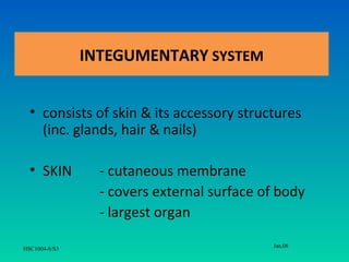
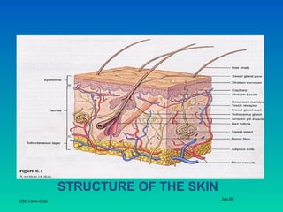
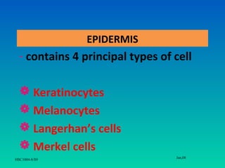
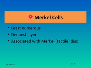
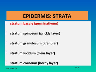
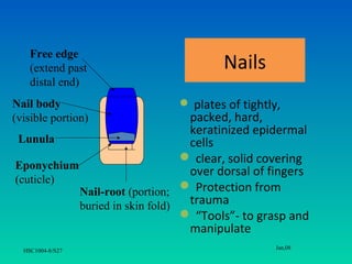
Integumentary System
                  S.S.MOORTHY SEMENCHALAM
               M.Sc. Comm Health (Occ Health) UKM
                       B.HSc. Nursing (Aust)
                        Dip Med Sc. (Moh)


                                                    Jan,08
HSC1004-8/S1
Learning Outcomes
After completing this system, students should be able to:
i. Identify the functions of skin
ii.   List the layers of epidermis and cells that compose them
iii. State the composition of papillary & reticular layers of dermis
iv. Discuss the structure & functions of skin appendages (hair, glands
    and nail)


                                                             Jan,08
HSC1004-8/S2
INTEGUMENTARY SYSTEM


  • consists of skin & its accessory structures
    (inc. glands, hair & nails)

  • SKIN         - cutaneous membrane
                 - covers external surface of body
                 - largest organ
                                            Jan,08
HSC1004-8/S3
SKIN FUNCTIONS
• protection - against invasion of
  microorganisms, water loss and dehydration.
• defense - contains macrophages, lymph nodes
  and other structures which identify pathogens
  and provide first line of defense against them.
• sensation - the skin contains sense organs for
  light touch, pressure, temperature, and pain.


                                        Jan,08
HSC1004-8/S4
Cont.
• secretion - the skin secretes the precursor to
  Vitamin D (this is then activated by processing
  in the liver and kidney), and melanin.
• thermoregulation - by diverting blood into or
  away from the skin the body can release or
  conserve heat.
• excretion – small amount of waste are lost
  through the skin


                                        Jan,08
HSC1004-8/S5
STRUCTURE OF THE SKIN
                                       Jan,08
HSC1004-8/S6
The layers/ strata of the skin….
   EPIDERMIS
      superficial
      thinner portion of epithelial tissue
   DERMIS
     connective tissue
      deeper, thicker
   SUBCUTANEOUS LAYER
     adipose & areolar tissue
                                              Jan,08
HSC1004-8/S7
SUBCUTANEOUS
                  TISSUE


                              Jan,08
HSC1004-8/S8
EPIDERMIS
  - contains 4 principal types of cell

   Keratinocytes
   Melanocytes
   Langerhan’s cells
   Merkel cells
                                  Jan,08
HSC1004-8/S9
 Keratinocytes
•    arranged in 4-5 layers
•    90% of epidermal cells
•    Produce:
(i)  Protein keratin (tough, fibrous)
     - protection
(ii) Lamellar granules
     - water-repellant sealant

                                        Jan,08
HSC1004-8/S10
 Melanocytes

• Produce melanin (brown-black pigment);
  contributes to skin color
• Everyone have same number
• shield DNA from being damaged by UV


                                    Jan,08
HSC1004-8/S11
 Langerhan’s cells

• Originate from bone marrow
• Participate in immune response
• Site of invasion of HIV


                                      Jan,08
HSC1004-8/S12
 Merkel Cells
  • Least numerous
  • Deepest layer
  • Associated with Merkel (tactile) disc


                                     Jan,08
HSC1004-8/S13
EPIDERMIS: STRATA
 stratum basale (germinativum)

 stratum spinosum (prickly layer)

 stratum granulosum (granular)

 stratum lucidum (clear layer)

 stratum corneum (horny layer)
                                     Jan,08
HSC1004-8/S14
 Stratum basale
• deepest layer
• single row of cuboidal/ columnar of actively dividing
  keratinocytes
• melanocytes, Langerhan’s cells & Merkel cells

 Stratum spinosum
• 8- 10 layers of keratinocytes
• projections of melanocytes & langerhan’s cells
                                             Jan,08
HSC1004-8/S15
 Stratum granulosum

  • 3-5 layers of flattened keratinocytes that
    undergo apoptosis
  • degeneration of nuclei and organelles


** apoptosis- genetically programmed cell death


                                                  Jan,08
HSC1004-8/S16
 Stratum lucidum
 present only in skin of fingertips, palms & soles
 3-5 rows of clear, flat, dead keratinocytes

                  Stratum corneum
• 25-30 rows of dead, flat keratinocytes
• continously shed & replaced by cells of deeper
layer
• protection against abrasion and penetration
                                             Jan,08
HSC1004-8/S17
Jan,08
HSC1004-8/S18
DERMIS
• connective tissue; collagen & elastics fibers
• 2 layers
     Papillary layer
       - superficial portion (1/5)
       - loose fibers
       - contains dermal palpillae that house capillaries, corpuscles of touch &
       free nerve endings
     Reticular layer
      - deeper portion (4/5)
      - dense connective tissue
      - spaces between fibers; adipose cells, hair follicles, nerves, sebaceous
        glands
                                                                    Jan,08
  HSC1004-8/S19
SUBCUTANEOUS LAYER
•   Subcutaneous tissue
•   Deep to dermis
•   Storage depot for fat
•   Anchors skin to underlying tissue
•   Blood vessels & nerves ending: Pacinian
    corpuscle (sensitive to pressure)


                                         Jan,08
HSC1004-8/S20
SKIN COLOR
•      3 pigments
(i) Melanin (pale-yellow-tan-black)
(ii) Carotene (yellow-orange)
    - Precursor of Vit. A
    - Subcutaneous & fatty area
(iii) Hemoglobin (red)
    - O2-carrying pigment

                                      Jan,08
HSC1004-8/S21
Melanin
 • different skin color due to amount of
   pigment produce
 • freckles: accumulation of melanin
 • > UV exposure, > melanin production,
      > protective against UV radiation


                                      Jan,08
HSC1004-8/S22
APPENDAGES OF SKIN
                        Hair

                        Nail

                       Glands
                    Sweat glands
                   Sebaceous glands
                                       Jan,08
HSC1004-8/S23
Hair
Functions:
• warmth
• protection (scalp injury, sun)
• shield from foreign particles (e.g. eyelashes)
• filters (nostrils)


                                         Jan,08
HSC1004-8/S24
Structure of the hair

• Shaft – project from surface
• Root- embedded in dermis
• Bulb- base of root; enclosed in hair
  follicle; associated with arrector pili
  muscle
                                         Jan,08
 HSC1004-8/S25
Structure of the hair


                                    Jan,08
HSC1004-8/S26
Free edge
    (extend past                       Nails
    distal end)
Nail body                      plates of tightly,
(visible portion)                packed, hard,
                                 keratinized epidermal
 Lunula                          cells
                                 clear, solid covering
Eponychium
                                 over dorsal of fingers
(cuticle)
           Nail-root (portion;  Protection from
           buried in skin fold)  trauma
                                 “Tools”- to grasp and
                                 manipulate
                                                 Jan,08
  HSC1004-8/S27
Sweat (Sudoriferous) glands
   •       everywhere; except nipples & external
           genitilia
   •       types:
             a. Eccrine sweat glands
             b. Apocrine sweat glands
             c. Ceruminous glands
             d. Mammary glands
                                           Jan,08
HSC1004-8/S28
Eccrine sweat glands
• abundant, with odorless secretion
• most numerous (palms, soles, forehead),
  originate in subcutaneous layer
• produce sweat (99% water)
• excrete salts, vitamin C, antibodies, metabolic
  wastes, lactic acid
• thermoregulation (lower body temperature)
• autonomic nervous system
                                          Jan,08
HSC1004-8/S29
Apocrine sweat glands
   less numerous, secretions that develop odors
   axillary, groin
   ducts empty into hair follicles
   true sweat and fatty acids
   viscous, milky, bacteria food
   active during puberty
   sympathetic nervous system (pain, stress)
                                         Jan,08
HSC1004-8/S30
Ceruminous glands
• modified apocrine glands
• secrete cerumen (ear wax)
• external auditory canal

 Mammary glands
 ■ modified sweat glands
 ■ secrete milk
 ■ breast
                                    Jan,08
HSC1004-8/S31
Sebaceous (oil) glands
• groups of specialized epithelial cells
• secrete oil or sebum
• everywhere, except palms & soles
• usually secrete into hair follicles
• lubricates hair and skin
      - softens dead cells (pliability)
      - slows water loss
      - bactericidal
• stimulated by hormones (androgens)
                                             Jan,08
    HSC1004-8/S32
Wound healing
 events that repairs the skin to its normal (or
 near-normal) structure & function
 2 kinds (depending on depth of injury):
     - EPIDERMAL wound healing
           - affects only epidermis
     - DEEP wound healing
           - penetrates dermis

                                         Jan,08
HSC1004-8/S33
Scar formation- fibrosis
  • Hyperthropic scar
    - scar elevated above normal epidermal
    surface; but within boundaries of original
    wound

  • Keloid scar
    - extend beyond boundaries into the normal
    surrounding tissue

                                                 Jan,08
HSC1004-8/S34
Jan,08
HSC1004-8/S35
Differences between scar and
                 normal skin tissue
 • Collagen fibers more densely arranged in
   scar tissue
 • Less blood vessel, unequal number of hairs,
   skin gland & sensory structure
 • More lighter in color (due to arrangement
   of collagen & scarcity of blood vessel)
                                       Jan,08
HSC1004-8/S36
Thank you….

                Questions please!!

                                 Jan,08
HSC1004-8/S37

4. intergumentary

  • 1.
    Integumentary System S.S.MOORTHY SEMENCHALAM M.Sc. Comm Health (Occ Health) UKM B.HSc. Nursing (Aust) Dip Med Sc. (Moh) Jan,08 HSC1004-8/S1
  • 2.
    Learning Outcomes After completingthis system, students should be able to: i. Identify the functions of skin ii. List the layers of epidermis and cells that compose them iii. State the composition of papillary & reticular layers of dermis iv. Discuss the structure & functions of skin appendages (hair, glands and nail) Jan,08 HSC1004-8/S2
  • 3.
    INTEGUMENTARY SYSTEM • consists of skin & its accessory structures (inc. glands, hair & nails) • SKIN - cutaneous membrane - covers external surface of body - largest organ Jan,08 HSC1004-8/S3
  • 4.
    SKIN FUNCTIONS • protection- against invasion of microorganisms, water loss and dehydration. • defense - contains macrophages, lymph nodes and other structures which identify pathogens and provide first line of defense against them. • sensation - the skin contains sense organs for light touch, pressure, temperature, and pain. Jan,08 HSC1004-8/S4
  • 5.
    Cont. • secretion -the skin secretes the precursor to Vitamin D (this is then activated by processing in the liver and kidney), and melanin. • thermoregulation - by diverting blood into or away from the skin the body can release or conserve heat. • excretion – small amount of waste are lost through the skin Jan,08 HSC1004-8/S5
  • 6.
    STRUCTURE OF THESKIN Jan,08 HSC1004-8/S6
  • 7.
    The layers/ strataof the skin…. EPIDERMIS  superficial  thinner portion of epithelial tissue DERMIS connective tissue  deeper, thicker SUBCUTANEOUS LAYER adipose & areolar tissue Jan,08 HSC1004-8/S7
  • 8.
    SUBCUTANEOUS TISSUE Jan,08 HSC1004-8/S8
  • 9.
    EPIDERMIS -contains 4 principal types of cell  Keratinocytes  Melanocytes  Langerhan’s cells  Merkel cells Jan,08 HSC1004-8/S9
  • 10.
     Keratinocytes • arranged in 4-5 layers • 90% of epidermal cells • Produce: (i) Protein keratin (tough, fibrous) - protection (ii) Lamellar granules - water-repellant sealant Jan,08 HSC1004-8/S10
  • 11.
     Melanocytes • Producemelanin (brown-black pigment); contributes to skin color • Everyone have same number • shield DNA from being damaged by UV Jan,08 HSC1004-8/S11
  • 12.
     Langerhan’s cells •Originate from bone marrow • Participate in immune response • Site of invasion of HIV Jan,08 HSC1004-8/S12
  • 13.
     Merkel Cells • Least numerous • Deepest layer • Associated with Merkel (tactile) disc Jan,08 HSC1004-8/S13
  • 14.
    EPIDERMIS: STRATA  stratumbasale (germinativum)  stratum spinosum (prickly layer)  stratum granulosum (granular)  stratum lucidum (clear layer)  stratum corneum (horny layer) Jan,08 HSC1004-8/S14
  • 15.
     Stratum basale •deepest layer • single row of cuboidal/ columnar of actively dividing keratinocytes • melanocytes, Langerhan’s cells & Merkel cells  Stratum spinosum • 8- 10 layers of keratinocytes • projections of melanocytes & langerhan’s cells Jan,08 HSC1004-8/S15
  • 16.
     Stratum granulosum • 3-5 layers of flattened keratinocytes that undergo apoptosis • degeneration of nuclei and organelles ** apoptosis- genetically programmed cell death Jan,08 HSC1004-8/S16
  • 17.
     Stratum lucidum present only in skin of fingertips, palms & soles  3-5 rows of clear, flat, dead keratinocytes  Stratum corneum • 25-30 rows of dead, flat keratinocytes • continously shed & replaced by cells of deeper layer • protection against abrasion and penetration Jan,08 HSC1004-8/S17
  • 18.
  • 19.
    DERMIS • connective tissue;collagen & elastics fibers • 2 layers  Papillary layer - superficial portion (1/5) - loose fibers - contains dermal palpillae that house capillaries, corpuscles of touch & free nerve endings  Reticular layer - deeper portion (4/5) - dense connective tissue - spaces between fibers; adipose cells, hair follicles, nerves, sebaceous glands Jan,08 HSC1004-8/S19
  • 20.
    SUBCUTANEOUS LAYER • Subcutaneous tissue • Deep to dermis • Storage depot for fat • Anchors skin to underlying tissue • Blood vessels & nerves ending: Pacinian corpuscle (sensitive to pressure) Jan,08 HSC1004-8/S20
  • 21.
    SKIN COLOR • 3 pigments (i) Melanin (pale-yellow-tan-black) (ii) Carotene (yellow-orange) - Precursor of Vit. A - Subcutaneous & fatty area (iii) Hemoglobin (red) - O2-carrying pigment Jan,08 HSC1004-8/S21
  • 22.
    Melanin • differentskin color due to amount of pigment produce • freckles: accumulation of melanin • > UV exposure, > melanin production, > protective against UV radiation Jan,08 HSC1004-8/S22
  • 23.
    APPENDAGES OF SKIN  Hair  Nail  Glands  Sweat glands  Sebaceous glands Jan,08 HSC1004-8/S23
  • 24.
    Hair Functions: • warmth • protection(scalp injury, sun) • shield from foreign particles (e.g. eyelashes) • filters (nostrils) Jan,08 HSC1004-8/S24
  • 25.
    Structure of thehair • Shaft – project from surface • Root- embedded in dermis • Bulb- base of root; enclosed in hair follicle; associated with arrector pili muscle Jan,08 HSC1004-8/S25
  • 26.
    Structure of thehair Jan,08 HSC1004-8/S26
  • 27.
    Free edge (extend past Nails distal end) Nail body  plates of tightly, (visible portion) packed, hard, keratinized epidermal Lunula cells  clear, solid covering Eponychium over dorsal of fingers (cuticle) Nail-root (portion;  Protection from buried in skin fold) trauma  “Tools”- to grasp and manipulate Jan,08 HSC1004-8/S27
  • 28.
    Sweat (Sudoriferous) glands • everywhere; except nipples & external genitilia • types: a. Eccrine sweat glands b. Apocrine sweat glands c. Ceruminous glands d. Mammary glands Jan,08 HSC1004-8/S28
  • 29.
    Eccrine sweat glands •abundant, with odorless secretion • most numerous (palms, soles, forehead), originate in subcutaneous layer • produce sweat (99% water) • excrete salts, vitamin C, antibodies, metabolic wastes, lactic acid • thermoregulation (lower body temperature) • autonomic nervous system Jan,08 HSC1004-8/S29
  • 30.
    Apocrine sweat glands  less numerous, secretions that develop odors  axillary, groin  ducts empty into hair follicles  true sweat and fatty acids  viscous, milky, bacteria food  active during puberty  sympathetic nervous system (pain, stress) Jan,08 HSC1004-8/S30
  • 31.
    Ceruminous glands • modifiedapocrine glands • secrete cerumen (ear wax) • external auditory canal Mammary glands ■ modified sweat glands ■ secrete milk ■ breast Jan,08 HSC1004-8/S31
  • 32.
    Sebaceous (oil) glands •groups of specialized epithelial cells • secrete oil or sebum • everywhere, except palms & soles • usually secrete into hair follicles • lubricates hair and skin - softens dead cells (pliability) - slows water loss - bactericidal • stimulated by hormones (androgens) Jan,08 HSC1004-8/S32
  • 33.
    Wound healing  eventsthat repairs the skin to its normal (or near-normal) structure & function  2 kinds (depending on depth of injury): - EPIDERMAL wound healing - affects only epidermis - DEEP wound healing - penetrates dermis Jan,08 HSC1004-8/S33
  • 34.
    Scar formation- fibrosis • Hyperthropic scar - scar elevated above normal epidermal surface; but within boundaries of original wound • Keloid scar - extend beyond boundaries into the normal surrounding tissue Jan,08 HSC1004-8/S34
  • 35.
  • 36.
    Differences between scarand normal skin tissue • Collagen fibers more densely arranged in scar tissue • Less blood vessel, unequal number of hairs, skin gland & sensory structure • More lighter in color (due to arrangement of collagen & scarcity of blood vessel) Jan,08 HSC1004-8/S36
  • 37.
    Thank you…. Questions please!! Jan,08 HSC1004-8/S37