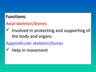
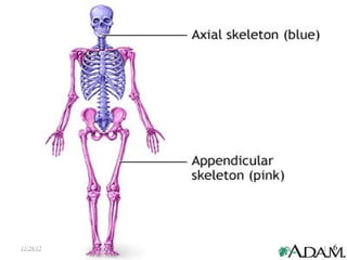
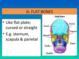
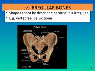
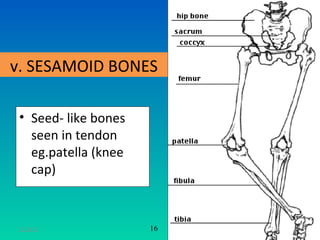
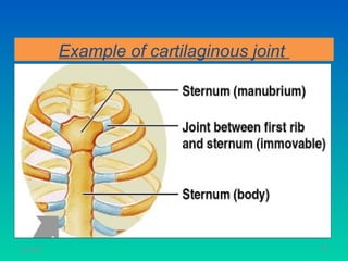
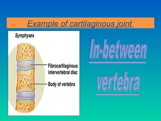
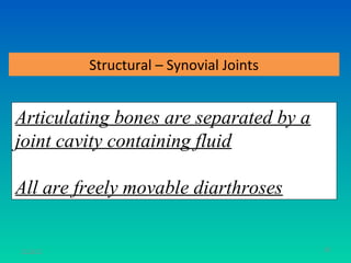
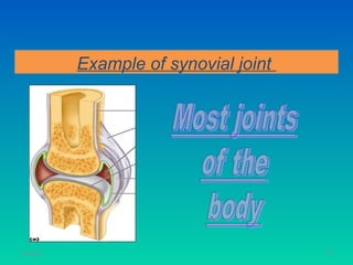
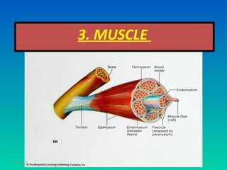
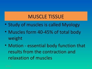
SKELETAL MUSCLE
               S.S.MOORTHY SEMENCHALAM
            M.Sc. Comm Health (Occ Health) UKM
                    B.HSc. Nursing (Aust)
                     Dip Med Sc. (Moh)


11/26/12                                         1
Learning Objective
At the end of these session students should be able to:
• State the 2 classifications of bones
• List the functions of bones
• Name the types of bones
• List the composition of bone
• List the types of joints
• Described 3 types of muscle tissue
• State the characteristics & structure of muscle tissue
• State the relations of muscle and movement

11/26/12                                                   2
1. BONE
           Axial Skeleton – forms the long axis of the body
               eg. bones of the skull, vertebral column,
                    rib cage, hyoid bone


                         Classification of Bones

           Appendicular Skeleton – relating to an appendage, 126 bones
              eg. bones of the upper and lower limbs,
              shoulder girdle, hip girdle
11/26/12                                                                 3   4
Functions:
Axial skeleton/bones
 Involved in protecting and supporting of
   the body and organs
Appendicular skeleton/bones
 Help in movement


11/26/12                                 4   5
11/26/12   5   6
Functions of Bones
• Support – forms the framework (central axis
  of body); gives shape, supports the body
• Protection – provide protective case for brain,
  spinal cord & organs ( heart & lungs)
• Movement – provide levers for muscle
• Mineral storage – reservoir for minerals; e.g.
  calcium & phosphorus
• Blood cell formation – haematopoiesis occurs
  within the marrow cavities of bones
• Attachment for muscles & tendons
11/26/12                                            6   7
Number of Bones
There are 206 bones in our whole body
• Upper limbs - 64
• Lower limbs - 62
• Vertebrae - 33 ( C7,T12,L5,S5,CO4 )
• Skull       - 22
• Ribs        - 24
• Sternum      -1
11/26/12                                7   8
Long
                        bones


  Sesamoid                                Short
    bones               Types             bones
                       of bones


           Irregular               Flat
             bones                bones

11/26/12                                          8   9
11/26/12   9 10
i. LONG BONES

• Bones of the extremities
• Cylindrically in shape (shaft) with
  expanded end
• E.g. femur,humerus, tibia, fibula

11/26/12                                10   11
11/26/12   11   12
ii. SHORT LONG BONES & SHORT BONES

• Describes the bones of the hand & foot
• E.g. metatarsal, metacarpal,carpals, tarsals


11/26/12                                         13   13
iii. FLAT BONES

• Like flat plate;
  curved or straight
• E.g. sternum,
  scapula & parietal


 11/26/12                      14   14
iv. IRREGULAR BONES
• Shape cannot be described because it is irregular
• E.g. vertebrae, pelvic bone


11/26/12                                          15 15
v. SESAMOID BONES

 • Seed- like bones
   seen in tendon
   eg.patella (knee
   cap)


 11/26/12             16   17
GROSS ANATOMY OF BONES

• External features of bones – projections,
  depression & openings
  - as sites for muscle, ligament & tendon
  attachment
  - as surface joints
  - conduits for blood vessels & nerves


11/26/12                                      18 17
Internal features:
• Compact bone –
  dense outer layer &
  hard
• Spongy bone less
  hard (cancellous
  bone) – honeycomb
  of trabeculae filled
  with bone marrow


11/26/12                 19 18
Structure of Typical Long Bone
• Diaphysis
  - tubular shaft; forming
   the axis of long bones
   - composed of compact
   bone + central medullary
   canal (houses fatty yellow
   bone marrow)
• Epiphysis
   - expanded end of long
   bones
   - spongy bones inside
11/26/12                                     19
                                            20
Con`t
  Thin layer of articular
   cartilage covers end of
   epiphysis
  Long bone that are still
   growing has an
   epiphyseal plate or
   growth plate
  When the bone growth
   stops, the epiphyseal
   plate is replaced by
   bone- epiphyseal line
11/26/12                      21   20
Structure of Typical Long Bone:
                     Periosteum
                           double-layered
                            protective membrane
                           supplied with nerve
                            fibers, blood, and
                            lymphatic vessels
                            entering the bone via
                            nutrient foramina
                           Secured to underlying
                            bone by Sharpey’s
                            fibers (collagen)
11/26/12                                        22   21
AXIAL SKELETON

           1. Skull
           2. Vertebral column
           3. Thoracic cage


11/26/12                         23   28
1. Skull

• Consist of 2 parts
  1. Cranium – contain the brain
  2. The face
• Fused/ joined bones – not moveable
• Mandible bone – moveable


11/26/12                               24 29
Consist of 11 bones   5. Sphenoid bone
1. Frontal bone       6. Nasal bone
2. Parietal bone      7. Lacrimal bone
3. Temporal bone      8. Ethmoid bone
4. Occipital bone     9. Zygomatic bone
                      10.Maxilla bone
                      11.Mandible bone

 11/26/12                                 25 30
Frontal
             Nasal
                        Parietal
                      Temporal

zygomatic              occipital


                     Mandible
                     Maxilla
  11/26/12                     26   31
Function of the skull
1. Cranial bones protect the brain
2. Bone eye sockets protect the eye
3. Temporal bone protects the ear
4. Gives resonance of voice due to the air filled
   cavities called sinuses
5. Keeps the air passage open to facilitate
   breathing
11/26/12                                            27   32
2. Vertebral Column
•       Consist of 24 movable bones
•       Sacrum & coccyx
•       Separated by intervertebral discs (cartilage)
      i. 7 cervical
      ii. 12 thoracic
      iii. 5 lumbar
      iv. 5 sacrum
      v. 4 coccyx

11/26/12                                                28   33
11/26/12   29 34
Functions of vertebral column
1. Protects the spinal cord.
2. Houses 31 pairs of spinal nerves going
   through the foramina
3. Vertebrae articulate with the ribs, forming
   joints which move during respiration.

 11/26/12                                   30 35
3. Thoracic cage

• Formed by 12 thoracic vertebrae
• 12 pairs of ribs
• 1 sternum/ breast bone
• The ribs are attached to the sternum by costal
  cartilage
• Two pairs of ribs (rib 11 & 12) –called as floating ribs-
  no anterior attachment
11/26/12                                                  31   36
Functions of thoracic cage
1) Protects heart, lungs, major blood vessels
2) Forms joints between the upper limbs and the
   axial skeleton.
3) Manubrium (upper part of sternum),
   articulates with the clavicle bones
4) Gives attachment to the respiratory muscles
   to enables respiration

11/26/12                                     32 37
The Sternum (breast bone )


11/26/12                                33
                                         38
APPENDICULAR SKELETON

1. Shoulder girdle and the upper limb/extremity
2. Pelvis girdle and the lower limb/extremity
3. Arches of the foot


11/26/12                                     34 39
11/26/12   35   40
Ligaments

• A band of fibrous tissue that connects bones
  or cartilages
• Function: support and strengthen joints


11/26/12                                          43
                                                 36
11/26/12   37   44
2. JOINTS


11/26/12               38
Joints = Articulations

Articulation – site where two or more
bones meet

Two Fundamental Functions of Joints:
    Allow the skeleton to have mobility
    Hold the skeleton together

11/26/12                                  39
Classification of Joints: Structural


                  Fibrous
                Cartilaginous
                  Synovial
11/26/12                                          40
Classification of Joints: Functional

Fibrous joints – immovable
Cartilaginous joints – slightly movable
Synovial joints – freely movable


 11/26/12                                          41
Structural - Fibrous Joints


-The bones are joined by fibrous tissues

- No joint cavity

- Most are immovable (synarthroses)

11/26/12                                 42
Example of fibrous joint


11/26/12                              43
Example of fibrous joint


11/26/12                              44
Example of fibrous joint


11/26/12                              45
Structural – Cartilaginous Joints


- Articulating bones are joined by
cartilage
- No joint cavity

11/26/12                                       46
Example of cartilaginous joint


11/26/12                                    47
Example of cartilaginous joint


11/26/12                                    48
Example of cartilaginous joint


11/26/12                                    49
Structural – Synovial Joints


Articulating bones are separated by a
joint cavity containing fluid

All are freely movable diarthroses


11/26/12                                  50
Example of synovial joint


11/26/12                               51
Synovial Joints: General Structures


11/26/12                                         52
Synovial Joints: Friction-Reducing Structures


11/26/12                                                   53
Synovial Joints: Range of Motion

    Nonaxial – slipping movements only
    Uniaxial – movement in one plane
    Biaxial – movement in two planes
    Multiaxial – movement around all planes

           Three General Types of Movements:

                 Gliding (slipping)
                      Angular
                     Rotation
11/26/12                                       54
Gliding (slipping) movements

One flat bone surface glides or slips over another bone


11/26/12                                            55
Angular movements


 Involve change of angle between bones


11/26/12                             56
Example of angular movement


 Flexion
- bending movement
that decreases the
angle of the joint
11/26/12                                 57
Example of angular movement
 Extension
-straightening movement that increases
the angle of the joint


11/26/12                                 58
Example of angular movement


     Dorsiflexion &
    Plantar flexion
    - Up and down
    movement of the
      foot
11/26/12                                 59
Example of angular movement
• Abduction
  — movement of a limb away from the midline or
  median plane

• Adduction
  — movement of a limb toward the midline or
  median plane

• Circumduction
  — movement of a limb describing a cone in space
11/26/12                                            60
Rotation movements


Turning of a bone around its own axis

           Example:
           Hip and shoulder joints

11/26/12                                61
Special Movements
                 Supination and Pronation
           – movements of radius around the ulna –


11/26/12                                             62
Special Movements


Inversion and Eversion


                         Protraction and Retraction

11/26/12                                       63
Special Movements


Elevation and
Depression


                            Opposition


 11/26/12                                64
Types of Synovial Joints-Diarthrotic

                  Six Major Categories:


              Plane             Condyloid
              Hinge              Saddle
              Pivot         Ball-and-Socket


11/26/12                                          65
Types of Synovial Joints - Hinge
  Motion is along a single plane (only permits
  flexion and extension)

  Resembles action of a hinge

  Examples:
  elbow joint
  knee joint                                     66
11/26/12
Ball & socket


                      Example :
                     SHOULDER JOINT
                     HIP JOINT


11/26/12                          67
Condyloid
• Oval shaped condyle

• Angular motion but not rotation

Example : Occipital condyles with atlas;
         metacarpals & metatarsals with
           palanges

11/26/12                                   68
Saddle
• Articulating surfaces of both bones (concave &
  convex regions)
• Permits wide range of movement

Example:
       Carpometacarpal joint of thump is
         the only saddle joint in the body

11/26/12                                       69
Pivot

• Rounded or conical surface of one bone fits
  into a ring of bone or tendon
• For rotation

 Example:
        Joint between the atlas and axis;
        proximal radioulnar joint
11/26/12                                        70
Gliding/Plane
• Flat or lightly curved surfaces
• Moving against each other
• Sliding or twisting without circular movement

Example: Between carpals (wrist) &
         tarsals ( ankle)

11/26/12                                          71
3. MUSCLE


11/26/12               72
MUSCLE TISSUE
• Study of muscles is called Myology
• Muscles form 40-45% of total body
  weight
• Motion - essential body function that
  results from the contraction and
  relaxation of muscles
11/26/12                                  73
Cont..
• Posture maintaining both during rest and at activity
• Heat generated by skeletal muscle contraction
  produce 80% of all the body heat
• Beating of heart is specific function of cardiac muscle
• Smooth muscle throughout body help in movement
  of secretions, waste products, etc.


11/26/12                                                74
TYPE OF MUSCLE TISSUE

• 3 Types of muscle in the body
  - skeletal muscle
  - cardiac muscle
  - smooth muscle


11/26/12                           75
Types- skeletal muscle
• 40 % of body weight
• It is voluntary as it can be made to contract and
  relax at conscious level
• Also called as striated muscle
• Eg: diaphragm is structurally voluntary but it
  works automatically with breathing
• Upper part of esophagus is voluntary but is not
  under conscious control
11/26/12                                          76
Skeletal muscle tissue
Over 600 muscle in body
Skeletal muscle tissue has three
  components (epimysium, perimysium,
  endomysium)
- Connective tissue component
- Nerve and blood supply
- Muscle tissue proper
11/26/12                               77
11/26/12   78
11/26/12   79
Blood supply
• Skeletal muscle fibers are well supplied by
  nerve and vessels which help in contraction,
  supply energy and transfer of waste products
• Generally an artery and two veins accompany
  the nerve that penetrate skeletal muscle


11/26/12                                         80
Skeletal muscle and movement
• It produces movement by exerting force
  on tendons which in turn pulls on bones
  or other structures like skin

• They cross at least one joint and are
  attached to articulating bone that form
  joint.
• Abundant blood & nerve supply
11/26/12                                    81
Group actions
- Skeletal muscle act in groups
- They are arrange in opposing pairs in joints
  like flexor and extensors
- Muscle that produce desired movement is
  called prime movers / agonist
- Muscle moving opposite are relaxing are
  antagonist
11/26/12                                         82
11/26/12   83
• MOST SKELETAL MUSCLES WORK IN PAIRS.

           • When one muscle or set of muscles CONTRACTS,
             the other RELAXES.


11/26/12                                                84
• The Muscles of the upper arm are a good
example of this dual action:
ANTAGONISTIC MUSCLES.

FLEXOR, A MUSCLE THAT BENDS A
JOINT.
EXTENSOR, A MUSCLE THAT
STRAIGHTENS A JOINT.
11/26/12                                    85
A. When the BICEPS CONTRACTS, it
   FLEXES the elbow joint

 B. When the TRICEPS CONTRACTS, it
   EXTENDS the elbow joint

 C. A controlled movement requires
  contraction by both muscles.
11/26/12                             86
SMOOTH MUSCLE
•    Smooth muscle cell are small
•    Not striated
•    Contract more slowly
•    Is under involuntary control. And they to
     function as a unit and contract at the same
     time. e.g intestine


11/26/12                                           87
CARDIAC MUSCLE

• Share some characteristic with both smooth
  and skeletal muscle
• Is under involuntary control and are
  influenced by hormon


11/26/12                                       88
11/26/12   89
Thank you
           Questions please


11/26/12                      90

5. skeletal muscle system

  • 1.
    SKELETAL MUSCLE S.S.MOORTHY SEMENCHALAM M.Sc. Comm Health (Occ Health) UKM B.HSc. Nursing (Aust) Dip Med Sc. (Moh) 11/26/12 1
  • 2.
    Learning Objective At theend of these session students should be able to: • State the 2 classifications of bones • List the functions of bones • Name the types of bones • List the composition of bone • List the types of joints • Described 3 types of muscle tissue • State the characteristics & structure of muscle tissue • State the relations of muscle and movement 11/26/12 2
  • 3.
    1. BONE Axial Skeleton – forms the long axis of the body eg. bones of the skull, vertebral column, rib cage, hyoid bone Classification of Bones Appendicular Skeleton – relating to an appendage, 126 bones eg. bones of the upper and lower limbs, shoulder girdle, hip girdle 11/26/12 3 4
  • 4.
    Functions: Axial skeleton/bones  Involvedin protecting and supporting of the body and organs Appendicular skeleton/bones  Help in movement 11/26/12 4 5
  • 5.
  • 6.
    Functions of Bones •Support – forms the framework (central axis of body); gives shape, supports the body • Protection – provide protective case for brain, spinal cord & organs ( heart & lungs) • Movement – provide levers for muscle • Mineral storage – reservoir for minerals; e.g. calcium & phosphorus • Blood cell formation – haematopoiesis occurs within the marrow cavities of bones • Attachment for muscles & tendons 11/26/12 6 7
  • 7.
    Number of Bones Thereare 206 bones in our whole body • Upper limbs - 64 • Lower limbs - 62 • Vertebrae - 33 ( C7,T12,L5,S5,CO4 ) • Skull - 22 • Ribs - 24 • Sternum -1 11/26/12 7 8
  • 8.
    Long bones Sesamoid Short bones Types bones of bones Irregular Flat bones bones 11/26/12 8 9
  • 9.
  • 10.
    i. LONG BONES •Bones of the extremities • Cylindrically in shape (shaft) with expanded end • E.g. femur,humerus, tibia, fibula 11/26/12 10 11
  • 11.
    11/26/12 11 12
  • 13.
    ii. SHORT LONGBONES & SHORT BONES • Describes the bones of the hand & foot • E.g. metatarsal, metacarpal,carpals, tarsals 11/26/12 13 13
  • 14.
    iii. FLAT BONES •Like flat plate; curved or straight • E.g. sternum, scapula & parietal 11/26/12 14 14
  • 15.
    iv. IRREGULAR BONES •Shape cannot be described because it is irregular • E.g. vertebrae, pelvic bone 11/26/12 15 15
  • 17.
    v. SESAMOID BONES • Seed- like bones seen in tendon eg.patella (knee cap) 11/26/12 16 17
  • 18.
    GROSS ANATOMY OFBONES • External features of bones – projections, depression & openings - as sites for muscle, ligament & tendon attachment - as surface joints - conduits for blood vessels & nerves 11/26/12 18 17
  • 19.
    Internal features: • Compactbone – dense outer layer & hard • Spongy bone less hard (cancellous bone) – honeycomb of trabeculae filled with bone marrow 11/26/12 19 18
  • 20.
    Structure of TypicalLong Bone • Diaphysis - tubular shaft; forming the axis of long bones - composed of compact bone + central medullary canal (houses fatty yellow bone marrow) • Epiphysis - expanded end of long bones - spongy bones inside 11/26/12 19 20
  • 21.
    Con`t Thinlayer of articular cartilage covers end of epiphysis Long bone that are still growing has an epiphyseal plate or growth plate When the bone growth stops, the epiphyseal plate is replaced by bone- epiphyseal line 11/26/12 21 20
  • 22.
    Structure of TypicalLong Bone: Periosteum double-layered protective membrane supplied with nerve fibers, blood, and lymphatic vessels entering the bone via nutrient foramina Secured to underlying bone by Sharpey’s fibers (collagen) 11/26/12 22 21
  • 23.
    AXIAL SKELETON 1. Skull 2. Vertebral column 3. Thoracic cage 11/26/12 23 28
  • 24.
    1. Skull • Consistof 2 parts 1. Cranium – contain the brain 2. The face • Fused/ joined bones – not moveable • Mandible bone – moveable 11/26/12 24 29
  • 25.
    Consist of 11bones 5. Sphenoid bone 1. Frontal bone 6. Nasal bone 2. Parietal bone 7. Lacrimal bone 3. Temporal bone 8. Ethmoid bone 4. Occipital bone 9. Zygomatic bone 10.Maxilla bone 11.Mandible bone 11/26/12 25 30
  • 26.
    Frontal Nasal Parietal Temporal zygomatic occipital Mandible Maxilla 11/26/12 26 31
  • 27.
    Function of theskull 1. Cranial bones protect the brain 2. Bone eye sockets protect the eye 3. Temporal bone protects the ear 4. Gives resonance of voice due to the air filled cavities called sinuses 5. Keeps the air passage open to facilitate breathing 11/26/12 27 32
  • 28.
    2. Vertebral Column • Consist of 24 movable bones • Sacrum & coccyx • Separated by intervertebral discs (cartilage) i. 7 cervical ii. 12 thoracic iii. 5 lumbar iv. 5 sacrum v. 4 coccyx 11/26/12 28 33
  • 29.
    11/26/12 29 34
  • 30.
    Functions of vertebralcolumn 1. Protects the spinal cord. 2. Houses 31 pairs of spinal nerves going through the foramina 3. Vertebrae articulate with the ribs, forming joints which move during respiration. 11/26/12 30 35
  • 31.
    3. Thoracic cage •Formed by 12 thoracic vertebrae • 12 pairs of ribs • 1 sternum/ breast bone • The ribs are attached to the sternum by costal cartilage • Two pairs of ribs (rib 11 & 12) –called as floating ribs- no anterior attachment 11/26/12 31 36
  • 32.
    Functions of thoraciccage 1) Protects heart, lungs, major blood vessels 2) Forms joints between the upper limbs and the axial skeleton. 3) Manubrium (upper part of sternum), articulates with the clavicle bones 4) Gives attachment to the respiratory muscles to enables respiration 11/26/12 32 37
  • 33.
    The Sternum (breastbone ) 11/26/12 33 38
  • 34.
    APPENDICULAR SKELETON 1. Shouldergirdle and the upper limb/extremity 2. Pelvis girdle and the lower limb/extremity 3. Arches of the foot 11/26/12 34 39
  • 35.
    11/26/12 35 40
  • 36.
    Ligaments • A bandof fibrous tissue that connects bones or cartilages • Function: support and strengthen joints 11/26/12 43 36
  • 37.
    11/26/12 37 44
  • 38.
  • 39.
    Joints = Articulations Articulation– site where two or more bones meet Two Fundamental Functions of Joints: Allow the skeleton to have mobility Hold the skeleton together 11/26/12 39
  • 40.
    Classification of Joints:Structural Fibrous Cartilaginous Synovial 11/26/12 40
  • 41.
    Classification of Joints:Functional Fibrous joints – immovable Cartilaginous joints – slightly movable Synovial joints – freely movable 11/26/12 41
  • 42.
    Structural - FibrousJoints -The bones are joined by fibrous tissues - No joint cavity - Most are immovable (synarthroses) 11/26/12 42
  • 43.
    Example of fibrousjoint 11/26/12 43
  • 44.
    Example of fibrousjoint 11/26/12 44
  • 45.
    Example of fibrousjoint 11/26/12 45
  • 46.
    Structural – CartilaginousJoints - Articulating bones are joined by cartilage - No joint cavity 11/26/12 46
  • 47.
    Example of cartilaginousjoint 11/26/12 47
  • 48.
    Example of cartilaginousjoint 11/26/12 48
  • 49.
    Example of cartilaginousjoint 11/26/12 49
  • 50.
    Structural – SynovialJoints Articulating bones are separated by a joint cavity containing fluid All are freely movable diarthroses 11/26/12 50
  • 51.
    Example of synovialjoint 11/26/12 51
  • 52.
    Synovial Joints: GeneralStructures 11/26/12 52
  • 53.
    Synovial Joints: Friction-ReducingStructures 11/26/12 53
  • 54.
    Synovial Joints: Rangeof Motion Nonaxial – slipping movements only Uniaxial – movement in one plane Biaxial – movement in two planes Multiaxial – movement around all planes Three General Types of Movements: Gliding (slipping) Angular Rotation 11/26/12 54
  • 55.
    Gliding (slipping) movements Oneflat bone surface glides or slips over another bone 11/26/12 55
  • 56.
    Angular movements Involvechange of angle between bones 11/26/12 56
  • 57.
    Example of angularmovement Flexion - bending movement that decreases the angle of the joint 11/26/12 57
  • 58.
    Example of angularmovement Extension -straightening movement that increases the angle of the joint 11/26/12 58
  • 59.
    Example of angularmovement Dorsiflexion & Plantar flexion - Up and down movement of the foot 11/26/12 59
  • 60.
    Example of angularmovement • Abduction — movement of a limb away from the midline or median plane • Adduction — movement of a limb toward the midline or median plane • Circumduction — movement of a limb describing a cone in space 11/26/12 60
  • 61.
    Rotation movements Turning ofa bone around its own axis Example: Hip and shoulder joints 11/26/12 61
  • 62.
    Special Movements Supination and Pronation – movements of radius around the ulna – 11/26/12 62
  • 63.
    Special Movements Inversion andEversion Protraction and Retraction 11/26/12 63
  • 64.
  • 65.
    Types of SynovialJoints-Diarthrotic Six Major Categories: Plane Condyloid Hinge Saddle Pivot Ball-and-Socket 11/26/12 65
  • 66.
    Types of SynovialJoints - Hinge Motion is along a single plane (only permits flexion and extension) Resembles action of a hinge Examples: elbow joint knee joint 66 11/26/12
  • 67.
    Ball & socket Example : SHOULDER JOINT HIP JOINT 11/26/12 67
  • 68.
    Condyloid • Oval shapedcondyle • Angular motion but not rotation Example : Occipital condyles with atlas; metacarpals & metatarsals with palanges 11/26/12 68
  • 69.
    Saddle • Articulating surfacesof both bones (concave & convex regions) • Permits wide range of movement Example: Carpometacarpal joint of thump is the only saddle joint in the body 11/26/12 69
  • 70.
    Pivot • Rounded orconical surface of one bone fits into a ring of bone or tendon • For rotation Example: Joint between the atlas and axis; proximal radioulnar joint 11/26/12 70
  • 71.
    Gliding/Plane • Flat orlightly curved surfaces • Moving against each other • Sliding or twisting without circular movement Example: Between carpals (wrist) & tarsals ( ankle) 11/26/12 71
  • 72.
  • 73.
    MUSCLE TISSUE • Studyof muscles is called Myology • Muscles form 40-45% of total body weight • Motion - essential body function that results from the contraction and relaxation of muscles 11/26/12 73
  • 74.
    Cont.. • Posture maintainingboth during rest and at activity • Heat generated by skeletal muscle contraction produce 80% of all the body heat • Beating of heart is specific function of cardiac muscle • Smooth muscle throughout body help in movement of secretions, waste products, etc. 11/26/12 74
  • 75.
    TYPE OF MUSCLETISSUE • 3 Types of muscle in the body - skeletal muscle - cardiac muscle - smooth muscle 11/26/12 75
  • 76.
    Types- skeletal muscle •40 % of body weight • It is voluntary as it can be made to contract and relax at conscious level • Also called as striated muscle • Eg: diaphragm is structurally voluntary but it works automatically with breathing • Upper part of esophagus is voluntary but is not under conscious control 11/26/12 76
  • 77.
    Skeletal muscle tissue Over600 muscle in body Skeletal muscle tissue has three components (epimysium, perimysium, endomysium) - Connective tissue component - Nerve and blood supply - Muscle tissue proper 11/26/12 77
  • 78.
  • 79.
  • 80.
    Blood supply • Skeletalmuscle fibers are well supplied by nerve and vessels which help in contraction, supply energy and transfer of waste products • Generally an artery and two veins accompany the nerve that penetrate skeletal muscle 11/26/12 80
  • 81.
    Skeletal muscle andmovement • It produces movement by exerting force on tendons which in turn pulls on bones or other structures like skin • They cross at least one joint and are attached to articulating bone that form joint. • Abundant blood & nerve supply 11/26/12 81
  • 82.
    Group actions - Skeletalmuscle act in groups - They are arrange in opposing pairs in joints like flexor and extensors - Muscle that produce desired movement is called prime movers / agonist - Muscle moving opposite are relaxing are antagonist 11/26/12 82
  • 83.
  • 84.
    • MOST SKELETALMUSCLES WORK IN PAIRS. • When one muscle or set of muscles CONTRACTS, the other RELAXES. 11/26/12 84
  • 85.
    • The Musclesof the upper arm are a good example of this dual action: ANTAGONISTIC MUSCLES. FLEXOR, A MUSCLE THAT BENDS A JOINT. EXTENSOR, A MUSCLE THAT STRAIGHTENS A JOINT. 11/26/12 85
  • 86.
    A. When theBICEPS CONTRACTS, it FLEXES the elbow joint B. When the TRICEPS CONTRACTS, it EXTENDS the elbow joint C. A controlled movement requires contraction by both muscles. 11/26/12 86
  • 87.
    SMOOTH MUSCLE • Smooth muscle cell are small • Not striated • Contract more slowly • Is under involuntary control. And they to function as a unit and contract at the same time. e.g intestine 11/26/12 87
  • 88.
    CARDIAC MUSCLE • Sharesome characteristic with both smooth and skeletal muscle • Is under involuntary control and are influenced by hormon 11/26/12 88
  • 89.
  • 90.
    Thank you Questions please 11/26/12 90