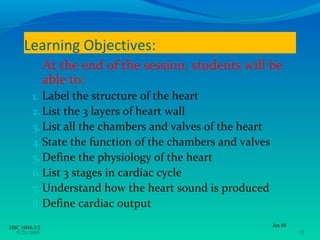
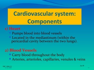
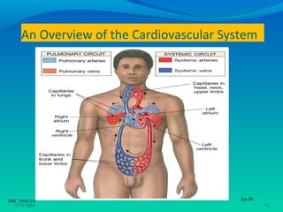
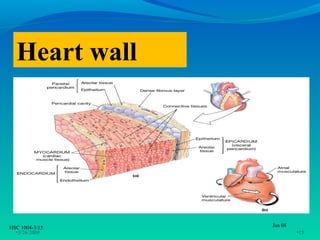
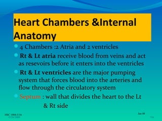
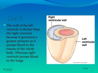
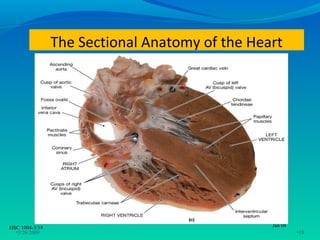
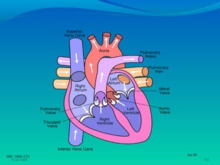
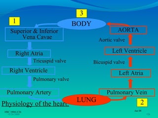
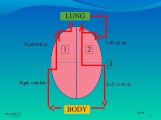
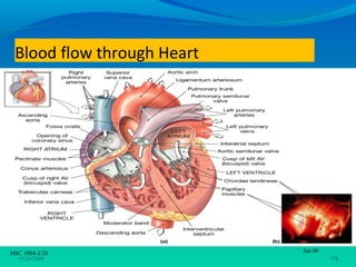
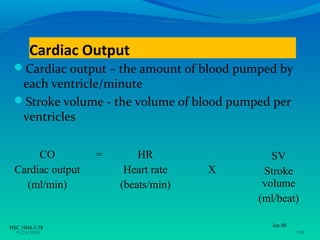
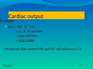
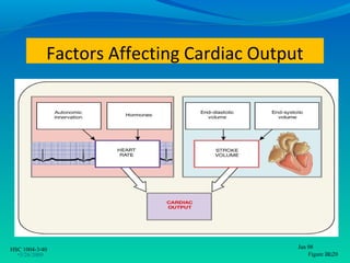
CARDIOVASCULAR SYSTEM
               (Anatomy & Physiology)

                     S.S.MOORTHY SEMENCHALAM
                  M.Sc. Comm Health (Occ Health) UKM
                          B.HSc. Nursing (Aust)
                           Dip Med Sc. (Moh)


HSC 1004-3/1                                           Jan 08
  •3/26/2009                                                    •1
Learning Objectives:
               At the end of the session, students will be
               able to:
         1. Label the structure of the heart
         2. List the 3 layers of heart wall
         3. List all the chambers and valves of the heart
         4. State the function of the chambers and valves
         5. Define the physiology of the heart
         6. List 3 stages in cardiac cycle
         7. Understand how the heart sound is produced
         8. Define cardiac output

HSC 1004-3/2                                                Jan 08
  •3/26/2009                                                         •2
Cardiovascular system:
                    Components
  1) Heart
        Pumps blood into blood vessels
        Located in the mediastinum (within the
           pericardial cavity between the two lungs).

  2) Blood Vessels
        Carry blood throughout the body
        Arteries, arterioles, capillaries, venules & veins

HSC 1004-3/3                                            Jan 08
  •3/26/2009                                                     •3
An Overview of the Cardiovascular System


HSC 1004-3/4                              Jan 08
  •3/26/2009                                       •4
Anatomy of heart

  • Lies in thoracic cavity, 2/3 is to the left of
    midline (mediastinum)
  • Size: about a size of closed fist
  • Cone-shaped with apex & base


HSC 1004-3/5                                         Jan 08
  •3/26/2009                                                  •5
Apex
     most inferior part of the heart,directed
          anteriorly and to the left
      situated at the level of 5th intercostal space,
          9cm to the left of the midline
     Base
     deep into sternum and extends to the 2nd
          intercostal space

HSC 1004-3/6                                      Jan 08
  •3/26/2009                                               •6
Base


                 Apex


HSC 1004-3/7   Jan 08
  •3/26/2009            •7
ANATOMY OF THE HEART
  PERICARDIUM
  Double layered closed sac that surrounds the heart and
   anchors it within the mediastinum
  Consists of:
     fibrous pericardium:
      - enclosing the heart & vessels
     serous pericardium
      - allow heart to move freely
      - parietal layer & visceral layer
      - between the 2 layers = pericardial cavity
    (filled with pericardial fluid  to reduce friction)
HSC 1004-3/8                                           Jan 08
  •3/26/2009                                                    •8
Membranes around heart


HSC 1004-3/9                Jan 08
  •3/26/2009                         •9
The Superficial Anatomy of the Heart
    The heart consists of four chambers
          Two atria and two ventricles separated by
                septum

    Major blood vessels of the heart include
          Inferior vena cava
          Superior vena cava
          Aorta
          Pulmonary trunk
          Blood vessels
HSC 1004-3/10                                          Jan 08
  •3/26/2009                                                    •10
The Superficial Anatomy of the Heart


HSC 1004-3/11                   Jan 08
  •3/26/2009                        Figure •11
                                           20.3a
The Superficial Anatomy of the Heart


HSC 1004-3/12                   Jan 08
  •3/26/2009                      Figure 20.3b, c
                                           •12
The Heart Wall
     Composed of 3 layers
           Epicardium: Outermost, serous membrane forming
            the smooth outer surface consists of blood vessels
            that nourish the heart wall.
           Myocardium: Middle, muscular layer,responsible
            for the ability of the heart to contract (cardiac
            muscle tissue)
           Endocardium: Innermost, endothelium(simple
            squamous epithelium overlying connective
            tissue.Also forms the valves of the heart.


HSC 1004-3/13                                                Jan 08
  •3/26/2009                                                          •13
Connective Tissues
      Connective tissue fibers of the
          heart
          Provide physical support
          Distribute the force of
           contraction
          Prevent overexpansion
HSC 1004-3/14                            Jan 08
  •3/26/2009                                      •14
Heart wall


HSC 1004-3/15   Jan 08
  •3/26/2009             •15
Heart Chambers &Internal
      Anatomy
      4 Chambers :2 Atria and 2 ventricles
      Rt & Lt atria receive blood from veins and act
       as resevoirs before it enters into the ventricles
      Rt & Lt ventricles are the major pumping
       system that forces blood into the arteries and
       flow through the circulatory system
      Septum : wall that divides the heart to the Lt
                & Rt side
HSC 1004-3/16                                        Jan 08
  •3/26/2009                                                  •16
Con`t
The wall of the left
    ventricle is thicker than
    the right ventricle
    because it generates a
    greater pressure as it
    pumps blood to the
    tissues of the whole
    body. Whereas right
    ventricle pumps blood
    to the lungs.

HSC 1004-3/17                   Jan 08
  •3/26/2009                             •17
The Sectional Anatomy of the Heart


HSC 1004-3/18                                   Jan 08
  •3/26/2009                                             •18
Heart chambers and valves
HEART VALVES
Atrioventriculars valves (AV) – located between the Rt
 atrium and Rt ventricle and Lt atrium and Lt ventricles
The AV between Rt atrium and Rt ventricle – TRICUSPID
 VALVE
The AV between Lt atrium and Lt ventricles – BICUSPID or
 MITRAL VALVE
The AV valves are connected to the heart wall by thin ,strong
 connective tissue – CHORDAE TENDINAE
The aorta and pulmonary trunks has – SEMILUNAR
 VALVES (aortic semilunar valve and pulmonary semilunar
 valve)

HSC 1004-3/19                                          Jan 08
  •3/26/2009                                                    •19
Functions of valves
            •AV (atrio-ventricular) valves prevent
            backflow of blood from the ventricles to
            the atria
            •Semilunar (SL) valves prevent backflow
            into the ventricles from the pulmonary
            trunk and aorta


HSC 1004-3/20                                      Jan 08
  •3/26/2009                                                •20
Valves of the Heart


HSC 1004-3/21                Jan 08
  •3/26/2009                          •21
Structures leading to & from heart:
       Superior & inferior vena cava – carry blood from the
        body to the Rt atrium
       Pulmonary artery- arises from the Rt ventricle,spilt
        into the Rt and Lt pulmonary arteries,carry blood to
        the lungs ( pulmonary circulation)
       Pulmonary vein –carries blood from the lung to the Lt
        atrium
       Aorta- carries blood from the Lt ventricle to the body
        (systemic circulation)

HSC 1004-3/22                                             Jan 08
  •3/26/2009                                                       •22
HSC 1004-3/23   Jan 08
  •3/26/2009             •23
Pathway of blood through the
     Heart
      2 Coronary arteries supply blood to
       the heart- Lt and Rt coronary
       arteries
      Cardiac veins drain blood from the
       heart & send it to the body.

HSC 1004-3/24                           Jan 08
  •3/26/2009                                     •24
Coronary Circulation


HSC 1004-3/25              Jan 08
  •3/26/2009                        •25
3
       1                           BODY
     Superior & Inferior                            AORTA
         Vena Cavae                         Aortic valve

         Right Atria                              Left Ventricle
                 Tricuspid valve          Bicuspid valve
    Right Ventricle                                  Left Atria
                 Pulmonary valve

  Pulmonary Artery                             Pulmonary Vein
                                    LUNG                        2
Physiology of the heart:
 HSC 1004-3/26                                             Jan 08
   •3/26/2009                                                       •26
LUNG


                Right atrium                  Left atrium
                               1          2

                                               3

           Right ventricle                    Left ventricle


HSC 1004-3/27
                                   BODY                        Jan 08
  •3/26/2009                                                            •27
Blood flow through Heart


HSC 1004-3/28               Jan 08
  •3/26/2009                         •28
Cardiac muscle cell types
     Autorhythmic cells: Specialized muscle cells
       -Self depolarizing
       -Form the conduction system and carry impulse

       Contractile cells:
       - Cells that contract in response to impulse
       - Branched fibers
       - Connected to one another by gap junctions.


HSC 1004-3/29                                         Jan 08
  •3/26/2009                                                   •29
Cardiac Muscle Cells


HSC 1004-3/30             Jan 08
  •3/26/2009                       •30
Conduction System of the Heart
      Contraction of the atria and ventricles
          is coordinated by specialized cardiac
          muscle in the wall of heart that forms
          the conduction system of the heart


HSC 1004-3/31                                  Jan 08
  •3/26/2009                                            •31
The Conduction System
The conduction system includes:
      Sinoatrial (SA) node (pace maker of the heart)
      Inter nodal pathway
      Atrioventricular (AV) node
      AV Bundle (Left and right branches)
      Purkinje fibers
      ☺Conduction system sets sinus rhythm
          (Responsible for heart beat)

HSC 1004-3/32                                           Jan 08
  •3/26/2009                                                     •32
Impulse Conduction through the heart

                          SA node (pacemaker)
                                       impulse
                Travels through atria by inter-nodal pathway

                          Spreads to AV node
                 Travels through AV bundle (Bundle of
                                  His)

   Distributed throughout ventricles by Purkinje fibers
HSC 1004-3/33                                             Jan 08
  •3/26/2009                                                       •33
HSC 1004-3/34   Jan 08
  •3/26/2009             •34
The cardiac cycle
     The event takes place within a single beat
     Begins with the onset of cardiac muscle
      contraction and ends with the beginning
      of the next contraction
     During a cardiac cycle
           Each heart chamber goes through systole
            and diastole
           Lasts for 0.8 sec (75beats/min)

HSC 1004-3/35                                   Jan 08
  •3/26/2009                                             •35
Stages of Cardiac cycle
                Atrial systole
                Ventricular systole
                Ventricular diastole


HSC 1004-3/36                           Jan 08
  •3/26/2009                                     •36
Heart sounds
Auscultation – listening to heart sound via stethoscope
Two main heart sounds
      S1 – “lubb” caused by the closing of the AV
       valves
      S2 – “dupp” caused by the closing of the SL
       valves (shorter & higher pitched)


HSC 1004-3/37                                      Jan 08
  •3/26/2009                                                •37
Cardiac Output
 Cardiac output – the amount of blood pumped by
  each ventricle/minute
 Stroke volume - the volume of blood pumped per
  ventricles


       CO          =       HR                SV
  Cardiac output        Heart rate   X     Stroke
    (ml/min)           (beats/min)         volume
                                          (ml/beat)

HSC 1004-3/38                                Jan 08
  •3/26/2009                                          •38
Cardiac output
Example…
   CO = HR X              SV
                 = 75 X 70 ml/min
                 = 5250 ml/min
                 = 5.25 L/min

   All factors that control HR and SV will influence CO


 HSC 1004-3/39                                       Jan 08
   •3/26/2009                                                 •39
Factors Affecting Cardiac Output


HSC 1004-3/40                              Jan 08
  •3/26/2009                                   Figure •40
                                                      20.20
Thank you

                Questions please….


HSC 1004-3/41                        Jan 08
  •3/26/2009                                  •41

6. cvs 1

  • 1.
    CARDIOVASCULAR SYSTEM (Anatomy & Physiology) S.S.MOORTHY SEMENCHALAM M.Sc. Comm Health (Occ Health) UKM B.HSc. Nursing (Aust) Dip Med Sc. (Moh) HSC 1004-3/1 Jan 08 •3/26/2009 •1
  • 2.
    Learning Objectives: At the end of the session, students will be able to: 1. Label the structure of the heart 2. List the 3 layers of heart wall 3. List all the chambers and valves of the heart 4. State the function of the chambers and valves 5. Define the physiology of the heart 6. List 3 stages in cardiac cycle 7. Understand how the heart sound is produced 8. Define cardiac output HSC 1004-3/2 Jan 08 •3/26/2009 •2
  • 3.
    Cardiovascular system: Components 1) Heart Pumps blood into blood vessels Located in the mediastinum (within the pericardial cavity between the two lungs). 2) Blood Vessels Carry blood throughout the body Arteries, arterioles, capillaries, venules & veins HSC 1004-3/3 Jan 08 •3/26/2009 •3
  • 4.
    An Overview ofthe Cardiovascular System HSC 1004-3/4 Jan 08 •3/26/2009 •4
  • 5.
    Anatomy of heart • Lies in thoracic cavity, 2/3 is to the left of midline (mediastinum) • Size: about a size of closed fist • Cone-shaped with apex & base HSC 1004-3/5 Jan 08 •3/26/2009 •5
  • 6.
    Apex most inferior part of the heart,directed anteriorly and to the left  situated at the level of 5th intercostal space, 9cm to the left of the midline Base deep into sternum and extends to the 2nd intercostal space HSC 1004-3/6 Jan 08 •3/26/2009 •6
  • 7.
    Base Apex HSC 1004-3/7 Jan 08 •3/26/2009 •7
  • 8.
    ANATOMY OF THEHEART PERICARDIUM Double layered closed sac that surrounds the heart and anchors it within the mediastinum Consists of:  fibrous pericardium: - enclosing the heart & vessels  serous pericardium - allow heart to move freely - parietal layer & visceral layer - between the 2 layers = pericardial cavity (filled with pericardial fluid  to reduce friction) HSC 1004-3/8 Jan 08 •3/26/2009 •8
  • 9.
    Membranes around heart HSC1004-3/9 Jan 08 •3/26/2009 •9
  • 10.
    The Superficial Anatomyof the Heart The heart consists of four chambers Two atria and two ventricles separated by septum Major blood vessels of the heart include Inferior vena cava Superior vena cava Aorta Pulmonary trunk Blood vessels HSC 1004-3/10 Jan 08 •3/26/2009 •10
  • 11.
    The Superficial Anatomyof the Heart HSC 1004-3/11 Jan 08 •3/26/2009 Figure •11 20.3a
  • 12.
    The Superficial Anatomyof the Heart HSC 1004-3/12 Jan 08 •3/26/2009 Figure 20.3b, c •12
  • 13.
    The Heart Wall Composed of 3 layers Epicardium: Outermost, serous membrane forming the smooth outer surface consists of blood vessels that nourish the heart wall. Myocardium: Middle, muscular layer,responsible for the ability of the heart to contract (cardiac muscle tissue) Endocardium: Innermost, endothelium(simple squamous epithelium overlying connective tissue.Also forms the valves of the heart. HSC 1004-3/13 Jan 08 •3/26/2009 •13
  • 14.
    Connective Tissues Connective tissue fibers of the heart Provide physical support Distribute the force of contraction Prevent overexpansion HSC 1004-3/14 Jan 08 •3/26/2009 •14
  • 15.
    Heart wall HSC 1004-3/15 Jan 08 •3/26/2009 •15
  • 16.
    Heart Chambers &Internal Anatomy 4 Chambers :2 Atria and 2 ventricles Rt & Lt atria receive blood from veins and act as resevoirs before it enters into the ventricles Rt & Lt ventricles are the major pumping system that forces blood into the arteries and flow through the circulatory system Septum : wall that divides the heart to the Lt & Rt side HSC 1004-3/16 Jan 08 •3/26/2009 •16
  • 17.
    Con`t The wall ofthe left ventricle is thicker than the right ventricle because it generates a greater pressure as it pumps blood to the tissues of the whole body. Whereas right ventricle pumps blood to the lungs. HSC 1004-3/17 Jan 08 •3/26/2009 •17
  • 18.
    The Sectional Anatomyof the Heart HSC 1004-3/18 Jan 08 •3/26/2009 •18
  • 19.
    Heart chambers andvalves HEART VALVES Atrioventriculars valves (AV) – located between the Rt atrium and Rt ventricle and Lt atrium and Lt ventricles The AV between Rt atrium and Rt ventricle – TRICUSPID VALVE The AV between Lt atrium and Lt ventricles – BICUSPID or MITRAL VALVE The AV valves are connected to the heart wall by thin ,strong connective tissue – CHORDAE TENDINAE The aorta and pulmonary trunks has – SEMILUNAR VALVES (aortic semilunar valve and pulmonary semilunar valve) HSC 1004-3/19 Jan 08 •3/26/2009 •19
  • 20.
    Functions of valves •AV (atrio-ventricular) valves prevent backflow of blood from the ventricles to the atria •Semilunar (SL) valves prevent backflow into the ventricles from the pulmonary trunk and aorta HSC 1004-3/20 Jan 08 •3/26/2009 •20
  • 21.
    Valves of theHeart HSC 1004-3/21 Jan 08 •3/26/2009 •21
  • 22.
    Structures leading to& from heart: Superior & inferior vena cava – carry blood from the body to the Rt atrium Pulmonary artery- arises from the Rt ventricle,spilt into the Rt and Lt pulmonary arteries,carry blood to the lungs ( pulmonary circulation) Pulmonary vein –carries blood from the lung to the Lt atrium Aorta- carries blood from the Lt ventricle to the body (systemic circulation) HSC 1004-3/22 Jan 08 •3/26/2009 •22
  • 23.
    HSC 1004-3/23 Jan 08 •3/26/2009 •23
  • 24.
    Pathway of bloodthrough the Heart 2 Coronary arteries supply blood to the heart- Lt and Rt coronary arteries Cardiac veins drain blood from the heart & send it to the body. HSC 1004-3/24 Jan 08 •3/26/2009 •24
  • 25.
    Coronary Circulation HSC 1004-3/25 Jan 08 •3/26/2009 •25
  • 26.
    3 1 BODY Superior & Inferior AORTA Vena Cavae Aortic valve Right Atria Left Ventricle Tricuspid valve Bicuspid valve Right Ventricle Left Atria Pulmonary valve Pulmonary Artery Pulmonary Vein LUNG 2 Physiology of the heart: HSC 1004-3/26 Jan 08 •3/26/2009 •26
  • 27.
    LUNG Right atrium Left atrium 1 2 3 Right ventricle Left ventricle HSC 1004-3/27 BODY Jan 08 •3/26/2009 •27
  • 28.
    Blood flow throughHeart HSC 1004-3/28 Jan 08 •3/26/2009 •28
  • 29.
    Cardiac muscle celltypes Autorhythmic cells: Specialized muscle cells -Self depolarizing -Form the conduction system and carry impulse Contractile cells: - Cells that contract in response to impulse - Branched fibers - Connected to one another by gap junctions. HSC 1004-3/29 Jan 08 •3/26/2009 •29
  • 30.
    Cardiac Muscle Cells HSC1004-3/30 Jan 08 •3/26/2009 •30
  • 31.
    Conduction System ofthe Heart Contraction of the atria and ventricles is coordinated by specialized cardiac muscle in the wall of heart that forms the conduction system of the heart HSC 1004-3/31 Jan 08 •3/26/2009 •31
  • 32.
    The Conduction System Theconduction system includes: Sinoatrial (SA) node (pace maker of the heart) Inter nodal pathway Atrioventricular (AV) node AV Bundle (Left and right branches) Purkinje fibers ☺Conduction system sets sinus rhythm (Responsible for heart beat) HSC 1004-3/32 Jan 08 •3/26/2009 •32
  • 33.
    Impulse Conduction throughthe heart SA node (pacemaker) impulse Travels through atria by inter-nodal pathway Spreads to AV node Travels through AV bundle (Bundle of His) Distributed throughout ventricles by Purkinje fibers HSC 1004-3/33 Jan 08 •3/26/2009 •33
  • 34.
    HSC 1004-3/34 Jan 08 •3/26/2009 •34
  • 35.
    The cardiac cycle The event takes place within a single beat Begins with the onset of cardiac muscle contraction and ends with the beginning of the next contraction During a cardiac cycle Each heart chamber goes through systole and diastole Lasts for 0.8 sec (75beats/min) HSC 1004-3/35 Jan 08 •3/26/2009 •35
  • 36.
    Stages of Cardiaccycle Atrial systole Ventricular systole Ventricular diastole HSC 1004-3/36 Jan 08 •3/26/2009 •36
  • 37.
    Heart sounds Auscultation –listening to heart sound via stethoscope Two main heart sounds S1 – “lubb” caused by the closing of the AV valves S2 – “dupp” caused by the closing of the SL valves (shorter & higher pitched) HSC 1004-3/37 Jan 08 •3/26/2009 •37
  • 38.
    Cardiac Output Cardiacoutput – the amount of blood pumped by each ventricle/minute Stroke volume - the volume of blood pumped per ventricles CO = HR SV Cardiac output Heart rate X Stroke (ml/min) (beats/min) volume (ml/beat) HSC 1004-3/38 Jan 08 •3/26/2009 •38
  • 39.
    Cardiac output Example… CO = HR X SV = 75 X 70 ml/min = 5250 ml/min = 5.25 L/min All factors that control HR and SV will influence CO HSC 1004-3/39 Jan 08 •3/26/2009 •39
  • 40.
    Factors Affecting CardiacOutput HSC 1004-3/40 Jan 08 •3/26/2009 Figure •40 20.20
  • 41.
    Thank you Questions please…. HSC 1004-3/41 Jan 08 •3/26/2009 •41