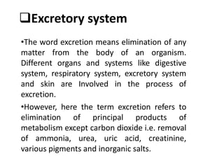
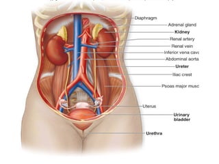
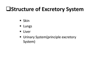
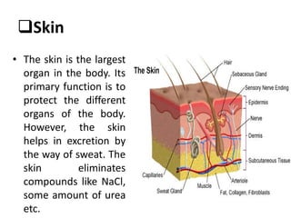
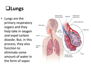
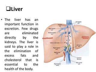
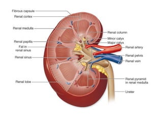
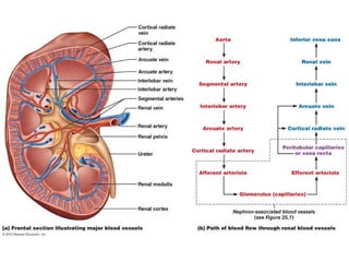
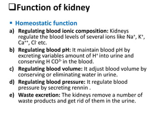
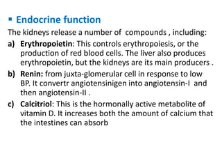
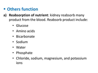
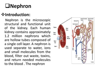
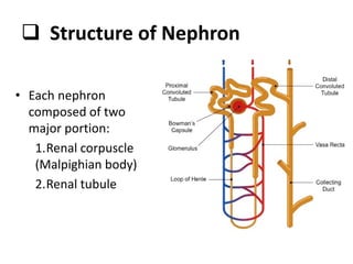
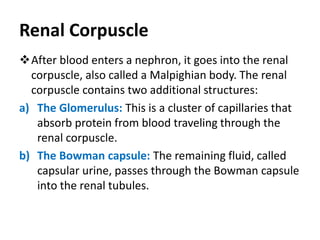
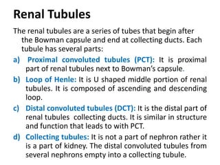
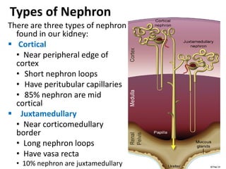
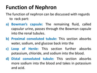
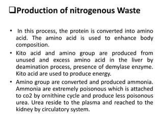
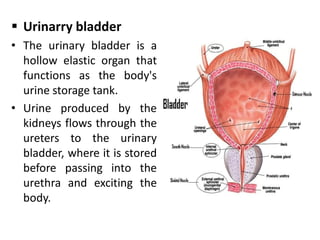
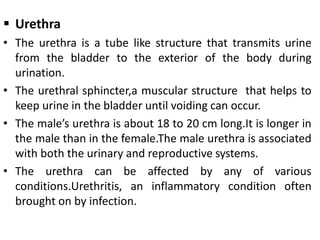
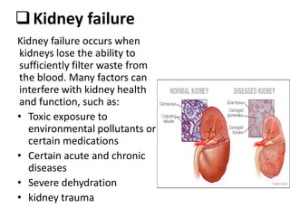
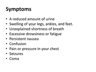
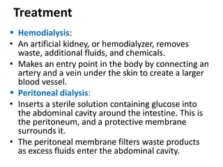
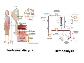
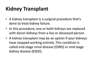
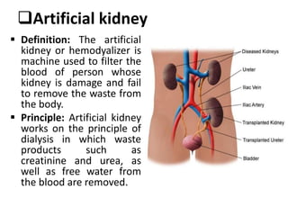
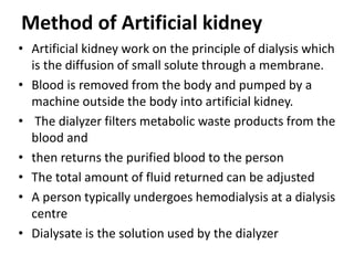
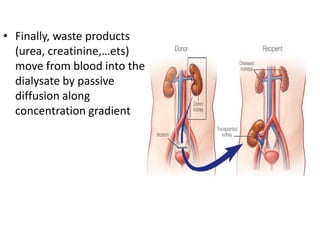
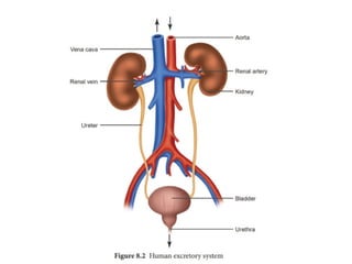
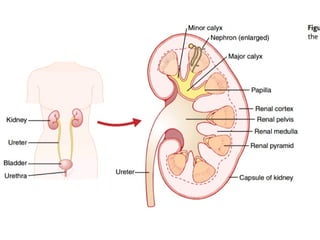
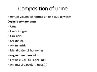
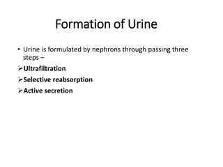
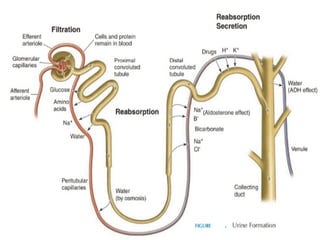
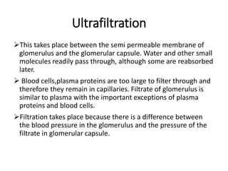
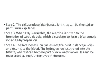
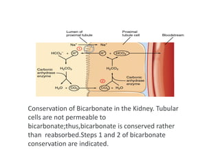
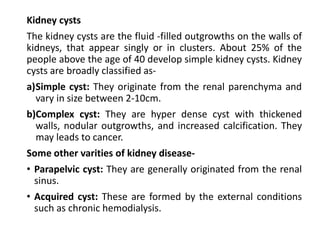
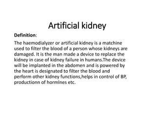
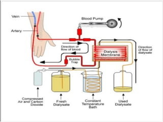
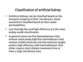
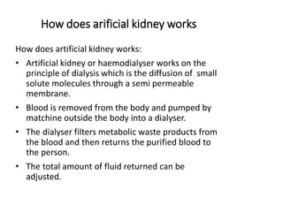
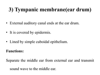
The excretory system is a passive biological system that removes excess, unnecessary materials from the body fluids of an organism, so as to help maintain internal chemical homeostasis and prevent damage to the body. The dual function of excretory systems is the elimination of the waste products of metabolism and to drain the body of used up and broken down components in a liquid and gaseous state. In humans and other amniotes (mammals, birds and reptiles) most of these substances leave the body as urine and to some degree exhalation, mammals also expel them through sweating.
Only the organs specifically used for the excretion are considered a part of the excretory system. In the narrow sense, the term refer to the urinary system. However, as excretion involves several functions that are only superficially related, it is not usually used in more formal classifications of anatomy or function.
As most healthy functioning organs produce metabolic and other wastes, the entire organism depends on the function of the system. Breaking down of one of more of the systems is a serious health condition, for example kidney failure.
Human kidney,structure and functions of kidneyAnand P P
human kidney structural and functions.different types of structural components present in kidney and each structure having definite functions.structural and functional aspects of kidney.
Human kidney,structure and functions of kidneyAnand P P
human kidney structural and functions.different types of structural components present in kidney and each structure having definite functions.structural and functional aspects of kidney.
A powerpoint on the Human Excretory System, intended for the SA Grade 11 Life Sciences Syllabus. Includes information on kidneys, osmoregulation, nephrons, excretion, etc. Hope it helps :)
these slides are prepared to understand digestive system IN EASY WAY Important links- NOTES- https://mynursingstudents.blogspot.com/ youtube channel https://www.youtube.com/c/MYSTUDENTSU... CHANEL PLAYLIST- ANATOMY AND PHYSIOLOGY-https://www.youtube.com/playlist?list=PL93S13oM2gAPM3VTGVUXIeswKJ3XGaD2p COMMUNITY HEALTH NURSING- https://www.youtube.com/playlist?list=PL93S13oM2gAPyslPNdIJoVjiXEDTVEDzs CHILD HEALTH NURSING- https://www.youtube.com/playlist?list=PL93S13oM2gANcslmv0DXg6BWmWN359Gvg FIRST AID- https://www.youtube.com/playlist?list=PL93S13oM2gAMvGqeqH2ZTklzFAZhOrvgP HCM- https://www.youtube.com/playlist?list=PL93S13oM2gAM7mZ1vZhQBHWbdLnLb-cH9 FUNDAMENTALS OF NURSING- https://www.youtube.com/playlist?list=PL93S13oM2gAPFxu78NDLpGPaxEmK1fTao COMMUNICABLE DISEASES- https://www.youtube.com/playlist?list=PL93S13oM2gAOWo4IwNjLU_LCuhRN0ZLeb ENVIRONMENTAL HEALTH- https://www.youtube.com/playlist?list=PL93S13oM2gAPkI6LvfS8Zu1nm6mZi9FK6 MSN- https://www.youtube.com/playlist?list=PL93S13oM2gAOdyoHnDLAoR_o8M6ccqYBm HINDI ONLY- https://www.youtube.com/playlist?list=PL93S13oM2gAN4L-FJ3s_IEXgZCijGUA1A ENGLISH ONLY- https://www.youtube.com/playlist?list=PL93S13oM2gAMYv2a1hFcq4W1nBjTnRkHP facebook profile- https://www.facebook.com/suresh.kr.lrhs/ FACEBOOK PAGE- https://www.facebook.com/My-Student-S... facebook group NURSING NOTES- https://www.facebook.com/groups/24139... FOR MAKING EASY NOTES YOU CAN ALSO VISIT MY BLOG – BLOGGER- https://mynursingstudents.blogspot.com/ Instagram- https://www.instagram.com/mystudentsu... Twitter- https://twitter.com/student_system?s=08
#pancreas, #gallbladder ,#liver ,#BORN,#ASSESSMENT, #APPEARENCE,#PULSE,#GRIMACE,#REFLEX,#RESPIRATION,#RESUSCITATION,#NEWBORN,#BABY,#VIRGINIA, #APGAR, #OXYGEN,#CYANOSIS,#OPTICNERVE, #SARACHNA,#MYSTUDENTSUPPORTSYSTEM, #rashes,#nursingclasses, #communityhealthnursing,#ANM, #GNM, #BSCNURING,#NURSINGSTUDENTS, #WHO,#NURSINGINSTITUTION,#COLLEGEOFNURSING,#nursingofficer,#COMMUNITYHEALTHOFFICER
The urinary system's function is to filter blood and create urine as a waste by-product. The organs of the urinary system include the kidneys, renal pelvis, ureters, bladder and urethra. The body takes nutrients from food and converts them to energy.
This presentation is all about Renal System and it's Physiological Processes with complete description.
This is a presentation file for medical students of all disciplines.
What is Excretion ?, organ used, types of regulation, modes of excretion, explanation of each, human excretory system & function, kidney and its parts, mechanism of urine formation, Dialysis
Excretory system and its parts with detailsরেজা তানজিল
this is a group presentation about the excretory system
The excretory system is a passive biological system that removes excess, unnecessary materials from the body fluids of an organism, so as to help maintain internal chemical homeostasis and prevent damage to the body.
A preservative is a substance or a chemical that is added to products such as food products, beverages, pharmaceutical drugs, paints, biological samples, cosmetics, wood, and many other products to prevent decomposition by microbial growth or by undesirable chemical changes. In general, preservation is implemented in two modes, chemical and physical. Chemical preservation entails adding chemical compounds to the product. Physical preservation entails processes such as refrigeration or drying.[1] Preservative food additives reduce the risk of foodborne infections, decrease microbial spoilage, and preserve fresh attributes and nutritional quality. Some physical techniques for food preservation include dehydration, UV-C radiation, freeze-drying, and refrigeration. Chemical preservation and physical preservation techniques are sometimes combined.
Organic chemistry is a subdiscipline of chemistry that studies the structure, properties and reactions of organic compounds, which contain carbon in covalent bonding.[1] Study of structure determines their chemical composition and formula. Study of properties includes physical and chemical properties, and evaluation of chemical reactivity to understand their behavior. The study of organic reactions includes the chemical synthesis of natural products, drugs, and polymers, and study of individual organic molecules in the laboratory and via theoretical (in silico) study.
The range of chemicals studied in organic chemistry includes hydrocarbons (compounds containing only carbon and hydrogen) as well as compounds based on carbon, but also containing other elements,[1][2][3] especially oxygen, nitrogen, sulfur, phosphorus (included in many biochemicals) and the halogens. Organometallic chemistry is the study of compounds containing carbon–metal bonds.
In addition, contemporary research focuses on organic chemistry involving other organometallics including the lanthanides, but especially the transition metals zinc, copper, palladium, nickel, cobalt, titanium and chromium.
Organic chemistry with its history and effectরেজা তানজিল
Organic chemistry is a subdiscipline of chemistry that studies the structure, properties and reactions of organic compounds, which contain carbon in covalent bonding.[1] Study of structure determines their chemical composition and formula. Study of properties includes physical and chemical properties, and evaluation of chemical reactivity to understand their behavior. The study of organic reactions includes the chemical synthesis of natural products, drugs, and polymers, and study of individual organic molecules in the laboratory and via theoretical (in silico) study.
The range of chemicals studied in organic chemistry includes hydrocarbons (compounds containing only carbon and hydrogen) as well as compounds based on carbon, but also containing other elements,[1][2][3] especially oxygen, nitrogen, sulfur, phosphorus (included in many biochemicals) and the halogens. Organometallic chemistry is the study of compounds containing carbon–metal bonds.
In addition, contemporary research focuses on organic chemistry involving other organometallics including the lanthanides, but especially the transition metals zinc, copper, palladium, nickel, cobalt, titanium and chromium.
Falsely-solid, or, semisolid is the physical term for something whose state lies between a solid and a liquid. While similar to solids in some respects, such as having the ability to support their own weight and hold their shapes, a quasi-solid also shares some properties of liquids, such as conforming in shape to something applying pressure to it and the ability to flow under pressure. The words quasi-solid, semisolid, and semiliquid may be used interchangeably.
Quasi-solids and semisolids are also known as amorphous solids because at the microscopic scale they have a disordered structure unlike the more common crystalline solids.
Oxidation reduction reactions and reactantsরেজা তানজিল
Redox (reduction–oxidation, pronunciation: /ˈrɛdɒks/ redoks or /ˈriːdɒks/ reedoks[1]) is a type of chemical reaction in which the oxidation states of atoms are changed. Redox reactions are characterized by the transfer of electrons between chemical species, most often with one species (the reducing agent) undergoing oxidation (losing electrons) while another species (the oxidizing agent) undergoes reduction (gains electrons).[2] The chemical species from which the electron is stripped is said to have been oxidized, while the chemical species to which the electron is added is said to have been reduced. In other words:
Oxidation is the loss of electrons or an increase in the oxidation state of an atom by another atom, an ion, or a molecule.
Reduction is the gain of electrons or a decrease in the oxidation state of an atom by another atom, an ion, or a molecule.
For example, during the combustion of wood, electrons are transferred from carbon atoms in the wood to oxygen atoms in the air. The oxygen atoms undergo reduction, gaining electrons, while the carbon atoms undergo oxidation, losing electrons. Thus oxygen is the oxidizing agent and carbon is the reducing agent in this reaction.[3]
Although oxidation reactions are commonly associated with the formation of oxides from oxygen molecules, oxygen is not necessarily included in such reactions, as other chemical species can serve the same function.[3]
Redox reactions can occur relatively slowly, as in the formation of rust, or much more rapidly, as in the case of burning fuel. There are simple redox processes, such as the oxidation of carbon to yield carbon dioxide (CO2) or the reduction of carbon by hydrogen to yield methane (CH4), and more complex processes such as the oxidation of glucose (C6H12O6) in the human body.
Solid is one of the four fundamental states of matter (the others being liquid, gas, and plasma). The atoms in a solid are closely packed together and contain the least amount of kinetic energy. A solid is characterized by structural rigidity and resistance to a force applied to the surface. Unlike a liquid, a solid object does not flow to take on the shape of its container, nor does it expand to fill the entire available volume like a gas. The atoms in a solid are bound to each other, either in a regular geometric lattice (crystalline solids, which include metals and ordinary ice), or irregularly (an amorphous solid such as common window glass). Solids cannot be compressed with little pressure whereas gases can be compressed with little pressure because the molecules in a gas are loosely packed.
The branch of physics that deals with solids is called solid-state physics, and is the main branch of condensed matter physics (which also includes liquids). Materials science is primarily concerned with the physical and chemical properties of solids. Solid-state chemistry is especially concerned with the synthesis of novel materials, as well as the science of identification and chemical composition.
An aerosol is a suspension of fine solid particles or liquid droplets in air or another gas.[1] Aerosols can be natural or anthropogenic. Examples of natural aerosols are fog, dust, forest exudates and geyser steam. Examples of anthropogenic aerosols are haze, particulate air pollutants and smoke.[dubious – discuss][1] The liquid or solid particles have diameters typically <1 μm; larger particles with a significant settling speed make the mixture a suspension, but the distinction is not clear-cut. In general conversation, aerosol usually refers to an aerosol spray that delivers a consumer product from a can or similar container. Other technological applications of aerosols include dispersal of pesticides, medical treatment of respiratory illnesses, and combustion technology.[2] Diseases can also spread by means of small droplets in the breath, also called aerosols (or sometimes bioaerosols).[3]
Aerosol science covers generation and removal of aerosols, technological application of aerosols, effects of aerosols on the environment and people, and other topics.[1]
An excipient is a substance formulated alongside the active ingredient of a medication,[1] included for the purpose of long-term stabilization, bulking up solid formulations that contain potent active ingredients in small amounts (thus often referred to as "bulking agents", "fillers", or "diluents"), or to confer a therapeutic enhancement on the active ingredient in the final dosage form, such as facilitating drug absorption,[2][3] reducing viscosity,[4] or enhancing solubility.[5] Excipients can also be useful in the manufacturing process, to aid in the handling of the active substance concerns such as by facilitating powder flowability or non-stick properties, in addition to aiding in vitro stability such as prevention of denaturation or aggregation over the expected shelf life. The selection of appropriate excipients also depends upon the route of administration and the dosage form, as well as the active ingredient and other factors. A comprehensive classification system based on structure-property-application relationships has been proposed for excipients used in parenteral medications.[6][7]
Pharmaceutical regulations and standards require that all ingredients in drugs, as well as their chemical decomposition products, be identified and shown to be safe. Often, more excipient is found in a final drug formulation than active ingredient, and practically all marketed drugs contain excipients.[1]:1 As with new drug substances and dosage forms thereof, novel excipients themselves can be patented; sometimes, however, a particular formulation involving them is kept as a trade secret instead (if not easily reverse-engineered).[citation needed]
The Excipients Industry Forum as a multi stake-holder forum and information hub for the wider excipients industry and the public. It’s goal is to promote open dialogue and partnership between the public, policy makers and the pharmaceutical industry.[8]
The Federation of International Pharmaceutical Excipients Council (IPEC),[9] a pharmaceutical regulatory non-profit, develops, implements, and promotes global use of appropriate quality, safety, and functionality standards for pharmaceutical excipients and excipient delivery systems. IPEC-Americas, along with its counterparts in Europe, China, and Japan serves as a primary international resource on excipients for its members, governments, and public audiences. IPEC works in collaboration with ExcipientFest to present topics ranging from regulatory affairs to research and development, often featuring speakers from FDA and other pharmaceutical organizations
organic chemistry, amines (/əˈmiːn, ˈæmiːn/,[1][2] UK also /ˈeɪmiːn/)[3] are compounds and functional groups that contain a basic nitrogen atom with a lone pair. Amines are formally derivatives of ammonia, wherein one or more hydrogen atoms have been replaced by a substituent such as an alkyl or aryl group[4] (these may respectively be called alkylamines and arylamines; amines in which both types of substituent are attached to one nitrogen atom may be called alkylarylamines). Important amines include amino acids, biogenic amines, trimethylamine, and aniline; see Category:Amines for a list of amines. Inorganic derivatives of ammonia are also called amines, such as monochloramine (NClH2).[5]
The substituent -NH2 is called an amino group.[6]
Compounds with a nitrogen atom attached to a carbonyl group, thus having the structure R–CO–NR′R″, are called amides and have different chemical properties from amines.
Aldehyde ketone and their types and propertiesরেজা তানজিল
An aldehyde /ˈældɪhaɪd/ is a compound containing a functional group with the structure −CHO, consisting of a carbonyl center (a carbon double-bonded to oxygen) with the carbon atom also bonded to hydrogen and to an R group,[1] which is any generic alkyl or side chain. The group—without R—is the aldehyde group, also known as the formyl group. Aldehydes are common in organic chemistry, and many fragrances are or contain aldehydes.
The action of drugs on the human body is called pharmacodynamics, and what the body does with the drug is called pharmacokinetics. The drugs that enter the human tend to stimulate certain receptors, ion channels, act on enzymes or transporter proteins. As a result, they cause the human body to react in a specific way.
There are 2 different types of drugs:
Agonists - they stimulate and activate the receptors
Antagonists - they stop the agonists from stimulating the receptors
Once the receptors are activated, they either trigger a particular response directly on the body, or they trigger the release of hormones and/or other endogenous drugs in the body to stimulate a particular response.
Resonance in organic molecules and their typesরেজা তানজিল
resonance is a way of describing bonding in certain molecules or ions by the combination of several contributing structures (or forms,[1] also variously known as resonance structures or canonical structures) into a resonance hybrid (or hybrid structure) in valence bond theory. It has particular value for describing delocalized electrons within certain molecules or polyatomic ions where the bonding cannot be expressed by one single Lewis structure.
Pharmaceutical formulation, in pharmaceutics, is the process in which different chemical substances, including the active drug, are combined to produce a final medicinal product. The word formulation is often used in a way that includes dosage form.
The citric acid cycle (CAC) – also known as the TCA cycle (tricarboxylic acid cycle) or the Krebs cycle[1][2] – is a series of chemical reactions used by all aerobic organisms to release stored energy through the oxidation of acetyl-CoA derived from carbohydrates, fats, and proteins, into adenosine triphosphate (ATP) and carbon dioxide. In addition, the cycle provides precursors of certain amino acids, as well as the reducing agent NADH, that are used in numerous other reactions. Its central importance to many biochemical pathways suggests that it was one of the earliest established components of cellular metabolism and may have originated abiogenically.[3][4] Even though it is branded as a 'cycle', it is not necessary for metabolites to follow only one specific route; at least three segments of the citric acid cycle have been recognized.[5]
The name of this metabolic pathway is derived from the citric acid (a type of tricarboxylic acid, often called citrate, as the ionized form predominates at biological pH[6]) that is consumed and then regenerated by this sequence of reactions to complete the cycle. The cycle consumes acetate (in the form of acetyl-CoA) and water, reduces NAD+ to NADH, and produces carbon dioxide as a waste byproduct. The NADH generated by the citric acid cycle is fed into the oxidative phosphorylation (electron transport) pathway. The net result of these two closely linked pathways is the oxidation of nutrients to produce usable chemical energy in the form of ATP.
In eukaryotic cells, the citric acid cycle occurs in the matrix of the mitochondrion. In prokaryotic cells, such as bacteria, which lack mitochondria, the citric acid cycle reaction sequence is performed in the cytosol with the proton gradient for ATP production being across the cell's surface (plasma membrane) rather than the inner membrane of the mitochondrion. The overall yield of energy-containing compounds from the TCA cycle is three NADH, one FADH2, and one GTP.[7]
Organic chemistry is a subdiscipline of chemistry that studies the structure, properties and reactions of organic compounds, which contain carbon in covalent bonding.[1] Study of structure determines their chemical composition and formula. Study of properties includes physical and chemical properties, and evaluation of chemical reactivity to understand their behavior. The study of organic reactions includes the chemical synthesis of natural products, drugs, and polymers, and study of individual organic molecules in the laboratory and via theoretical (in silico) study.
The range of chemicals studied in organic chemistry includes hydrocarbons (compounds containing only carbon and hydrogen) as well as compounds based on carbon, but also containing other elements,[1][2][3] especially oxygen, nitrogen, sulfur, phosphorus (included in many biochemicals) and the halogens. Organometallic chemistry is the study of compounds containing carbon–metal bonds.
In addition, contemporary research focuses on organic chemistry involving other organometallics including the lanthanides, but especially the transition metals zinc, copper, palladium, nickel, cobalt, titanium and chromium
Enzymes /ˈɛnzaɪmz/ are both proteins and biological catalysts (biocatalysts). Catalysts accelerate chemical reactions. The molecules upon which enzymes may act are called substrates, and the enzyme converts the substrates into different molecules known as products. Almost all metabolic processes in the cell need enzyme catalysis in order to occur at rates fast enough to sustain life.[1]:8.1 Metabolic pathways depend upon enzymes to catalyze individual steps. The study of enzymes is called enzymology and a new field of pseudoenzyme analysis has recently grown up, recognising that during evolution, some enzymes have lost the ability to carry out biological catalysis, which is often reflected in their amino acid sequences and unusual 'pseudocatalytic' properties.[2][3]
Enzymes are known to catalyze more than 5,000 biochemical reaction types.[4] Other biocatalysts are catalytic RNA molecules, called ribozymes. Enzymes' specificity comes from their unique three-dimensional structures.
Like all catalysts, enzymes increase the reaction rate by lowering its activation energy. Some enzymes can make their conversion of substrate to product occur many millions of times faster. An extreme example is orotidine 5'-phosphate decarboxylase, which allows a reaction that would otherwise take millions of years to occur in milliseconds.[5][6] Chemically, enzymes are like any catalyst and are not consumed in chemical reactions, nor do they alter the equilibrium of a reaction. Enzymes differ from most other catalysts by being much more specific. Enzyme activity can be affected by other molecules: inhibitors are molecules that decrease enzyme activity, and activators are molecules that increase activity. Many therapeutic drugs and poisons are enzyme inhibitors. An enzyme's activity decreases markedly outside its optimal temperature and pH, and many enzymes are (permanently) denatured when exposed to excessive heat, losing their structure and catalytic properties.
Some enzymes are used commercially, for example, in the synthesis of antibiotics. Some household products use enzymes to speed up chemical reactions: enzymes in biological washing powders break down protein, starch or fat stains on clothes, and enzymes in meat tenderizer break down proteins into smaller molecules, making the meat easier to chew.
In biology and biochemistry, a lipid is a biomolecule that is soluble in nonpolar solvents.[3] Non-polar solvents are typically hydrocarbons used to dissolve other naturally occurring hydrocarbon lipid molecules that do not (or do not easily) dissolve in water, including fatty acids, waxes, sterols, fat-soluble vitamins (such as vitamins A, D, E, and K), monoglycerides, diglycerides, triglycerides, and phospholipids.
The functions of lipids include storing energy, signaling, and acting as structural components of cell membranes.[4][5] Lipids have applications in the cosmetic and food industries as well as in nanotechnology.[6]
Scientists sometimes define lipids as hydrophobic or amphiphilic small molecules; the amphiphilic nature of some lipids allows them to form structures such as vesicles, multilamellar/unilamellar liposomes, or membranes in an aqueous environment. Biological lipids originate entirely or in part from two distinct types of biochemical subunits or "building-blocks": ketoacyl and isoprene groups.[4] Using this approach, lipids may be divided into eight categories: fatty acids, glycerolipids, glycerophospholipids, sphingolipids, saccharolipids, and polyketides (derived from condensation of ketoacyl subunits); and sterol lipids and prenol lipids (derived from condensation of isoprene subunits).[4]
Although the term "lipid" is sometimes used as a synonym for fats, fats are a subgroup of lipids called triglycerides. Lipids also encompass molecules such as fatty acids and their derivatives (including tri-, di-, monoglycerides, and phospholipids), as well as other sterol-containing metabolites such as cholesterol.[7] Although humans and other mammals use various biosynthetic pathways both to break down and to synthesize lipids, some essential lipids can't be made this way and must be obtained from the diet.
Alcohol phenol ether with types and propertiesরেজা তানজিল
In chemistry, alcohol is an organic compound that carries at least one hydroxyl functional group (−OH) bound to a saturated carbon atom.[2] The term alcohol originally referred to the primary alcohol ethanol (ethyl alcohol), which is used as a drug and is the main alcohol present in alcoholic beverages. An important class of alcohols, of which methanol and ethanol are the simplest members, includes all compounds for which the general formula is CnH2n+1OH. Simple monoalcohols that are the subject of this article include primary (RCH2OH), secondary (R2CHOH) and tertiary (R3COH) alcohols.
The suffix -ol appears in the IUPAC chemical name of all substances where the hydroxyl group is the functional group with the highest priority. When a higher priority group is present in the compound, the prefix hydroxy- is used in its IUPAC name. The suffix -ol in non-IUPAC names (such as paracetamol or cholesterol) also typically indicates that the substance is an alcohol. However, many substances that contain hydroxyl functional groups (particularly sugars, such as glucose and sucrose) have names which include neither the suffix -ol, nor the prefix hydroxy-.
Peptic ulcer disease (PUD) is a break in the inner lining of the stomach, the first part of the small intestine, or sometimes the lower esophagus.[1][7] An ulcer in the stomach is called a gastric ulcer, while one in the first part of the intestines is a duodenal ulcer.[1] The most common symptoms of a duodenal ulcer are waking at night with upper abdominal pain and upper abdominal pain that improves with eating.[1] With a gastric ulcer, the pain may worsen with eating.[8] The pain is often described as a burning or dull ache.[1] Other symptoms include belching, vomiting, weight loss, or poor appetite.[1] About a third of older people have no symptoms.[1] Complications may include bleeding, perforation, and blockage of the stomach.[2] Bleeding occurs in as many as 15% of cases.[2]
Common causes include the bacteria Helicobacter pylori and non-steroidal anti-inflammatory drugs (NSAIDs).[1] Other, less common causes include tobacco smoking, stress due to serious illness, Behcet disease, Zollinger-Ellison syndrome, Crohn disease, and liver cirrhosis.[1][3] Older people are more sensitive to the ulcer-causing effects of NSAIDs.[1] The diagnosis is typically suspected due to the presenting symptoms with confirmation by either endoscopy or barium swallow.[1] H. pylori can be diagnosed by testing the blood for antibodies, a urea breath test, testing the stool for signs of the bacteria, or a biopsy of the stomach.[1] Other conditions that produce similar symptoms include stomach cancer, coronary heart disease, and inflammation of the stomach lining or gallbladder inflammation.[1]
Diet does not play an important role in either causing or preventing ulcers.[9] Treatment includes stopping smoking, stopping use of NSAIDs, stopping alcohol, and taking medications to decrease stomach acid.[1] The medication used to decrease acid is usually either a proton pump inhibitor (PPI) or an H2 blocker, with four weeks of treatment initially recommended.[1] Ulcers due to H. pylori are treated with a combination of medications, such as amoxicillin, clarithromycin, and a PPI.[4] Antibiotic resistance is increasing and thus treatment may not always be effective.[4] Bleeding ulcers may be treated by endoscopy, with open surgery typically only used in cases in which it is not successful.[2]
Peptic ulcers are present in around 4% of the population.[1] New ulcers were found in around 87.4 million people worldwide during 2015.[5] About 10% of people develop a peptic ulcer at some point in their life.[10] Peptic ulcers resulted in 267,500 deaths in 2015, down from 327,000 in 1990.[6][11] The first description of a perforated peptic ulcer was in 1670, in Princess Henrietta of England.[2] H. pylori was first identified as causing peptic ulcers by Barry Marshall and Robin Warren in the late 20th century,[4] a discovery for which they received the Nobel Prize in 2005
Nucleic acid and its types with propertiesরেজা তানজিল
Nucleic acids are the biopolymers, or small biomolecules, essential to all known forms of life. The term nucleic acid is the overall name for DNA and RNA. They are composed of nucleotides, which are the monomers made of three components: a 5-carbon sugar, a phosphate group and a nitrogenous base. If the sugar is a compound ribose, the polymer is RNA (ribonucleic acid); if the sugar is derived from ribose as deoxyribose, the polymer is DNA (deoxyribonucleic acid).
Nucleic acids are the most important of all biomolecules. These are found in abundance in all living things, where they function to create and encode and then store information of every living cell of every life-form organism on Earth. In turn, they function to transmit and express that information inside and outside the cell nucleus—to the interior operations of the cell and ultimately to the next generation of each living organism. The encoded information is contained and conveyed via the nucleic acid sequence, which provides the 'ladder-step' ordering of nucleotides within the molecules of RNA and DNA.
Strings of nucleotides are bonded to form helical backbones—typically, one for RNA, two for DNA—and assembled into chains of base-pairs selected from the five primary, or canonical, nucleobases, which are: adenine, cytosine, guanine, thymine, and uracil. Thymine occurs only in DNA and uracil only in RNA. Using amino acids and the process known as protein synthesis,[1] the specific sequencing in DNA of these nucleobase-pairs enables storing and transmitting coded instructions as genes. In RNA, base-pair sequencing provides for manufacturing new proteins that determine the frames and parts and most chemical processes of all life forms.
The French Revolution, which began in 1789, was a period of radical social and political upheaval in France. It marked the decline of absolute monarchies, the rise of secular and democratic republics, and the eventual rise of Napoleon Bonaparte. This revolutionary period is crucial in understanding the transition from feudalism to modernity in Europe.
For more information, visit-www.vavaclasses.com
Students, digital devices and success - Andreas Schleicher - 27 May 2024..pptxEduSkills OECD
Andreas Schleicher presents at the OECD webinar ‘Digital devices in schools: detrimental distraction or secret to success?’ on 27 May 2024. The presentation was based on findings from PISA 2022 results and the webinar helped launch the PISA in Focus ‘Managing screen time: How to protect and equip students against distraction’ https://www.oecd-ilibrary.org/education/managing-screen-time_7c225af4-en and the OECD Education Policy Perspective ‘Students, digital devices and success’ can be found here - https://oe.cd/il/5yV
How to Make a Field invisible in Odoo 17Celine George
It is possible to hide or invisible some fields in odoo. Commonly using “invisible” attribute in the field definition to invisible the fields. This slide will show how to make a field invisible in odoo 17.
Instructions for Submissions thorugh G- Classroom.pptxJheel Barad
This presentation provides a briefing on how to upload submissions and documents in Google Classroom. It was prepared as part of an orientation for new Sainik School in-service teacher trainees. As a training officer, my goal is to ensure that you are comfortable and proficient with this essential tool for managing assignments and fostering student engagement.
Operation “Blue Star” is the only event in the history of Independent India where the state went into war with its own people. Even after about 40 years it is not clear if it was culmination of states anger over people of the region, a political game of power or start of dictatorial chapter in the democratic setup.
The people of Punjab felt alienated from main stream due to denial of their just demands during a long democratic struggle since independence. As it happen all over the word, it led to militant struggle with great loss of lives of military, police and civilian personnel. Killing of Indira Gandhi and massacre of innocent Sikhs in Delhi and other India cities was also associated with this movement.
We all have good and bad thoughts from time to time and situation to situation. We are bombarded daily with spiraling thoughts(both negative and positive) creating all-consuming feel , making us difficult to manage with associated suffering. Good thoughts are like our Mob Signal (Positive thought) amidst noise(negative thought) in the atmosphere. Negative thoughts like noise outweigh positive thoughts. These thoughts often create unwanted confusion, trouble, stress and frustration in our mind as well as chaos in our physical world. Negative thoughts are also known as “distorted thinking”.
How to Create Map Views in the Odoo 17 ERPCeline George
The map views are useful for providing a geographical representation of data. They allow users to visualize and analyze the data in a more intuitive manner.
3. Excretory system
•The word excretion means elimination of any
matter from the body of an organism.
Different organs and systems like digestive
system, respiratory system, excretory system
and skin are Involved in the process of
excretion.
•However, here the term excretion refers to
elimination of principal products of
metabolism except carbon dioxide i.e. removal
of ammonia, urea, uric acid, creatinine,
various pigments and inorganic salts.
4.
5. Structure of Excretory System
Skin
Lungs
Liver
Urinary System(principle excretory
System)
6. Skin
• The skin is the largest
organ in the body. Its
primary function is to
protect the different
organs of the body.
However, the skin
helps in excretion by
the way of sweat. The
skin eliminates
compounds like NaCl,
some amount of urea
etc.
7. Lungs
• Lungs are the
primary respiratory
organs and they
help take in oxygen
and expel carbon
dioxide. But, in this
process, they also
function to
eliminate some
amount of water in
the form of vapor.
8. Liver
• The liver has an
important function in
excretion. Few drugs
are eliminated
directly by the
kidneys. The liver is
said to play a role in
the elimination of
excess fats and
cholesterol that is
essential to the
health of the body.
9. Liver and Waste
• The liver produces
urea and uric acid as a
by-product of the
breakdown of
proteins.
• Urea and uric acid are
sent to the kidneys to
be processed.
10.
11. Organs of Urinary System
Urinary system consist of:
• Two Kidneys
• Two Ureters
• A Urinary Bladder
• A Urethra
12. Primary organs: Kidneys
• Filter waste products from the
bloodstream.
• Convert the filtrate into urine.
Urinary tracts:
• Ureters
• Urinary bladder
• Urethra
13.
14. Introduction
• The kidneys are a pair of bean-shaped organs located
in the abdomen, on either side of the spine, under
the diaphragm present in all vertebrates.
• They are made of a large number of structural and
functional subunits called nephrons.
• They remove waste products from the body,
maintain balanced electrolyte levels, and regulate
blood pressure.
15.
16. External Structure
• Diameter: Kidneys are bean shaped organ,
about 11 cm long, 6 cm wide, 3 cm thick and
weight of 150 g.
• Coverings: Each kidney is enclosed by a thin
tough fibrous connective tissue called renal
capsule that protects it from the infection and
injuries.
• The renal capsule which is further enclosed by
another layer of fibrous membrane is called
renal fascia.
17.
18. • Location: The kidney lie
on the posterior
abdominal wall, one on
each side of the vertebral
column, behind the
peritoneum and below
the diaphragm.
• Position: It is situated at
the level of T12-L3. The
right kidney is usually
slightly lower than the
left, probably because of
the considerable space
occupied by the liver.
19. Internal Structure
• There are three major
regions of the kidney:
1) Renal cortex
2) Renal medulla
3) Renal pelvis
20. Renal Cortex
• The kidneys are surrounded by a renal cortex, a layer
of tissue that is also covered by renal fascia and the
renal capsule.
• The cortex provides a space for arterioles and
venules from the renal artery and vein to perfuse the
nephrons of the kidney.
• Erythropotein, a hormone necessary for the
synthesis of new red blood cells, is also produced in
the renal cortex.
21. Renal Medulla
• The medulla is the inner region of the parenchyma of
the kidney.
• The medulla consists of the renal pyramids, which
are triangle structures. There are 8-18 renal pyramids
in each kidney.
• The apex of each pyramid is called renal papilla.
Each papilla projects into a small depression, called a
minor calyx. Several minor calyces unite to form a
major calyx.
• The renal medulla contains the majority of the length
of nephrons the main functional unit of kidney.
22. Renal Pelvis
• The renal pelvis is a funnel-shaped space in the
innermost part of the kidney.
• The renal pelvis contains the hilum where blood
vessels and nerves enter and exit the kidney
• The first part of the renal pelvis contains the. cup-
shaped spaces that collect fluid Called calyces.
• The renal pelvis connects the kidney to the rest of
the body.
23.
24. Renal Circulation
• Two blood vessel is responsible for renal
circulation:
1) Renal artery: This brings oxygenated blood
from the heart to the kidney for filtration.
2) Renal vein: This carries filtered blood from
the kidneys back to the heart.
25.
26. Function of kidney
Homeostatic function
a) Regulating blood ionic composition: Kidneys
regulate the blood levels of several ions like Na+, K+,
Ca++, Cl- etc.
b) Regulating blood pH: It maintain blood pH by
excreting variables amount of H+ into urine and
conserving H CO3- in the blood.
c) Regulating blood volume: It adjust blood volume by
conserving or eliminating water in urine.
d) Regulating blood pressure: It regulate blood
pressure by secreting rennin .
e) Waste excretion: The kidneys remove a number of
waste products and get rid of them in the urine.
27. Endocrine function
The kidneys release a number of compounds , including:
a) Erythropoietin: This controls erythropoiesis, or the
production of red blood cells. The liver also produces
erythropoietin, but the kidneys are its main producers .
b) Renin: from juxta-glomerular cell in response to low
BP. It convertr angiotensinigen into angiotensin-I and
then angiotensin-II .
c) Calcitriol: This is the hormonally active metabolite of
vitamin D. It increases both the amount of calcium that
the intestines can absorb
28. Others function
a) Reabsorption of nutrient: kidney reabsorb many
product from the blood. Reabsorb product include:
• Glucose
• Amino acids
• Bicarbonate
• Sodium
• Water
• Phosphate
• Chloride, sodium, magnesium, and potassium
ions
29. b) Elimination: Kidney eliminate the drug and some
toxic substances from the blood.
c) Excretion: kidney excrete many metabolic waste
product from the body. Such as urea, uric acid,
creatininie and creatine etc.
d) Urine formation: Kidney help to form urine from
the nitrogenous waste product with the help of
nephrone.
30.
31. Nephron
Introduction:
Nephron is the microscopic
structural and functional unit
of the kidney. Each human
kidney contains approximately
1.2 million nephrons which
are hollow tubes composed of
a single cell layer. A nephron is
used separate to water, ions
and small molecules from the
blood, filter out waste, toxins,
and return needed molecules
to the blood . The nephron
32. Structure of Nephron
• Each nephron
composed of two
major portion:
1.Renal corpuscle
(Malpighian body)
2.Renal tubule
33. Renal Corpuscle
After blood enters a nephron, it goes into the renal
corpuscle, also called a Malpighian body. The renal
corpuscle contains two additional structures:
a) The Glomerulus: This is a cluster of capillaries that
absorb protein from blood traveling through the
renal corpuscle.
b) The Bowman capsule: The remaining fluid, called
capsular urine, passes through the Bowman capsule
into the renal tubules.
34. Renal Tubules
The renal tubules are a series of tubes that begin after
the Bowman capsule and end at collecting ducts. Each
tubule has several parts:
a) Proximal convoluted tubules (PCT): It is proximal
part of renal tubules next to Bowman’s capsule.
b) Loop of Henle: It is U shaped middle portion of renal
tubules. It is composed of ascending and descending
loop.
c) Distal convoluted tubules (DCT): It is the distal part of
renal tubules collecting ducts. It is similar in structure
and function that leads to with PCT.
d) Collecting tubules: It is not a part of nephron rather it
is a part of kidney. The distal convoluted tubules from
several nephrons empty into a collecting tubule.
35.
36. Types of Nephron
There are three types of nephron
found in our kidney:
Cortical
• Near peripheral edge of
cortex
• Short nephron loops
• Have peritubular capillaries
• 85% nephron are mid
cortical
Juxtamedullary
• Near corticomedullary
border
• Long nephron loops
• Have vasa recta
• 10% nephron are juxtamedullary
37. Function of Nephron
The function of nephron can be discussed with regurds
to rack part:
a) Bowman’s capsule: The remaining fluid, called
capsular urine, passes through the Bowman capsule
into the renal tubules.
b) Proximal convoluted tubule: This section absorbs
water, sodium, and glucose back into the
c) Loop of Henle: This section further absorbs
potassium, chloride, and sodium into the blood.
d) Distal convoluted tubule: This section absorbs
more sodium into the blood and takes in potassium
and acid.
38.
39. The physiology of excretion can be discussed in
two headings:
Production of nitrogenous waste product
Mechanism of urine formation
40. Production of nitrogenous Waste
• In this process, the protein is converted into amino
acid. The amino acid is used to enhance body
composition.
• Kito acid and amino group are produced from
unused and excess amino acid in the liver by
deamination process, presence of demylase enzyme.
Kito acid are used to produce energy.
• Amino group are converted and produced ammonia.
Ammonia are extremely poisonous which is attached
to co2 by ornithine cycle and produce less poisonous
urea. Urea reside to the plasma and reached to the
kidney by circulatory system.
41. • Amino acid kito acid + amino group
• Amino group + hydrogen ion ammonia
• 2ammonia + CO2 urea + CO2
42. Mechanism of urine formation
Urine is continually formed
by each nephron and the
processes involved in the
formation of urine are:
1) Glomerular ultrafiltration
2) Tubular Reabsorption
3) Tubular Secretion
43. Glomerular filtration
• Glomerular filtration is the first step in making urine.
• It is the process that our kidneys use to filter excess
fluid and waste products.
• It occurs in the renal corpuscle of nephron. The renal
corpuscle is divided into bowman’s capsule and
glomerulus.
• The filtrate is deposited in the glomerulus. This
filtrate is called glomerulus filtrate.
44. Tubular Reabsorption
Reabsorption in Proximal Convoluted Tubule:
• Proximal convoluted tubule is responsible for the
reabsorption of water, glucose, sodium phosphate
and bicarbonates.in this process, all glucose, 80%
water, 50% sodium bicarbonate, chloride, calcium,
sodium chloride etc. are actively reabsorbed.
Reabsorption in distal convoluted tubule:
• Urea, uric acid, phosphate is actively reabsorbed
this substance is called low threshold substance.
But sulphate and creatinine are not reabsorbed this
substance is called non threshold substance.
45. Reabsorption in Henle‘s loop:
• Water is reabsorbed mainly in the descending
limb and thin segment of the ascending limb.
• Sodium. calcium. chloride. magnesium and
potassium are actively reabsorb., it, the thick
segment of the ascending limb.
Reabsorption in collection tubule:
• As urine (isotonic) passes into the collecting
tubule it becomes once more hypertonic.
• Sodium. potassium and chloride reabsorption
and Hydrogen ion secretion.
• Thus the urine formed contains 96% of water, 2%
urea and 2% of the metabolic products.
46.
47. Tubular secretion
• Tubular secretion is the opposite process of
reabsorption. This secretion is caused mainly by
active transport and passive diffusion.
1) Active transport—the movement of molecules
via ATPase pumps that transport the substance
through the renal epithelial cell into the lumen
of the nephron.
2) Passive diffusion—the movement of molecules
from the peritubular capillaries to the intersitial
fluid within the nephron.
48.
49. Introduction
• Urine is a yellowish liquid by-
product of the body secreted by
the kidneys through a process
called urination and excreted
through the urethra.
• Urine is an aqueous solution of
greater than 95% water. Other
constituents include urea,chlorine,
sodium, potassium, creatine and
dissolved ions,and inorganic and
organic compounds. Urea is a
non‐toxic molecule made of toxic
ammonia and carbon dioxide.Any
abnormal constituents found in
urine are indication of disease.
50. Composition of urine
Chemical Concentration in g/100 ml urine
Water 95
Urea 2
Uric acid 0.03
Creatinine 0.1
Ammonia 0.05
Sodium 0.6
Patassium 0.15
Megnesium 0.01
Calcium 0.015
Phosphate 0.12
Sulphate 0.18
Protein -------
Glucose -------
51. Properties of urine
• Color: Typically yellow-amber, but varies according to
recent diet and the concentration of the urine.
• Smell: Generally fresh urine has a mild smell but aged
urine has a stronger odor similar to that of ammonia.
• PH: The pH of normal urine is generally in the range
4.6‐8.
• Density: The density of normal urine ranges from 0.001
to 0.035.
• Turbidity: The turbidity of the urine sample is reported
as clear, slightly cloudy, cloudy, opaque or flocculent.
52. Micturition
Introduction: Micturition is the process of
expelling of the urine from the bladder.
Stages of micturition:
1) Resting or filling stage
2) Voiding stage
53. 1) Resting or Filling Stage
• It is in this phase of the bladder that the urine is
transported from the kidneys via the ureters into the
bladder.
2) Voiding Stage
• During this stage, both the urinary bladder and the
urethra come into play together.
• The urethra is controlled by two sets of muscles: The
internal and external urethral sphincters. The
internal sphincter is a smooth muscle whereas the
external one is skeletal. Both these sphincters are in
a contracted state during the filling stage.
54.
55.
56. Urine Storage and Elimination
There are some part of the urinary system that take on
the storage and elimination of urine.
Ureters
• The ureter is a tube that carries urine from the kidney
to the urinary bladder. They are about 25-30 cm long
with a diameter of approximately 3mm.There are two
ureters, one attached to each kidney.
• The wall urerters is composed of three concentric
tunics:
i) Mucosa
ii) Muscularis
iii) Adventitia
57.
58. Urinarry bladder
• The urinary bladder is a
hollow elastic organ that
functions as the body's
urine storage tank.
• Urine produced by the
kidneys flows through the
ureters to the urinary
bladder, where it is stored
before passing into the
urethra and exciting the
body.
59. Urethra
• The urethra is a tube like structure that transmits urine
from the bladder to the exterior of the body during
urination.
• The urethral sphincter,a muscular structure that helps to
keep urine in the bladder until voiding can occur.
• The male’s urethra is about 18 to 20 cm long.It is longer in
the male than in the female.The male urethra is associated
with both the urinary and reproductive systems.
• The urethra can be affected by any of various
conditions.Urethritis, an inflammatory condition often
brought on by infection.
60.
61. Kidney failure
Kidney failure occurs when
kidneys lose the ability to
sufficiently filter waste from
the blood. Many factors can
interfere with kidney health
and function, such as:
• Toxic exposure to
environmental pollutants or
certain medications
• Certain acute and chronic
diseases
• Severe dehydration
• kidney trauma
62. Symptoms
• A reduced amount of urine
• Swelling of your legs, ankles, and feet.
• Unexplained shortness of breath
• Excessive drowsiness or fatigue
• Persistent nausea
• Confusion
• Pain or pressure in your chest
• Seizures
• Coma
63. Treatment
Hemodialysis:
• An artificial kidney, or hemodialyzer, removes
waste, additional fluids, and chemicals.
• Makes an entry point in the body by connecting an
artery and a vein under the skin to create a larger
blood vessel.
Peritoneal dialysis:
• Inserts a sterile solution containing glucose into
the abdominal cavity around the intestine. This is
the peritoneum, and a protective membrane
surrounds it.
• The peritoneal membrane filters waste products
as excess fluids enter the abdominal cavity.
65. Kidney Transplant
• A kidney transplant is a surgical procedure that’s
done to treat kidney failure.
• In this procedure, one or both kidneys are replaced
with donor kidneys from a live or deceased person.
• A kidney transplant may be an option if your kidneys
have stopped working entirely. This condition is
called end-stage renal disease (ESRD) or end-stage
kidney disease (ESKD).
66. Artificial kidney
Definition: The artificial
kidney or hemodyalizer is
machine used to filter the
blood of person whose
kidney is damage and fail
to remove the waste from
the body.
Principle: Artificial kidney
works on the principle of
dialysis in which waste
products such as
creatinine and urea, as
well as free water from
the blood are removed.
67. Method of Artificial kidney
• Artificial kidney work on the principle of dialysis which
is the diffusion of small solute through a membrane.
• Blood is removed from the body and pumped by a
machine outside the body into artificial kidney.
• The dialyzer filters metabolic waste products from the
blood and
• then returns the purified blood to the person
• The total amount of fluid returned can be adjusted
• A person typically undergoes hemodialysis at a dialysis
centre
• Dialysate is the solution used by the dialyzer
68. • Finally, waste products
(urea, creatinine,…ets)
move from blood into the
dialysate by passive
diffusion along
concentration gradient
69. Other Renal Diseases
• Diabetic nephropathy: In people with diabetic
nephropathy, damage occurs to the capillaries of the
kidney as a result of long-term diabetes.
• Kidney stones: Stones can form as a solid build-up of
minerals in the kidney. They can cause intense pain
and might affect kidney function if they block the
ureter.
• Kidney hydronephrosis: This means "water on the
kidney.“ It usually occurs when an obstruction
prevents urine from leaving the kidney, causing
intense pain.
70. • Kidney infections: These tend to result from bacteria
in the bladder that transfer to the kidneys. Kidney
infections are more common in women than in men,
as well as in women who are pregnant. The infection
often responds well to antibiotics.
• Nephrotic syndrome: Damage to the kidney function
causes protein levels in the urine to increase. This
results in a protein shortage throughout the body,
which draws water into the tissues.
71.
72. Regulation of urine concentration
• Urine volume and concentration is regulated through the
same processes that regulate blood volume.
• Antidiuretic hormone (ADH)—produced by the posterior
pituitary gland —increases the amount of water
reabsorbed in the distal convoluted tubule and collecting
duct.
• Osmoreceptors in the hypothalamus signal the posterior
pituitary gland to increase ADH secretion when plasma
osmolarity becomes too high.
• ADH causes decreased urine volume and decreased
plasma osmolarity.
• A diuretic increases urine volume and increases plasma
osmolarity.
• Common diuretics include alcohol, water, caffeine, and
many medications, and they generally function as diuretics
via different mechanisms.
73. • Sodium is one of the body's electrolytes, which
are minerals that the body needs in relatively
large amounts. Electrolytes carry an electric
charge when dissolved in body fluids such as
blood.
• The concentration of sodium in the blood may be
1) Too low (hyponatremia)
2) Too high (hypernatremia)
Regulation of Sodium in Blood
74. • When blood volume or sodium concentration
becomes too low, the sensors trigger mechanisms to
increase blood volume. These mechanisms include
the following:
• The kidneys stimulate the adrenal glandsto secrete
the hormone aldosterone. Aldosterone causes the
kidneys to retain sodium and to excrete potassium.
When sodium is retained, less urine is produced,
eventually causing blood volume to increase.
• The pituitary gland secretes vasopressin (sometimes
called antidiuretic hormone). Vasopressin causes the
kidneys to conserve water.
Regulating Blood Volume
75. Regulation of pH in Blood
Role of the kidneys:
• The kidneys are able to affect blood pH by excreting
excess acids or bases. The kidneys have some ability
to alter the amount of acid or base that is excreted.
• By buffer system
• By converting Hydrogen ion to bicarbonate.
76.
77. Steps of converting Hydrogen ion to
bicarbonate.
• Step 1: Sodium ions are reabsorbed from the filtrate in
exchange for H+ by an antiport mechanism in the
apical membranes of cells lining the renal tubule.
• Step 2: The cells produce bicarbonate ions that can be
shunted to peritubular capillaries.
• Step 3: When CO2 is available, the reaction is driven to
the formation of carbonic acid, which dissociates to
form a bicarbonate ion and a hydrogen ion.
• Step 4: The bicarbonate ion passes into the peritubular
capillaries and returns to the blood. The hydrogen ion
is secreted into the filtrate, where it can become part
of new water molecules and be reabsorbed as such, or
removed in the urine
79. Introduction
Excretory System is a passive biological system that removes
excess, unnecessary materials from an organism.
The term excretory system refers to those organs that are
used strictly for the elimination and excretion of these
broken-down componentsof metabolism that leave the body
in the form of urine, sweat, or feces.
Excretion is the removal of metabolic wastes from the body,
including toxic chemicals, excess water, carbon dioxide and
salts.
It is responsible for the elimination of waste produced by
homeostasis.
80.
81. Function of the Excretory System
• Metabolic and toxic wastes removal
• Collect water and filter body fluids
• Helps maintain Homeostasis (stable internal state of the
body)
• Eradicates harmful chemical buildsup and maintain a steady
• Balanced chemical concentration in the body
82. Metabolic waste
Metabolic wastes or excretements are substances left over
from metabolic processes (such as cellular respiration) which
cannot be used by the organism (they are surplus or toxic),
and must therefore be excreted.
This includes nitrogen compounds, water, CO2, phosphates,
sulphates, etc. Animals treat these compounds as excretes.
84. Liver
• It is the largest gland of the body.
• The liver is not an excretory organ, but produces many
products which are excreted elsewhere.
• Toxins and drugs as well as alcohol, is broken down In the
liver.
• The main function of liver is deamination. It is a process by
which some proteins and other nitrogenous compounds are
broken down in the liver. As a result, urea is formed.
• The liver also produces bile.
• Hence, an excess of smoking, medication and alcohol is
extremely harmful to the liver.
85. Lungs
• A pair of cone-shaped, porous and spongy, located in the
chest that are composed of bronchioles, alveoli, blood
vessels, lymphatic, nerves and elastic connective tissues.
• As an excretory organ, it releases carbon dioxide and some
water vapor as a waste product through respiration.
86. Large intestine
• The large intestine, or the colon, is the organ that removes
solid waste from the body.
• Its function is not only to remove solid waste but to collect
water from the waste that can be reused.
• Bile pigments, from the break down of haemoglobin, and
cholestrol are synthesised in the liver.
• They pass into the small intestine as bile and are finally
excreted in the faeces as bile salts, from the colon.
• Mucus and bacteria are too excreted through the colon.
87. Skin
• Skin is the largest organ of the human body.
• Skin secretes sweat which is a mixture of three metabolic
wastes: water, salts, & urea through the sweat glands.
• As the water in the sweat is excreted, heat is lost and the
body is cooled.
• Sweat is a form of excretion as it rids the body of waste, as
well as a form of secretion as it maintains the body
temperature.
• It also has sebaceous glands that produce oil that help
protect skin and keep it soft and pliable.
88. Urinary System
Introduction : The Urinary system also known as renal system or
urinary tract . Urinary system consists of the organs muscles,
tubes and nerves that are responsible for transporting and
storing urine. The purpose of urinary tract is to eliminate waste
from the body, regulate blood volume and blood pressure,
control levels of electrolytes and regulate blood PH.
89. Functions of urinary system
• Storage of urine:The urinary bladder is expandable,muscular
sac that can store as much as 1 litre of urine.
• Blood volume regulation: kidneys controll the volume of
interstitial fluid and blood under the irection of certain
hormones.
• Regulation of erythrocyte production: The kidneys secrete
erythropoitine ,which stimulates the production of red blood
cells by hematopoiteic stem cells in the bone marrow.
90. Functions of urinary system
• Regulation of ions in blood- kidneys maintain the ionic
balance by Na,K reabsorption and excretion in nephron.
• Regulation on PH- kidneys maintain PH balance be
excretion of variable amount of H+ in the urine and
conserve od bicarbonate ion.
• Removing waste products from blood stream.
91. Components of Urinary System
• The main components of urinary system are :
A pair of kidneys ( Main excretory organ)
Uterus
Urinary bladder
Urethra
92. Kidneys and their Structure
• Kidneys are the main excretory organ of urinary system.
The fluid waste of the body urine is formulated here. The
kidneys perform this function by filtering the plasma and
removing substances from the filtrate at variable rates,
depending on the needs of the body . Ultimately , the
kidneys “clear” unwanted substances from filtrate by
excretion while returning substances that are needed back
to the blood.
93.
94. Description of the structure of kidneys
Kidneys lies on the posterior abdominal wall one on each
side of the vertebral column, behind the peritoneum and
bellow the diaphram.
They extend from the level of 12th thoracic vertebra to the
3rd lumber vertebra inside the lower rib cage.
Kidneys are bean shaped organ, about 11cm long and 6cm
wide, 3cm thick and weight 150gm.
95. Structure of kidney
They are embaded in, and held in position by a mass of fat. A
sheath of fibrous connective tissue, the renal fascia, encloses
the kidney.
Gross structure :
An outer fibrous capsule, surrounding the kidney
The cortex reddish –brown layer of tissue
The medulla, the innermost layer, having conical shaped
straitions ,the renal pyramids
96. Gross structure
The space of the renal pelvis is flunnel shaped joined with
ureter.
The outer border of pelvis is divided into open ended
pouches called major calyces which extend downward and
divide into minor calyces.
97. Functions of Kidney
• The functions of kidney is shortly described bellow :
Excretion of metabolic waste products and foreign chemicals.
Regulation of water and electrolyte balances
Regulation of body fluid osmotic and electrolyte
concentrations
Regulation of atrial pressure .
Regulation of acid-base balance.
Regulation of erythrocyte production
Secretion, metabolism and excretion of hormones.
98. Kidney function tests
Why Test Renal Function:
*To asses the functional capacity of kidney.
*Early detection of possible renal impairment.
*Severity and progression of the impairment.
*Monitor response to treatment.
*Monitor the safe and effective use of drugs which
are excreted in the urine.
99. What to examine:
Renal function tests are divided into the following:
*Urine analysis
*Blood examination
*Glomerular function test
*Tubular function test
Urine analysis:
*Urine examination is an extremely valuable and
most easily performed test for the evaluation of renal
function.
*It includes physical or macroscopic
examination,chemicall examination and microscopic
examination.
100. Nephron
• Nephron are the functional unit of kidney, also known as
Microscopic Structure of kidney. Each human kidney contains
about 800,000 to 1000,000 nephrons ,each of which is capable
of forming urine. Kidney cannot regenerate nephrons. After 40
years of age, the number of functional nephrons usually
decreases about 10 percent every 10 years.
• There are three types of nephron
I. Cortical nephron
II. Mid cortical nephron
III. Juxtamedullary nephron
101.
102. Description of Nephrons structure
• Each nephron contains mainly two parts
Renal corpasule – which is consists of
i. Glomerulus : A net like structure made from blood
capillaries , in which blood ultrafiltration occurs.
ii. Bowman’s capsule : Reserves the filtered fluid.
103. Renal tubule – which is consists of
i. Proximal convoluted tubule : lies in the cortex of kidney.
ii. Loop of Henle : lies in the both renal cortex medulla.Each
loop consists of a ascending and a descending limb.
iii. Distal convoluted tubule : lies in the renal cortex.
The distal tubule is followed by connecting tubule and the
cortical connecting duct.
104. Urine
• By-product or fluid secreted by the kidnyes, transported by
the ureter to the urinary bladder and voided through
urethra.
• Fluids and materials being filtered by the kidneys come from
the blood anf interstitial fluids.
• Average amount of urine excreted in 24 hours 40-60 ounces.
105. Characteristics of urine
• Color: clear yellow to yellow orange
Pigment urochrome
• Odour: aromatic odor
Urinoid
• Transparency: clear and transparent
• PH: range of 4.8-7.5
• Specific gravity: 1.008-1.030
Biochemical use:
• Fertilizer
• In ancient use( used urine as a bleaching agent for cleaning
clothes)
106. Composition of urine
• 95% of volume of normal urine is due to water
Organic components:
• Urea
• Urobilinogen
• Uric acid
• Creatinine
• Amino acids
• Metabolites of hormones
Inorganic components:
• Cations: Na+, K+, Ca2+, NH+
• Anions: Cl-, SO4(2-), Hco3(_)
107. Formation of Urine
• Urine is formulated by nephrons through passing three
steps –
Ultrafiltration
Selective reabsorption
Active secretion
108.
109. Ultrafiltration
This takes place between the semi permeable membrane of
glomerulus and the glomerular capsule. Water and other small
molecules readily pass through, although some are reabsorbed
later.
Blood cells,plasma proteins are too large to filter through and
therefore they remain in capillaries. Filtrate of glomerulus is
similar to plasma with the important exceptions of plasma
proteins and blood cells.
Filtration takes place because there is a difference between
the blood pressure in the glomerulus and the pressure of the
filtrate in glomerular capsule.
110. Ultrafiltration
• The efferent arteriole is narrower than afferent arteriole,
therefore a capillary osmotic pressure occurs in glomerulas
about 55 mmHG.The pressure is opposite to the osmotic
pressure of blood about 30mmHG and filtration hydrostatic
pressure 15 mmHG.
• The net filtration pressure is:
• 55-(30+15)=10 mmHG.
111. Selective Reabsorption
Most reabsorbtion takes place from the filtrate back to the
blood in proximal convoluted tubule as their walls are lined
with microvilli to increase the surface area for reabsorption.
Water,electrolites and organic substance such as glucose is
reabsorbed in proximal tubule.
60 – 70% filtrate reaches the medullary loop and sodium ,
chloride especially water is reabsorbed here.
Only 15-20% of the original filtrate reaches the distal tubule
and the composition of the filtreate is now very different.
More electrolyte is reabsorbed here, so the entering the
collecting duct is actually quite dilute.
112. Active secrection
Substances not required and foreign materials e.g.drugs
including penicellin and aspirin,may not be entirely filtrated.
Such substances are cleared by secretion from the capillaries
into the filtrate within the convoluted tubules. Tubular
secretion of hydrogen ions is important in maintaining normal
blood PH.
Thus urine is formulated by attaining these three steps in
Nephron .
113. Ureter
• Introduction: The ureter is a tube that carries urine from
the kidney to the urinary bladder. There are two ureters,
one attached to each kidney . The upper half of
the ureter is located in the abdomen and the lower half
is located in the pelvic area.
114. Structure of ureter
• In the adult, the ureters are usually 25–30 cm (10–12 in)
long and around 3–4 mm (0.12–0.16 in) in diameter.
• The tube has thick walls composed of a fibrous, a
muscular, and a mucus coat, which are able to
contract.
115. Functions of ureter
• Transport urine from respective kidneys to the urinary
bladder
• Connects the urinary bladder and the kidneys.
Ureter disorder:
• Duplication of the ureter: a congenital (from birth)
condition in which two ureters form on the same kidney.
• Ureteropelvic junction obstruction: this occurs when the
connection between the kidney and ureter is blocked,
preventing urine from exiting the kidney.
• Ureterovesical junction obstruction: When the connection
between the ureter and bladder is blocked.
116. Urinary bladder
The urinary bladder is a hollow elastic organ that funtions as the
body’s urine storage tank.Urine produced by the kidneys flows
through the ureters to the urinary bladder, where is it srored before
passing inti the urethra and exiting the body.
Urine is made in the kidneys and travels down two tubes called
ureter to the bladder.The bladder stores urine,allowing urination to
be infrequent and vol-
untary normal capacity of the blader is 400 to 600 mL.
117. Bladder Disease:
Many conditions can affect your
bladder. Some common Ones are -
• Cystitis - inflammation of the bladder,
often from an infection.
118. Urethra
Urethra, duct that transmits urine from the bladder to the
exterior of the body during urination. The urethra is held
closed by the urethral sphincter, a muscular structure that
helps keep urine in the bladder until voiding can occur.
119. Funtions of urethra
In females-
• transportation of urine out of the body.
• prevention of urine reflux.
• protection against pathogenic bacteria
In males- the urethra has four functions –
• the expulsion of urine.
• the expulsion of sperm.
• the prevention of either of these fluids from traveling back
into the lower urinary tract.
• protection against bacteria entering from outside of the body.
120. Urethral Disorders
Urethral problems may happen due to aging,illness, or injury.They
includes –
• Urethral cancer - a rare cancer that happens more often in men.
• Uretl stricture - a narrowing of the opening of the urethra.
• Urethritis - inflammation of the urethra, sometimes caused by
infection.
Urethral problems may cause pain or difficulty passing urine. You may
also have bleeding or discharge from the urethra.
121. Renal Regulation of Acid –Base Balance
The kidneys helps to control acid-base balance by
excreting hydrogen ions and generating bicarbonate that
helps maintain. blood plasma pH within a anormal
range.Protein buffer systems work predominantly inside cells.
Bicarbo-nate ions, HCO3
–, found in the filtrate, are essential
to the bicarbonate buffer system,yet the cells of the tubule
are not permeable to bicarbonate ions. The steps involved
in supplying bicarbonate in the system are summarized
below:
• Step 1: Sodium ions are reabsorbed from the filtrate in
exchange for H+ by an antiport mechanism in the apical
membranes of cells lining the renal tubule.
122. • Step 2: The cells produce bicarbonate ions that can be shunted to
peritubular capillaries.
• Step 3: When CO2 is available, the reaction is driven to the
formation of carbonic acid, which dissociates to form a bicarbonate
ion and a hydrogen ion.
• Step 4: The bicarbonate ion passes into the peritubular capillaries
and returns to the blood. The hydrogen ion is secreted into the
filtrate, where it can become part of new water molecules and be
reabsorbed as such, or removed in the urine.
123. Conservation of Bicarbonate in the Kidney. Tubular
cells are not permeable to
bicarbonate;thus,bicarbonate is conserved rather
than reabsorbed.Steps 1 and 2 of bicarbonate
conservation are indicated.
125. Definition: Excretory system disease refer to the array
of disease that cause improper functioning of the
excretory system. Dysfunctions of the excretory may
lead to the retention of the toxic wastes that may give
rise to numerous complexities within the body.
Excretory system disease can be broadly categorized
into two types:
• Urinary disorders &
• Bowel problems.
Urinary disease: These are encompass both kidney
dysfunctions and urinary tract infections. These are
mostly mediated by microbial pathogens.
126. Bowel problems: These are caused by dysfunctions of the
sphincter muscles. Both types of excretory system disease
may be precipitated by autoimmune disorders that are often
present from birth.
The urinary disease will be discussed as detailed:
Urinary disease
These comprise kidney disease and urinary tract infections.
The kidney disease refer to the manufacturing of the kidney
due to infections, cysts or certain structural abnormalities.
Severe kidney disease can be divided into two main categories:
1. Acute kidney injury (AKI): In which there is an abrupt loss
of kidney function within a few days; the term acute renal
kidney injury where the kidneys may abruptly stop working
entirely or almost entirely, necessitating renal replacement
therapy such as dialysis.
127. 2. Chronic Kidney disease(CKD) : In which there is progressive
loss of function of more and more nephrons that gradually
decreases overall kidney function.
Within these two categories, there are many specific kidney
disease that can affect the kidney blood vessels, glomerul,
tubules and other parts of the urinary tract outside the kidney
including the ureters and bladder.
128. ACUTE KIDNEY INJURY:
The causes of AKI can be divided into 3 main categories;
1.AKI resulting from decreased blood supply to the kidneys.
This condition is often referred to as prerenal AKI to reflect
an abnormality originated outside of the kidneys. For
example, prerenal AKI can be a consequence of heart failure
with reduced cardiac output and low blood pressure.
2. Intrarenal AKI resulting from abnormalities within the
kidney itself, including those that affect the blood vessels,
glomeruli, or tubules.
3. Postrenal AKI, resulting from obstruction of the urinary
collecting system anywhere from the calyces to the out flow
from bladder.
129. Chronic kidney disease
CKD is usually defined as the presence of kidney damage or
decrease kidney function that persists of at least 3 months.
CKD is often associated with progressive and irreversible loss
of large number of functioning nephrons falls at least 70-75%
below the normal.
Some causesof CKD:
• Metabolic disorder
• Diabetes mellitus
• Obesity
• Hypertension
130. ACUTE KIDNEY INJURY CAUSE BY GLOMERULONEPHRITIS:
It is a type of intra renal AKI usually caused by an abnormal
immune reaction that damages the glomeruli. In about 95%
of patients with this disease damage to the glomeruli occur
1-3 weeks after an infection. It is not the infection itself that
damage the kidneys. Instead, over a few weeks, as
antibodies develop against the streptococcal antigen, the
antibodies and antigen react with each other to form an
insoluble immune complex that becomes entrapped in the
glomeruli, especially in the basement membrane portion of
the glomeruli.
CAUSES:
• Usually caused by certain types of group A beta
Streptococci
• Streptococcal infection in skin
• Viral infection such as HIV, hepatitis B and hepatitis C
• High blood pressure.
131. Symptoms
• Pink or cola-colored urinebfrom red blood cella in urine
• Foamy urine due to excess protein
• Fluid retention with evident in face, hands, feet's and
abdomen
• High boood pressure
132. Prevention:
There maybe no way to prevent most forms of
glomerulonephritis. However there are some steps that
might be beneficial:
• To prevent infections that can be lead to some forms of
glomerulonephritis, such as HIV and hepatitis.
• Follow safe-sex guidelines and avoid intravenous drug
use
• Control high blood pressure
• Control blood sugar to help prevent diabetic nephropathy
133. CHRONIC KIDNEY DISEASE LEADING TO END-STAGE RENEL
DISEASE:
In some cases, an initial insult to the kidney leads to
progressive deterioration of kidney function and further loss
of nephrons to the point when the person must receive
dialysis treatment or undergo transplantation with a
functional kidney to survive. This condition is referred to as
end-stage renal disease.
Causes of ESRD:
Diabetes mellitus
Hypertension
Glomerulonephritis
Polycystic kidney disease
Symptoms of ESRD:
• Vomiting
• Loss of appetite
134. Decrease mental sharpness
Swelling of feet and ankles
Chest pain, if fluid build up around the lining of the heart
Shortness of breath, if fluid builds up in the lungs
High blood pressure
PREVENTION:
• Loss weight if need
• Eat a balanced diet of nutritious, low sodium foods
• Control of blood pressure
• Control of blood sugar
• Don’t smoke or use tobacco products
135. Some others common urinary disease
Kidney stones:
The kidney stones are solidified masses of salts and minerals
that are normally present in urine. These usually form as
crystals due to the excess build up of minerals such as
calcium, oxalates, phosphates, or uric acid. Smaller kidney
stones may not cause many complications and may pass
easily through urine. The larger stones may cause bleeding
and painful urination. Kidney stones are of four types-
i. Calcium stones (calcium phosphate and calcium oxalate)
ii. Uric acid stones (uric acid and calcium)
iii.Struvite stones (kidney infections)
iv.Cysteine stones (excess amino acid)
136. The main causes of the formation of kidney stones are-
Excess build up of calcium, oxalate and uric acid
Disease like Renal Tubular acidosis, Gout or Cystic kidney
disease
An occurrence of Medullary Sponge Kidney
Clogged urinary tract by the accumulated wastes
Familial history of kidney diseases
Low intake of fluids
Diabetes type 2
SYMPTOMS:
Painful urination
See blood in urine
Fill a sharp pain in lower abdomen
Fill nausea and vomiting with pain
137. Urinary tract infections
The urinary tract infections encompass the group of
symptoms caused by microbial infections of the urinary
tract. When bacteria get into the bladder or kidney and
produce more bacteria in the urine, the cause a UTI.The
most common type of UTI is a bladder infection. Women
get UTIS more often than men. UTIS often treated with
antibiotics. They are mainly caused by-
Bacterial infections (E.coli)
Clogging of the urinary tract due to the build up of
wastes, the presence of stones or cysts
An enlargement prostate gland in men
Having sex with a partner having urinary infections
Past surgeries of urinary tract Excess intake of sodium in
regular diet
Dehydration
138. Symptoms of the urinary tract infections
The main symptoms of the urinary tract infections are as
follows :
• Frequent urge to urinate
• Painful urination
• Brownish discoloration of urine along with a strong odor
• Presence of blood In the urine
139. Kidney cysts
The kidney cysts are the fluid -filled outgrowths on the walls of
kidneys, that appear singly or in clusters. About 25% of the
people above the age of 40 develop simple kidney cysts. Kidney
cysts are broadly classified as-
a)Simple cyst: They originate from the renal parenchyma and
vary in size between 2-10cm.
b)Complex cyst: They are hyper dense cyst with thickened
walls, nodular outgrowths, and increased calcification. They
may leads to cancer.
Some other varities of kidney disease-
• Parapelvic cyst: They are generally originated from the renal
sinus.
• Acquired cyst: These are formed by the external conditions
such as chronic hemodialysis.
140. Bladder cancer
Bladder cancer develops when cells of the bladder begin to
grow abnormally. These cells develop mutations that cause
them grow out of control and not die. These abnormal cells
form a tumor.
Causes:
• Smoking and other tobacco use
• Exposure to chemical
• Past radiation exposure
• Chronic irritation of the lining og bladder
• Parasitic infections
It is not always clear what causes bladder cancer, and some
people with bladder cancer hane no obvious risk factors.
141. Symptoms
Bladder cancer signs and symptoms may include:
Blood in urine (hematuria)
Painful urination
Pelvic pain
Urine may bright red or cola colored
Back pain
Frequent urination
Prevention:
Although there is no guaranteed way to prevent bladder
cancer, one can takes steps to help reduce ones risk. For
instance:
• Don’t smoke
• Take caution around chemicals
142. Artificial kidney
Definition:
The haemodialyzer or artificial kidney is a matchine
used to filter the blood of a person whose kidneys are
damaged. It is the man made a device to replace the
kidney in case of kidney failure in humans.The device
will be implanted in the abdomen and is powered by
the heart is designated to filter the blood and
perform other kidney functions,helps in control of BP,
productionn of hormlnes etc.
143. Ways to build an artificial kidney:
There are 3 different ways. Such as:
1) Kidney-on-a-belt
2) The cyborg kidney
3) The dark horse kidney
144.
145. Classification of artificial kidney
• Arttificial kidneys can be classified based on the
transport property of their membranes. Some
researchers classified based on their water
permeabilities.
• Low flux,high flux and high efficiency are the most
widely usedd classification.
• In general sense low flux haemodialysers that
remove small solute,high flux haemodilysers that
remove middle molecule and low molecule weight
protein,high efficiency with haemodialysers that
either require short dialysis treatment time or
have a large membrane area.
146. How does arificial kidney works
How does artificial kidney works:
• Artificial kidney or haemodialyser works on the
principle of dialysis which is the diffusion of small
solute molecules through a semi permeable
membrane.
• Blood is removed from the body and pumped by
matchine outside the body into a dialyser.
• The dialyser filters metabolic waste products from
the blood and then returns the purified blood to
the person.
• The total amount of fluid returned can be
adjusted.
147. • A person typically undergoes hemodialysis at a dialysis
center.
•
• Dialysis is the solution used by the dialyzer.
•
• HD consists of perfusion of heparinized blood physiologic
salt solution on opposite sides of semipermeable
membrane.
148. Content
• Concept on ear
• Physiology of hearing
• Structure and function of organ of corti
• Transmission of sound
• Mechanism of hearing
• Mechanism of maintaining balance
• Disorders of ear
149. What is ear?
• The organ of hearing and balance in humans.
• An organ of hearing and equilibrium that detects
and analyzes sound by transduction and maintain
the sense of balance.
150. Functions of ear
There are two main functions of ear:
• Transmit and transduce sound to the brain
through the parts of the ear.
• Maintain our sense of balance.
151. Ear is divided into three main
regions:
• External ear
• Middle ear
• Inner ear
154. 1) Pinna
• It is a flap of elastic cartilage
• It is covered by skin
• Inferior portion is called lobule
• It opens into external auditory canal
Function:
Receive the sound wave and centralized it then
transmits to external auditory canal.
155.
156. 2)External auditory canal
• Curved tube, extends from pinna to ear drum.
• 2.5 cm long.
• external auditory canal contains a few hairs and
ceruminous glands .
• Ceruminous glands secrete the cerumen.
157. Functions :
• Through it the sound waves reach the tympanic
membrane vertically.
• The wax and hair located in it prevent the dust from
entering the ear.
• Maintains a favorable warmth and humidity in the
tympanic membrane.
158.
159. 3) Tympanic membrane(ear drum)
• External auditory canal ends at the ear drum.
• It is covered by epidermis.
• Lined by simple cuboidal epithelium.
Functions:
Separate the middle ear from external ear and transmit
sound wave to the middle ear.
161. 1)Auditory ossicles
• Malleus: Looks like a
hammer. In one side it
is attached with
tympanic membrane
and other side it is
attached with incus.
162. • Incus : looks like a
anvil. It adds malleus
and stapes.
Stapes: looks like
stirrup. Transmits
vibrations to the oval
window.
163. 2)Oval window
• The oval window is
the intersection of the
middle ear with the
inner ear and is
directly contracted by
the stapes.
164. • Oval window has two parts:
1. The upper hold is fenestra ovalis.
2. The lower hold is fenestra rotunda .
By the fenestra ovalis, sound transmits to
inner ear from middle ear. After the entering
of the sound wave to the cochlea , then it
comes out through fenestra rotunda.
165. 3)Eustachian tube
• It consists both bone and hyaline cartilage.
• It helps maintaining equal air pressure on the
two sides of ear drum.
• It is normally closed at pharyngeal end.
166.
167. Inner ear
• It is also called labyrinth.
• It sits within the temporal bone in a complex cavity
called bony labyrinth.
It is consist of:
1.Seculas: Organ of hearing
2.Utriculus: Organ of balance
168.
169. There are actually two labyrinths of inner ear.
• Membranous labyrinth
• Bony labyrinth
Bony labyrinth consists of :
1. Vestibule
2. three semicircular canal
3. Spirally coiled cochlea
Membranous labyrinth consists of :
1. Cochlear duct
2. Three semicircular duct
3. Saccule and utricle
170. • The inner ear has two main function:
1. Mechanical frequency analysis
2. Sensory transduction:
a) generated pressure waves which are
transformed into neural impulse.
171. Physiology of hearing:
• Pinna directs sound waves into external auditory
canal.
• When sound waves strike eardrum that cause
eardrum to vibrate.
• The central area of ear drum is connected to
malleus which starts to vibrate. The vibration is
transmitted from malleus to incus then to stepes.
172. • As a stepes moves back and forth it pushes the
membrane of oval window in and out.
• The movements of the oval window sets up fluid
pressure waves in the perilymph.
• So the vibrations are transmitted to “organ of
corti” through perilymph and endolymph.
• From the organ of corti the impulses are carried to
brain through 8th cranial nerve to auditory centers
of brain.
173. Organ of corti
What is organ of corti?
• The organ of corti is the hearing sense organ .
• It consists of supporting cells and hair cells.
• 2 groups of hear cells: inner and outer
174.
175. Function of organ of corti
• Receptor organ that generates nerve impulses in
response to vibration of the basilar membrane.
• Actual receptors are called “hair cells”
• Nerve fibers that are stimulated lead to the spiral
ganglion of corti which sends axons to the
cochlear nerve.
177. Transmission of sound
How do we hear?
• Sound waves travel into the ear canal until they rich the
ear drum.
• The eardrum passes the vibrations through the middle
ear bones or ossicles into the inner ear.
• The inner ear is shaped like a snail and is also called the
cochlea.
178. • Inside the cochlea, there are thousands of tiny
hair cells .
• Hair cells changed the vibrations into electrical
signals.
• This signal sent to the brain through the
hearing nerve.
• The brain tells you that you are hearing a
sound and what that sound is.
179. Mechanism of hearing
Broad classification:
• Mechanical conduction of sound
• Transduction of mechanical energy to
electrical impulses.
• Conduction of electrical impulses to brain
180. Mechanical conduction
• Pinna – determine origin of sound
• External Auditory Canal- in concert with pinna
increase sound pressure by 15 to 22 dB at
4000 Hz.
• Middle ear transformer mechanism-
1.Catenary lever
2. Ossicular lever
3.Hydraulic lever
181. Mechanical conduction(Acoustic
Transformer)
Middle ear transformer mechanism
1. Catenary lever- ear drum- 2 times gain in
sound pressure
2. Ossicular lever- ossicles-handle of malleus
1.3 times longer than long process of incus-
mechanical advantage of 1.3
3. Hydraulic fever- difference in surface
between TM (55 sq mm) and Stapes(3.2 sq
mm)- areal ratio- 14:1
182. Acoustic transduction
• By the lever effect sound waves enter the
liquid medium from air medium
• Thus by transduction sound waves are
converted to electrical waves by hair cells,
basilar membrane.
183. Conduction of electrical impulses to
brain
1. Hair cells peripheral
2. Cochlear nerve
3. Ventral and dorsal cochlear nucleus
4. Superior olivary nucleus
5. Lateral lemniscus
6. Inferior colliculus
7. Medial geniculate body
8. Auditory cortex
185. • The semicircular canal of the inner ear help
you with balance. When you move your head,
fluid inside the semicircular canals moves as
well. This movement of the fluid moves the
hairs of the canals, creating nerve impulses
that travel up to your brain and let it know that
your head is off balance.
186. Disorders of ear
Types of hearing loss:
1.Conductive hearing loss
-hearing loss caused by diseases found in the
external or middle ear.
2. Sensory hearing loss
-hearing loss caused by problems in the cochlea.
187. 3. Neural hearing loss
-hearing loss caused by damage to any neural
pathway that leads to auditory nerve then to
the brain.
4. Sensorineural hearing loss
- hearing loss arising from either problems in
the inner ear or neural pathway.
188. Diseases of the external ear
1. Boils
(furuncolosis or circumscribed otitis media)
Causes:
-Poor hygiene
-Bacterial infection
189. Symptoms:
• Presence of pustule in the skin of the outer ear canal.
• Pain when in the ear touched.
• Sometimes, there is presence of secretion.
Management:
• Draining of abscess from the boil
• Administration of warm compress to reduce pain and
inflammation
• Consult your doctor
190. 2. Fungi
Causes:
-improper ear hygiene resulting in loss of the
external auditory canal’s acidic property.
- fungal infection
Symptoms:
• Excessive itchiness
• Pain and discharge
Management:
• Advice patient to clean only the external part of
the ear canal with dry cotton.
191. Diseases of the middle ear
Otitis Media
Causes :
-OM develops if middle ear secretions are
unable to drain down the Eustachian tube.
- Fluid will build up and filled the middle ear.
193. Symptoms :
• Presence of fluid in the middle ear
• Absence of fever
• Ear discomfort is minimum
Complications :
• Conductive hearing loss
• Retraction of the ear drum
195. Symptoms :
• Inflammation of the ear drum
• Presence of pain
• Presence of foul-smelling secretions
• Accompanied with fever
Complications :
• Conductive hearing loss
• Perforation of the ear drum
197. Symptoms:
• Presence of foul smelling
• Absence of pain
• May have hearing loss
Complications:
• Conductive hearing loss
• May effect auditory ossicles
• Brain can also be effected
198. In ALL cases of Otitis Media,
Please CONSULT YOUR DOCTOR!
199. Diseases of the inner ear
1. Congenital
• Rubella
• Syphillis
• Hypoxia
• Jaundice
• Premature birth
200. 2. Acquired
• Age
• Viral illness
• Tumor
• Head trauma
• Bacterial meningitis