Download to read offline


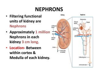
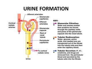


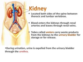
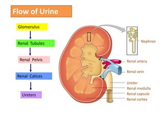
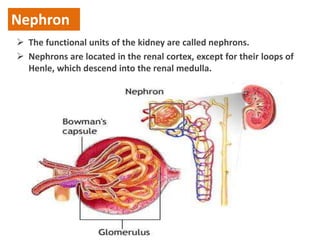
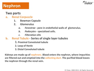
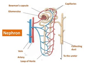
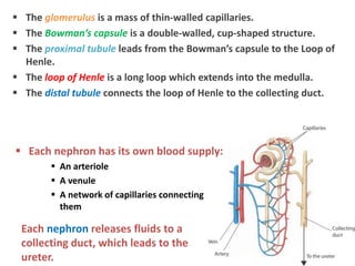
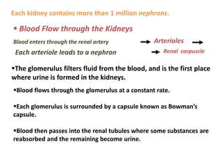

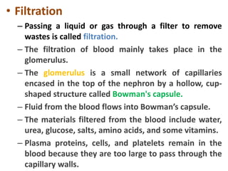
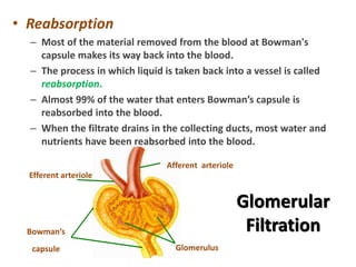
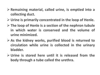


The genitourinary system consists of the kidneys, ureters, urinary bladder, and urethra. The kidneys filter waste from the blood to produce urine. Each kidney contains approximately 1 million nephrons, which are the functional filtering units of the kidney. In the nephrons, blood is filtered in the glomerulus and most of the filtrate is reabsorbed, with the remaining filtrate becoming urine. Urine travels from the kidneys through the ureters to the urinary bladder, where it is temporarily stored until urination through the urethra.


