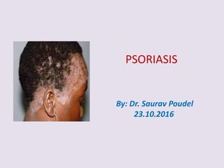
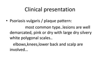
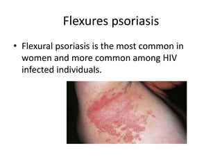
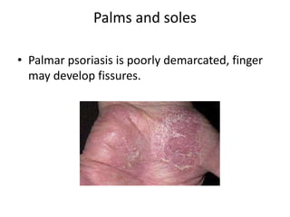
Psoriasis is a chronic, inflammatory skin condition characterized by well-circumscribed red patches covered with silvery scales. It is caused by a combination of genetic and environmental factors like stress, infection, and certain medications. Common types include plaque, guttate, scalp, and nail psoriasis. Symptoms include itching, burning, and discomfort. Diagnosis is usually made clinically but skin biopsy may show hyperkeratosis, parakeratosis, and inflammatory infiltrate. Treatment involves topical corticosteroids and vitamin D analogues for mild cases or phototherapy and systemic medications like methotrexate for severe cases. Complications can include secondary infection and psoriatic arthritis.
Invasive Squamous Cell Carcinoma (SCC)
SCC of the skin is a malignant tumor of keratinocytes, arising in the epidermis.
SCC usually arises in epidermal precancerous lesions and, depending on etiology and level of differentiation, varies in its aggressiveness.
The lesion is a plaque or a nodule with varying degrees of keratinization in the nodule and/or on the surface.
Thumb rule:
Undifferentiated SCC: is soft and has no hyperkeratosis;
Differentiated SCC: is hard on palpation and has hyperkeratosis.
Exposure:
Sunlight. Phototherapy, PUVA (oral psoralen + UVA). Excessive photochemotherapy can lead to promotion of SCC, particularly in patients with skin phototypes I and II or in patients with history of previous exposure to ionizing radiation or methotrexate treatment for psoriasis.
Lesions :
Indurated papule, plaque, or nodule ; adherent thick keratotic scale or hyperkeratosis ; when eroded or ulcerated, the lesion may have a crust in the center and a firm, hyperkeratotic, elevated margin
Clark levels
level I, intra-epidermal;
level II, invades papillary dermis;
level III fills papillary dermis;
level IV, invades reticular dermis;
level V, invades subcutaneous fat.
Erysipelas is a bacterial skin infection that usually affects the top most layer of the skin. Erysipelas is very rare, but requires immediate treatment. Erysipelas is often associated with other skin infection known as cellulitis, which affects the lower layers of the skin.
Aurafix Sakro İliak Korse ürünü ile kalça, bel ve basen bölgesindeki ağrıları gidermek için destek sağlayabilirsiniz. Ürünün detayları için http://www.portakalrengi.com/aurafix-sakro-iliak-korse adresini, diğer Aurafix ürünlerinin detayları içinse http://www.portakalrengi.com/aurafix adresini ziyaret edebilirsiniz.
history of TB,epidemiology, clinical features, lab diagnosis, treatment, MDR TB, XDR TB, TDR TB, and mechanism of drug resistant, methods of identification of resistant drugs
This is a presentation on cutaneous manifestations of tuberculosis. tuberculosis is a very important disease especially in the sub-Saharan region.
The pictures are not mine( from internet sites) and the study material majorly used was Fitzpatrick dermatology and extrapulmonary TB by Alper Senner. If anyone feels like some of the information is from their site and has been wrongly used do contact me via : lilacpreton12@gmail.com . This information is only for educational purposes.
Include infections of skin, subcutaneous tissue, fascia, and muscle, encompass a wide spectrum of clinical presentations, ranging from simple cellulitis to rapidly progressive necrotizing fasciitis.
Diagnosing the exact extent of the disease is critical for successful management of a patient of soft tissue infection
2. • Defination:
it is a chronic,recurrent,non-
infectious,inflammatory disorder of the skin
characterized by well circumscribed
erythematous dry scaling plaques covered by
silvery white scales distributed over the
extensor of the body
(scalp,knee,elbow,sacral regions).
It appears most often between 15 and 40
years.
4. • Pre disposing factor :
stress -90%
infection- RTI,UTI,HIV.
drugs: beta-blocker
lipid lowering agent
chloroquine
NSAIDs
hormonal factor
sunlight
trauma
family history
5. Clinical presentation
• Psoriasis vulgaris / plaque pattern:
most common type..lesions are well
demarcated, pink or dry with large dry silvery
white polygonal scales..
elbows,knees,lower back and scalp are
involved…
7. Guttate psoriasis
• Seen usually in children and adolscents…
may be first sign of the disease,often triggered
by streptococcal tonsillitis.
Numerous small round red macules come up
suddenly in the trunk..
17. • Auspitz’s sign: appearance of bleeding points
on forcible removal of the scales done by glass
slides.
18. • Koebner’s sign: development of isomorphic
lesion at the site of scratch,trauma,burn and
incision.
19. INVESTIGATION
• Routine investigation:
I. CBC- Normocytic Normochromic Anaemia,
Increased ESR,WBC raised.
II. Random blood sugar.
III. Urine routine examination.
IV. Chest x-ray.
20. Specific Investigation
• Skin bioposy:
• FINDINGS:
hyperkeratosis: increase thickness of stratum
corneum.
Parakeratosis: presence of nucleated cells in
s.corneum.
Micromunra abscess: collection of neutrophil in
s.corneum.
21. • Spongiosis : increased thickness of stratum
spinosum.
• Dilated and tortous capillary loops in the
dermal papillae.
• T-lymphocyte infiltrate in upper dermis.
22. General Management
• Assurance, explanation of disease.
• Aviodance of precipitating factor.
• Control of secondary infection.
24. Specific management
• Methotraxate:
Dose: 2.5 mg or 5 mg 12hrly, 3 doses in a week for 3-12 months
with folic acid supplementation. Duration of treatment depends on
patient’s condition.
* systemic retinoids: Acitrectin.
• antibiotic, antifungal.
• Photo chemotherapy: Psoralen and ultraviolet A.
During giving methotrexate & during treatment:
- complete blood count
- liver function test
- renal function test must be done.
** Methotrexate is contra-indicated in pregnancy.
** Never prescribe systemic steroid in psoriasis because it flares up
the condition.
25. complication
• Secondary bacterial infection.
• Exfoliative dermatitis
• Psoriatic arthropathy.
• Nail psoriasis leads to destruction of whole of
the nail.