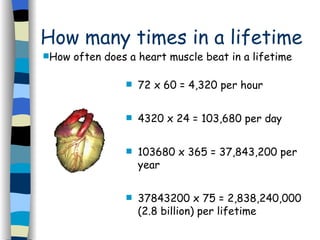
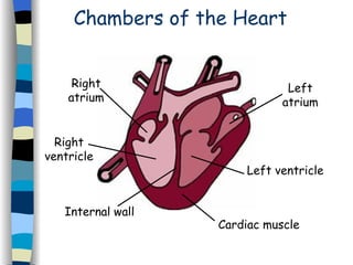
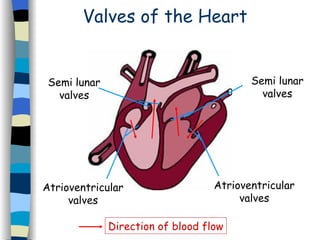
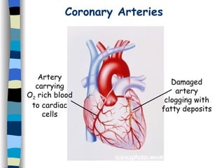
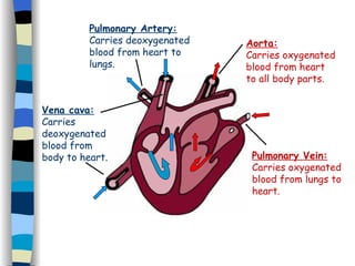
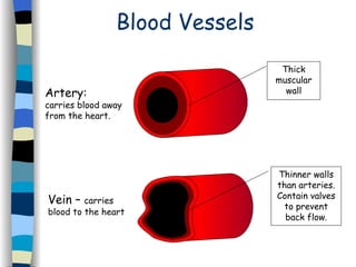
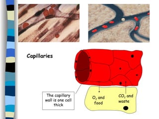
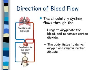
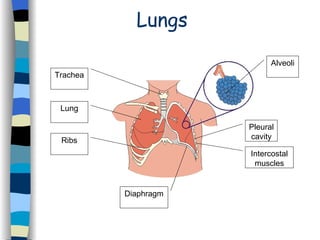
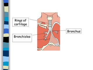
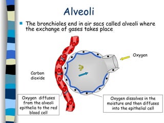
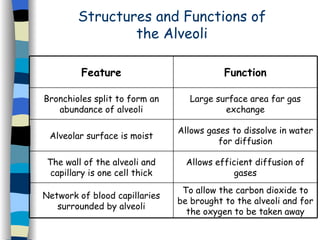
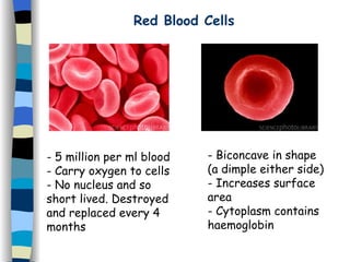
11.1 The Heart Today’s Learning Objectives: State the function of the heart. Name the type of tissue which makes up the walls of the heart. Name the four chambers of the heart and state their functions. Name the types of valve found in the heart. State the function of the valves in the heart. Name the blood vessels which supply heart tissue with blood. Describe what might happen if the blood supply to the heart is interrupted.
How many times in a lifetime 72 x 60 = 4,320 per hour  4320 x 24 = 103,680 per day  103680 x 365 = 37,843,200 per year  37843200 x 75 = 2,838,240,000 (2.8 billion) per lifetime How often does a heart muscle beat in a lifetime
Chambers of the Heart Right atrium Right ventricle Left atrium Left ventricle Internal wall Cardiac muscle
Valves of the Heart Atrioventricular valves Semi lunar valves Semi lunar valves Atrioventricular valves Direction of blood flow
Coronary Arteries Artery carrying O 2  rich blood to cardiac cells Damaged artery clogging with fatty deposits
11.2 Circulatory System Today’s Learning Objectives: Name the blood vessels which enter and leave the heart. Describe the path of blood flow through the heart. Explain why the left ventricle has a thicker wall than the right ventricle. State the function of arteries, veins and capillaries. State which type of blood vessel has a pulse. Name the main blood vessels that serve the liver, the alimentary canal and the kidneys.
Vena cava: Carries deoxygenated blood from body to heart. Pulmonary Artery: Carries deoxygenated blood from heart to lungs. Pulmonary Vein: Carries oxygenated blood from lungs to heart. Aorta: Carries oxygenated blood from heart to all body parts.
Blood Vessels Artery:  carries blood away from the heart. Vein –  carries blood to the heart Thick muscular wall Thinner walls than arteries. Contain valves to prevent back flow.
The capillary wall is one cell thick O 2  and food CO 2  and waste Capillaries
Direction of Blood Flow The circulatory system flows through the  Lungs to oxygenate the blood, and to remove carbon dioxide.  The body tissue to deliver oxygen and remove carbon dioxide. Capillaries in the lungs Capillaries in the body tissues
11.3 Gas Exchange Today’s Learning Objectives: Describe how oxygen reaches the gas exchange surfaces of the lungs. Name the structure in which gas exchange takes place in the lungs. Name the gases exchanged at the gas exchange surfaces in the lungs. Describe the exchange of oxygen between the alveoli and the blood. Describe the exchange of carbon dioxide between the blood and the alveoli. Describe the structural features which make gas exchange more efficient.
Lungs Trachea Lung Ribs Diaphragm Pleural cavity Intercostal muscles Alveoli
Bronchioles Rings of cartilage Bronchus
Alveoli The bronchioles end in air sacs called alveoli where the exchange of gases takes place Oxygen Carbon  dioxide Oxygen dissolves in the moisture and then diffuses into the epithelial cell Oxygen  diffuses from the alveoli epithelia to the red blood cell
Structures and Functions of the Alveoli To allow the carbon dioxide to be brought to the alveoli and for the oxygen to be taken away Network of blood capillaries surrounded by alveoli Allows efficient diffusion of gases The wall of the alveoli and capillary is one cell thick Allows gases to dissolve in water for diffusion Alveolar surface is moist Large surface area far gas exchange Bronchioles split to form an abundance of alveoli Function Feature
11.4 The Blood Today’s Learning Objectives: Identify and name the components of the blood. State the main function of the red blood cells. Describe the structural features of red blood cells that increase their efficiency in carrying oxygen. Name the pigment in red blood cells. Give two ways that carbon dioxide can be carried in the blood. Explain why only a small percentage of carbon dioxide is carried directly dissolved in the plasma. Describe how digested food products are carried in the blood.
Blood The average person has about 5 litres. Blood is made up of a pale yellow liquid called  plasma  which contains: -Blood cells (red/white) -Dissolved substances (glucose, amino acids, salts,  vitamins, hormones, urea etc.) -Blood proteins (e.g. antibodies)
Red Blood Cells 5 million per ml blood Carry oxygen to cells No nucleus and so short lived. Destroyed and replaced every 4 months Biconcave in shape (a dimple either side) Increases surface area Cytoplasm contains haemoglobin
White Blood Cells 5000 white blood cells per ml of blood Have nuclei but not haemoglobin Vital for a healthy immune system
11.5 Haemoglobin Today’s Learning Objectives: Describe the structure of haemoglobin. Explain the need for haemoglobin. Write the equation for the association and dissociation of haemoglobin and oxygen. Describe the circumstances where oxygen will associate with haemoglobin. Describe the circumstances where oxygen will dissociate from haemoglobin. Describe the colours of haemoglobin and oxyhaemoglobin.
Haemoglobin Protein chains (blue) Haem groups (pink) -Each haem group contains an atom of iron which allows an oxygen molecule to attach. -One haem group binds with one oxygen molecule, so each molecule of haemoglobon can carry four oxygen molecules.
When there is a high O 2  concentration (e.g in lungs) haemoglobin associates with O 2. When there is a low O 2  concentration (e.g in tissue) haemoglobin dissociates with O 2. oxyhaemoglobin haemoglobin  + oxygen haemoglobin  + oxygen oxyhaemoglobin
11.6 Body Defences Today’s Learning Objectives: What is meant by a pathogen. Describe some of the body’s first line defences. What is a macrophage. Describe the structure and function of a macrophage. Where are macrophages found in the body and what defence do they provide. What is meant by phagocytosis. Describe the stages of phagocytosis. State the role of lysosomes in phagocytosis.
A  pathogen  is an organism that can cause disease in the body. The body’s  first line defences  prevent the entry of these harmful organisms. First line defences can act as a barrier: skin, mucus First line defences can act to discourage pathogens: saliva, tears, acid, harmless bacteria First Line Defences
Second Line Defences These deal with pathogens that have made it past the first line defence. A macrophage is a special type of large white blood cell which can engulf and digest bacteria. Macrophages  engulf and digest  bacteria, dead cells, small particles and unwanted matter. This is a very  general response  and not specific against any one pathogen.
Phagocytosis Phagocytosis is the process by which white blood cells engulf and breakdown bacteria etc. http:// highered . mcgraw -hill.com/sites/0072495855/student_view0/chapter2/animation__phagocytosis.html
11.7 Antibody Production Today’s Learning Objectives: Describe lymphocytes and state their function. Define the term ‘ antigen ’. Define the term ‘ antibody ’. Describe the events which take place during the immune response. Draw diagrams to represent what happens in an antibody-antigen reaction. Explain the term ‘ passive immunity’  and ‘ active immunity’ . Describe how vaccines work.
General Immunity: -Non- specific. -Protects against a  wide variety  of microorganisms. - Phagocytosis  by macrophages is an example. Specific Immunity: - Antibodies  are produced by white blood cells. -The antibodies recognise and destroy  specific  microorganisms.
Producing Antibodies The surface of all cells and viruses have substances known as  antigens . These  antigens  are  specific  to the cell and act as an identity tag. A persons  own cells  have  self-antigens  which are not recognised as harmful. Bacteria and viruses  carry  non-self antigens  and are then recognised as potentially harmful.
There are millions of different  lymphocytes  (type of white blood cell). Each  lymphocyte  will recognise  one specific   non-self antigen. The  lymphocyte  will then produce an  antibody  which will specifically attack that  antigen. Antibodies (Y-shape structure) produced by a specific  lymphocyte  attack the   antigens. Bacteria or virus with  non-self antigens
Passive Immunity: The antibody is  passed on ready made  into the body e.g. from a mother to a new born via breast milk. This form of immunity is temporary. Active Immunity: Antibodies are produced in the body as a  response to disease antigens  or  vaccines. Vaccines  use  altered pathogens  that stimulate the immune system without causing disease!

11. Circulation

  • 1.
    11.1 The HeartToday’s Learning Objectives: State the function of the heart. Name the type of tissue which makes up the walls of the heart. Name the four chambers of the heart and state their functions. Name the types of valve found in the heart. State the function of the valves in the heart. Name the blood vessels which supply heart tissue with blood. Describe what might happen if the blood supply to the heart is interrupted.
  • 2.
    How many timesin a lifetime 72 x 60 = 4,320 per hour 4320 x 24 = 103,680 per day 103680 x 365 = 37,843,200 per year 37843200 x 75 = 2,838,240,000 (2.8 billion) per lifetime How often does a heart muscle beat in a lifetime
  • 3.
    Chambers of theHeart Right atrium Right ventricle Left atrium Left ventricle Internal wall Cardiac muscle
  • 4.
    Valves of theHeart Atrioventricular valves Semi lunar valves Semi lunar valves Atrioventricular valves Direction of blood flow
  • 5.
    Coronary Arteries Arterycarrying O 2 rich blood to cardiac cells Damaged artery clogging with fatty deposits
  • 6.
    11.2 Circulatory SystemToday’s Learning Objectives: Name the blood vessels which enter and leave the heart. Describe the path of blood flow through the heart. Explain why the left ventricle has a thicker wall than the right ventricle. State the function of arteries, veins and capillaries. State which type of blood vessel has a pulse. Name the main blood vessels that serve the liver, the alimentary canal and the kidneys.
  • 7.
    Vena cava: Carriesdeoxygenated blood from body to heart. Pulmonary Artery: Carries deoxygenated blood from heart to lungs. Pulmonary Vein: Carries oxygenated blood from lungs to heart. Aorta: Carries oxygenated blood from heart to all body parts.
  • 8.
    Blood Vessels Artery: carries blood away from the heart. Vein – carries blood to the heart Thick muscular wall Thinner walls than arteries. Contain valves to prevent back flow.
  • 9.
    The capillary wallis one cell thick O 2 and food CO 2 and waste Capillaries
  • 10.
    Direction of BloodFlow The circulatory system flows through the Lungs to oxygenate the blood, and to remove carbon dioxide. The body tissue to deliver oxygen and remove carbon dioxide. Capillaries in the lungs Capillaries in the body tissues
  • 11.
    11.3 Gas ExchangeToday’s Learning Objectives: Describe how oxygen reaches the gas exchange surfaces of the lungs. Name the structure in which gas exchange takes place in the lungs. Name the gases exchanged at the gas exchange surfaces in the lungs. Describe the exchange of oxygen between the alveoli and the blood. Describe the exchange of carbon dioxide between the blood and the alveoli. Describe the structural features which make gas exchange more efficient.
  • 12.
    Lungs Trachea LungRibs Diaphragm Pleural cavity Intercostal muscles Alveoli
  • 13.
    Bronchioles Rings ofcartilage Bronchus
  • 14.
    Alveoli The bronchiolesend in air sacs called alveoli where the exchange of gases takes place Oxygen Carbon dioxide Oxygen dissolves in the moisture and then diffuses into the epithelial cell Oxygen diffuses from the alveoli epithelia to the red blood cell
  • 15.
    Structures and Functionsof the Alveoli To allow the carbon dioxide to be brought to the alveoli and for the oxygen to be taken away Network of blood capillaries surrounded by alveoli Allows efficient diffusion of gases The wall of the alveoli and capillary is one cell thick Allows gases to dissolve in water for diffusion Alveolar surface is moist Large surface area far gas exchange Bronchioles split to form an abundance of alveoli Function Feature
  • 16.
    11.4 The BloodToday’s Learning Objectives: Identify and name the components of the blood. State the main function of the red blood cells. Describe the structural features of red blood cells that increase their efficiency in carrying oxygen. Name the pigment in red blood cells. Give two ways that carbon dioxide can be carried in the blood. Explain why only a small percentage of carbon dioxide is carried directly dissolved in the plasma. Describe how digested food products are carried in the blood.
  • 17.
    Blood The averageperson has about 5 litres. Blood is made up of a pale yellow liquid called plasma which contains: -Blood cells (red/white) -Dissolved substances (glucose, amino acids, salts, vitamins, hormones, urea etc.) -Blood proteins (e.g. antibodies)
  • 18.
    Red Blood Cells5 million per ml blood Carry oxygen to cells No nucleus and so short lived. Destroyed and replaced every 4 months Biconcave in shape (a dimple either side) Increases surface area Cytoplasm contains haemoglobin
  • 19.
    White Blood Cells5000 white blood cells per ml of blood Have nuclei but not haemoglobin Vital for a healthy immune system
  • 20.
    11.5 Haemoglobin Today’sLearning Objectives: Describe the structure of haemoglobin. Explain the need for haemoglobin. Write the equation for the association and dissociation of haemoglobin and oxygen. Describe the circumstances where oxygen will associate with haemoglobin. Describe the circumstances where oxygen will dissociate from haemoglobin. Describe the colours of haemoglobin and oxyhaemoglobin.
  • 21.
    Haemoglobin Protein chains(blue) Haem groups (pink) -Each haem group contains an atom of iron which allows an oxygen molecule to attach. -One haem group binds with one oxygen molecule, so each molecule of haemoglobon can carry four oxygen molecules.
  • 22.
    When there isa high O 2 concentration (e.g in lungs) haemoglobin associates with O 2. When there is a low O 2 concentration (e.g in tissue) haemoglobin dissociates with O 2. oxyhaemoglobin haemoglobin + oxygen haemoglobin + oxygen oxyhaemoglobin
  • 23.
    11.6 Body DefencesToday’s Learning Objectives: What is meant by a pathogen. Describe some of the body’s first line defences. What is a macrophage. Describe the structure and function of a macrophage. Where are macrophages found in the body and what defence do they provide. What is meant by phagocytosis. Describe the stages of phagocytosis. State the role of lysosomes in phagocytosis.
  • 24.
    A pathogen is an organism that can cause disease in the body. The body’s first line defences prevent the entry of these harmful organisms. First line defences can act as a barrier: skin, mucus First line defences can act to discourage pathogens: saliva, tears, acid, harmless bacteria First Line Defences
  • 25.
    Second Line DefencesThese deal with pathogens that have made it past the first line defence. A macrophage is a special type of large white blood cell which can engulf and digest bacteria. Macrophages engulf and digest bacteria, dead cells, small particles and unwanted matter. This is a very general response and not specific against any one pathogen.
  • 26.
    Phagocytosis Phagocytosis isthe process by which white blood cells engulf and breakdown bacteria etc. http:// highered . mcgraw -hill.com/sites/0072495855/student_view0/chapter2/animation__phagocytosis.html
  • 27.
    11.7 Antibody ProductionToday’s Learning Objectives: Describe lymphocytes and state their function. Define the term ‘ antigen ’. Define the term ‘ antibody ’. Describe the events which take place during the immune response. Draw diagrams to represent what happens in an antibody-antigen reaction. Explain the term ‘ passive immunity’ and ‘ active immunity’ . Describe how vaccines work.
  • 28.
    General Immunity: -Non-specific. -Protects against a wide variety of microorganisms. - Phagocytosis by macrophages is an example. Specific Immunity: - Antibodies are produced by white blood cells. -The antibodies recognise and destroy specific microorganisms.
  • 29.
    Producing Antibodies Thesurface of all cells and viruses have substances known as antigens . These antigens are specific to the cell and act as an identity tag. A persons own cells have self-antigens which are not recognised as harmful. Bacteria and viruses carry non-self antigens and are then recognised as potentially harmful.
  • 30.
    There are millionsof different lymphocytes (type of white blood cell). Each lymphocyte will recognise one specific non-self antigen. The lymphocyte will then produce an antibody which will specifically attack that antigen. Antibodies (Y-shape structure) produced by a specific lymphocyte attack the antigens. Bacteria or virus with non-self antigens
  • 31.
    Passive Immunity: Theantibody is passed on ready made into the body e.g. from a mother to a new born via breast milk. This form of immunity is temporary. Active Immunity: Antibodies are produced in the body as a response to disease antigens or vaccines. Vaccines use altered pathogens that stimulate the immune system without causing disease!