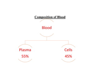
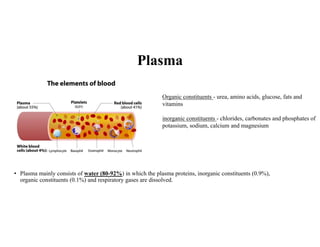
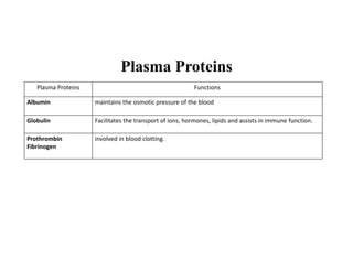
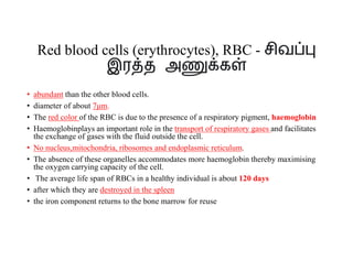
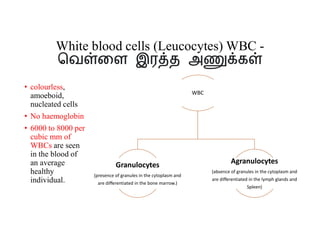
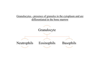
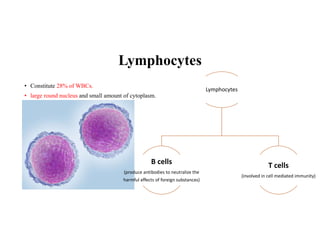
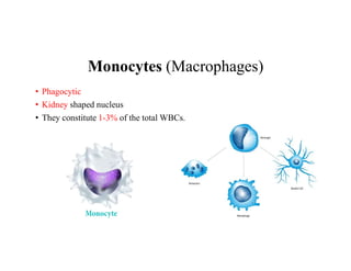
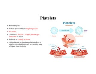
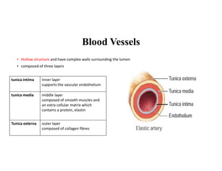
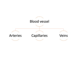
3. Plasma
• Plasma mainly consists of water (80-92%) in which the plasma proteins, inorganic constituents (0.9%),
organic constituents (0.1%) and respiratory gases are dissolved.
Organic constituents - urea, amino acids, glucose, fats and
vitamins
inorganic constituents - chlorides, carbonates and phosphates of
potassium, sodium, calcium and magnesium
4. Plasma Proteins
Plasma Proteins Functions
Albumin maintains the osmotic pressure of the blood
Globulin Facilitates the transport of ions, hormones, lipids and assists in immune function.
Prothrombin
Fibrinogen
involved in blood clotting.
5. Formed elements/ Cells
• Red blood cells (erythrocytes), RBC
• White blood cells (Leucocytes) WBC
• Platelets
6. Red blood cells (erythrocytes), RBC - சிவ
இர த அ க
• abundant than the other blood cells.
• diameter of about 7μm.
• The red color of the RBC is due to the presence of a respiratory pigment, haemoglobin
• Haemoglobinplays an important role in the transport of respiratory gases and facilitates
the exchange of gases with the fluid outside the cell.
• No nucleus,mitochondria, ribosomes and endoplasmic reticulum.
• The absence of these organelles accommodates more haemoglobin thereby maximising
the oxygen carrying capacity of the cell.
• The average life span of RBCs in a healthy individual is about 120 days
• after which they are destroyed in the spleen
• the iron component returns to the bone marrow for reuse
7. White blood cells (Leucocytes) WBC -
ெவ ைள இர த அ க
• colourless,
amoeboid,
nucleated cells
• No haemoglobin
• 6000 to 8000 per
cubic mm of
WBCs are seen
in the blood of
an average
healthy
individual.
WBC
Granulocytes
(presence of granules in the cytoplasm and
are differentiated in the bone marrow.)
Agranulocytes
(absence of granules in the cytoplasm and
are differentiated in the lymph glands and
Spleen)
9. Neutrophils
• heterophils or polymorphonuclear
• 3-4 lobes of nucleus connected with delicate threads
• constitute about 60%-65% of the total WBCs.
• They are phagocytic in nature and appear in large numbers in and around the
infected tissues
10. Eosinophils
• bilobed nucleus and the lobes are joined by thin strands.
• They are non-phagocytic
• constitute about 2-3% of the total WBCs.
• Eosinophils increase during certain types of parasitic infections and allergic
reactions.
11. Basophils
• less numerous than any other type of WBCs
• constituting 0.5%-1.0% of the total number of leucocytes.
• The cytoplasmic granules are large sized
• Basophils secrete substances such as heparin, serotonin and
histamines.
• They are also involved in inflammatory reactions.
13. Lymphocytes
• Constitute 28% of WBCs.
• large round nucleus and small amount of cytoplasm.
Lymphocytes
B cells
(produce antibodies to neutralize the
harmful effects of foreign substances)
T cells
(involved in cell mediated immunity)
15. Platelets
• thrombocytes
• that are produced from megakaryocytes
• No nuclei.
• contains 1, 50,000-3, 50,000 platelets per
cubic mm of blood.
• involved in clotting of blood.
• The reduction in platelet number can lead to
clotting disorders that result in excessive loss
of blood from the body.
16. Blood Vessels
• Hollow structure and have complex walls surrounding the lumen
• composed of three layers
tunica intima Inner layer
supports the vascular endothelium
tunica media middle layer
composed of smooth muscles and
an extra cellular matrix which
contains a protein, elastin
Tunica externa outer layer
composed of collagen fibres
18. Arteries
• The blood vessels that carry blood away from the heart are called arteries.
• The arteries usually lie deep inside the body.
• The walls of the arteries are thick, non collapsible to withstand high pressure.
• Valves are absent and have a narrow lumen.
• All arteries carry oxygenated blood, except the pulmonary artery. The largest
artery, the aorta (2.5 cm in diameter and 2 mm thick) branch into smaller arteries
and culminates into the tissues as feed arteries.
• In the tissues the arteries branches into arterioles.
19. Capillaries
• thin walled and consist of single layer of squamous epithelium.
• Tunica media and elastin fibres are absent.
• The capillary beds are the site for exchange of materials between blood and tissues.
• The walls of the capillaries are guarded by semilunar valves.
• The blood volume in the capillaries is high but the flow of blood is slow.
• Mixed blood (oxygenated and deoxygenated) is present in the capillaries.
20. Veins
• Veins have thinner walls and a larger lumen and hence can be easily stretched.
• They carry deoxygenated blood except, the pulmonary vein.
• The blood pressure is low and the lumen has a wide wall which is collapsible.
• Tunica media is thinner in veins than in arteries.
• Unidirectional flow of blood in veins is due to the presence of semilunar valves that prevents backflow of
blood.
• Blood samples are usually taken from the veins rather than artery because of low pressure in the veins.
21. Human Heart
• made of special type of muscle called the cardiac muscle.
• situated in the thoracic cavity and its apex portion is slightly tilted towards
left.
• It weighs about 300g in an adult.
• Heart is divided into four chambers, upper two small atrium and lower two
large ventricles.
• Layers Of Heart
• Myocardium– The myocardium includes heart muscle cells that make up the
bulk of heart wall and the middle layer.
• Epicardium – The outer layer of cells is known as epicardium and the second
layer which is membranous surrounds and protects the heart.
• Endocardium -The endocardium lines the inner wall of the heart
• The space present between the membranes is called pericardial space and is
filled with pericardial fluid.
22. Human Heart
• The two auricles are separated by inter
auricular septum
• The two ventricles are separated by inter
ventricular septum
• The separation of chambers avoids mixing
of oxygenated and deoxygenated blood
• The opening between the right atrium and
the right ventricle is guarded by the
tricuspid valve
• bicuspid or mitral valve guards the opening
between the left atrium and left ventricle
• The valves of the heart allows the blood to
flow only in one direction, i.e., from the
atria to the ventricles and from the
ventricles to the pulmonary artery or the
aorta.
• The opening of right and left ventricles into
the pulmonary artery and aorta are
guarded by aortic and pulmonary valves
and are called semilunar valves.
• Heart receives deoxygenated blood from
various parts of the body through the
inferior venacava and superior venacava
which open into the right auricle.
Oxygenated blood from lungs is drained
into the left auricle through four pulmonary
veins.