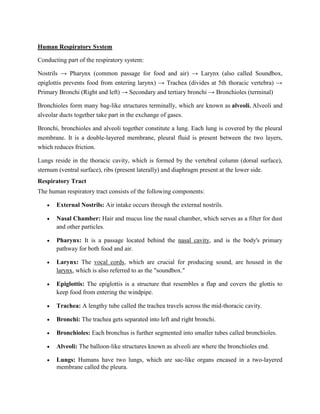
Respiration
We need energy to perform various activities. This energy is derived from the catabolism of
various components of food, e.g. proteins, carbohydrates, fats, etc. Oxygen is required for
catabolic processes and carbon dioxide is released in the process. So the body requires a
continuous exchange of gases, oxygen from the atmosphere is taken inside and carbon dioxide
produced is taken out. This process of gaseous exchange is called breathing or respiration.
The network of organs and tissues that makes up the human respiratory system aids in breathing.
This system's main job is to get more oxygen into the body and get more carbon dioxide out of it.
The human respiratory system is comprised of a group of organs and tissues that aids in
breathing. In addition to the lungs, the respiration process is aided by muscles and a complex
network of blood arteries.
 The only organs that can float on water are the lungs. Alveoli, which resemble small
balloons, are found in each of your lungs and are responsible for replacing the carbon
dioxide waste in your blood with oxygen.
 70% of the waste gases like carbon di oxide is expelled when you breathe.
 The alveoli are tiny air sacs located in the lungs where gas exchange takes place. The
adult lungs contain between 300 million and 500 million alveoli.
All the animals have different organs specialised for the gaseous exchange.
Mode of gaseous
exchange
Respiratory
organs
Examples
Cutaneous
respiration
Skin Through body surface- poriferans,
coelenterates, flatworms
Moist cuticle- Earthworms
Moist skin- Amphibians (frogs)
Tracheal Spiracles Arthropods (cockroaches)
Branchial
respiration
Gills Aquatic animals- arthropods,
molluscs and fishes
Pulmonary
respiration
Lungs Terrestrial vertebrates including
human
Human Respiratory System
Conducting part of the respiratory system:
Nostrils → Pharynx (common passage for food and air) → Larynx (also called Soundbox,
epiglottis prevents food from entering larynx) → Trachea (divides at 5th thoracic vertebra) →
Primary Bronchi (Right and left) → Secondary and tertiary bronchi → Bronchioles (terminal)
Bronchioles form many bag-like structures terminally, which are known as alveoli. Alveoli and
alveolar ducts together take part in the exchange of gases.
Bronchi, bronchioles and alveoli together constitute a lung. Each lung is covered by the pleural
membrane. It is a double-layered membrane, pleural fluid is present between the two layers,
which reduces friction.
Lungs reside in the thoracic cavity, which is formed by the vertebral column (dorsal surface),
sternum (ventral surface), ribs (present laterally) and diaphragm present at the lower side.
Respiratory Tract
The human respiratory tract consists of the following components:
 External Nostrils: Air intake occurs through the external nostrils.
 Nasal Chamber: Hair and mucus line the nasal chamber, which serves as a filter for dust
and other particles.
 Pharynx: It is a passage located behind the nasal cavity, and is the body's primary
pathway for both food and air.
 Larynx: The vocal cords, which are crucial for producing sound, are housed in the
larynx, which is also referred to as the "soundbox."
 Epiglottis: The epiglottis is a structure that resembles a flap and covers the glottis to
keep food from entering the windpipe.
 Trachea: A lengthy tube called the trachea travels across the mid-thoracic cavity.
 Bronchi: The trachea gets separated into left and right bronchi.
 Bronchioles: Each bronchus is further segmented into smaller tubes called bronchioles.
 Alveoli: The balloon-like structures known as alveoli are where the bronchioles end.
 Lungs: Humans have two lungs, which are sac-like organs encased in a two-layered
membrane called the pleura.
Process of Respiration in Humans
Pulmonary respiration (intake of atmospheric oxygen and release of CO2 rich alveolar air) →
Diffusion of O2 and CO2 across the alveolar membrane → Transportation of gases by blood →
Diffusion of O2 and CO2 between blood and tissues → Cellular respiration (catabolism of food
using O2 to release energy and CO2)
With the aid of the nostrils, the air is inhaled, and the tiny hair follicles inside them clean the air
in the nasal cavity. A collection of blood vessels that warm the air is also present in the hollow.
This air then travels through the pharynx, larynx, and trachea.
Goblet cells (secretory cells) and ciliated epithelial cells line the trachea and bronchi, releasing
mucus to moisten the air as it travels through the respiratory tract. Additionally, it stops any tiny
dust particles or pathogens from entering the nasal passages. The rising action of the motile cilia
transports the mucus and other foreign particles back to the buccal cavity, where they are
removed. The ascending motion of the motile cilia causes the mucus and other foreign particles
to be taken back to the buccal cavity, where they can either be coughed out or expelled (or
swallowed.)
The bronchioles and alveoli are the next places the air goes after entering the bronchus.
Mechanism of Breathing
1. Inspiration: Due to the lower intrapulmonary pressure, atmospheric air is drawn inside.
The diaphragm and intercostal muscles contract, resulting in an increase in lung volume.
The air enters the lungs when the sternum and ribs rise. Atmospheric air is taken inside
due to lower intrapulmonary pressure. The pulmonary volume is increased due to the
contraction of the diaphragm and intercostal muscles. Ribs and sternum lift up and the air
moves into the lungs.
2. Expiration: Alveolar air is spat out because the intrapulmonary pressure is higher than
the ambient pressure during expiration. When the diaphragm and intercostal muscles
relax, the pressure rises. The diaphragm, sternum, and ribs all come back to their original
locations. The abdominal muscles can aid in increasing the volume of inhalation and
expiration. The average person breathes 12–16 times each minute. A spirometer is a
device for measuring the amount of air breathed in and exhaled. Alveolar air is expelled
out due to high intrapulmonary pressure as compared to atmospheric pressure. The
pressure increases with the relaxation of the diaphragm and intercostal muscles. Ribs,
sternum and the diaphragm return to their normal positions.
Inspiration and expiration volume can be increased by the help of muscles in the abdomen.
Normal breathing rate is 12-16 times/ minute.
Spirometer- An apparatus for measuring the volume of inhaled and exhaled air.
3. Exchange of gases: The exchange of gases occurs across a gradient in pressure and
concentration. Between blood and tissues as well as between alveoli, there is gas
exchange. Exchange of gases is across pressure/ concentration gradient. Exchange of
gases occurs between alveoli and blood and between blood and tissues.
The difference in the partial pressure of CO2 (pCO2) and O2 (pO2) in alveolar tissues, blood and
other body tissues, results in diffusion of gases across membranes.
4. Oxygen transport: The blood carries O2 throughout the body. RBCs carry 97% of the
oxygen, while 3% of it is dissolved in plasma. O2 is transported through the blood. 97%
of oxygen is carried by RBCs and 3% dissolved in plasma. Each Haemoglobin molecule
can bind to 4 molecules of oxygen forming oxyhaemoglobin. In the alveoli,
oxyhaemoglobin formation is favoured due to higher pO2 and low pCO2 and
H+
concentration. The Oxygen dissociation curve is sigmoid. In tissues, oxyhaemoglobin
dissociates due to low pO2.
Transport of oxygen in human beings: The oxygen is transported in human beings by
hemoglobin present in the red blood cells from the air to the lungs. The oxygen molecules can
combine with the hemoglobin molecules easily. The molecules release the oxygen to the tissues
that are deficient in oxygen.
5. Carbon dioxide transport: The production of carbaminohemoglobin allows for the
transportation of CO2 via the blood. This method of transporting CO2 involves 20–25
percent of it, 70 percent of it as bicarbonate, and 7 percent of it as plasma-dissolved CO2.
CO2 is transported through the blood by the formation of carbaminohemoglobin. 20-
25% of CO2 is transported this way, 70% as bicarbonate and 7% as dissolved in plasma.
When there is high pCO2 and low pO2, CO2 binding to Hb is preferred. Enzyme carbonic
anhydrase, present in RBCs facilitates the transportation of CO2. CO2 gets diffused to the
blood from tissues as there is a high concentration of CO2 in tissues due to catabolic
activity. The CO2 forms HCO3
–
and H+
. In alveolar tissue, pCO2 is low so bicarbonate is
converted to CO2 and H2O. Carbonic anhydrase catalyses both the reactions
CO2 + H2O ⇄ H2CO3 ⇄ HCO3
–
+ H+
Transport of carbon dioxide in human beings: It is mostly transported from body tissues in
the dissolved form in our blood plasma to the lungs where it diffuses from blood to air in the
lungs and then is expelled out through nostrils.
Respiratory Volumes and Capacities
Tidal volume (TV)- 500 ml, the volume of air inspired or expired normally
Inspiratory reserve volume (IRV)- 2500 to 3000 ml, additional air by forcible inspiration. This
along with the tidal volume makes Inspiratory Capacity (IC), i.e. TV + IRV
Expiratory reserve volume (ERV)- 1000 to 1100 ml, additional air by forcible expiration. This
along with the tidal volume makes Expiratory Capacity (EC), i.e. TV + ERV
Residual volume (RV)- 1100 to 1200 ml, the air in the lungs after forcible expiration. Residual
volume and ERV together represent Functional residual capacity (FRC), i.e. the air remaining
in the lungs after normal expiration (ERV + RV)
Vital capacity (VC)- the maximum volume of air that can be breathed in or out after forced
expiration or inspiration (IRV + ERV + TV)
Total Lung capacity (TLC)- VC + RV
Regulation of Respiration
The respiratory rhythm centre (medulla region) and pneumotaxic centre (pons region) regulate
the respiration activity according to the demand.
Disorders of Respiratory System
Asthma: It is caused due to inflammation in the respiratory tract. Symptoms include wheezing
and difficulty in breathing.
Emphysema: It is caused due to damage in alveolar walls. Smoking is one of the major causes
of the condition. There is shortness of breath even at rest.
Occupational respiratory disorder: Workers, who are exposed to a lot of dust are prone to get
long term inflammation and leading to fibrosis and lung damage.
The Function of the Respiratory System
The human respiratory system serves the following purposes:
1. Inhalation and Exhalation: Breathing is aided by the respiratory system (also known as
pulmonary ventilation.) The pharynx, larynx, trachea, and lungs all pass the air that is
inhaled through the nose. Through the same channel, the exhaled air is returned.
Pulmonary ventilation is aided by changes in the volume and pressure of the lungs. Gases
are exchanged between the bloodstream and the lungs.
2. Exchange of Gases between Lungs and Bloodstream: Millions of tiny sacs called
alveoli allow oxygen and carbon dioxide to enter and leave the lungs, respectively. When
oxygen is inhaled, it diffuses into pulmonary capillaries, binds to haemoglobin, and then
moves through the bloodstream. The blood's carbon dioxide diffuses into the alveoli and
is released during breathing.
3. Exchange of Gases between Bloodstream and Body Tissues: When it reaches the
capillaries, the blood releases the oxygen that it has been carrying around the body since
it left the lungs. The bodily tissues receive oxygen diffusion through the capillary walls.
Additionally, the carbon dioxide diffuses into the circulation, where it is then transported
to the lungs for exhalation.
4. The Vibration of the Vocal Cords: The arytenoid cartilage is moved by the laryngeal
muscles while speaking. The vocal cords are pushed together by these cartilages. The air
that is exhaled causes the vocal cords to vibrate and produce sound when it passes
through them.
5. Olfaction: Some substances in the air bind to nasal cavities when it enters the nasal
canals during breathing, activating the cilia's nervous system receptors. The brain
transmits the impulses to the olfactory bulbs.
Oxygen dissociation curve
 The effect of carbon dioxide and acidity favor the formation of Oxyhaemoglobin at low
concentration of CO2 and H+ ion and causes the dissociation of Oxyhaemoglobin
releasing O2 at high concentration of CO2 and H+ ion.
 This shift in curve of oxyhaemoglobin due to concentration of carbondioxide at given
partial pressure of oxygen, is known as Bohr effect.
 The amount of Oxygen take up by Haemoglobin at particular time to from
Oxyhaemoglobin is called percentage saturation.
 The graph of percentage of O2 saturation of haemoglobin plotted against partial pressure
of Oxygen (PO2) is called Oxygen dissociation curve.
 The Oxygen dissociation curve is S-shaped (sigmoidal shape).
 The curve indicates that haemoglobin has high affinity to Oxygen.
 In human arterial blood have PO2 of about 95-100 mmHg, at this level percentage of O2
saturation of Hb is about 97 %. This indicates the formation of Oxyhaemoglobin is
favored.
 Similarly, the venous blood have PO2 of 40mmHg,at this level percentage of O2
saturation of Hb is about 70%.
Effect of Carbon-dioxide on Oxygen dissociation curve:
 The effect of CO2 on Oxygen dissociation curve is known as Bohr effect.
 It has been found that increase in concentration of CO2 decreases the amount of
oxyhaemoglobin formation.
 According to Bohr effect, for any particular partial pressure of Oxygen, the affinity of
Haemoglobin toward Oxygen decreases and favors dissociation of oxyhaemoglobin when
the partial pressure of carbondioxide increases.
 It means, higher CO2 concentration causes the dissociation of HbO2 releasing free O2.
 Increase in PCO2 shifts the O2 dissociation curve downwards. Higher PCO2 lowers the
affinity of haemoglobin for O2.
Fig. Oxygen dissociation curve of haemoglobin at different partial pressure of CO2
 Bohr effect is very important physiological phenomenon, because uptake of oxygen in
lungs and its releases in the tissue is regulated by the concentration of CO2 and H+ ion as
well as the partial pressure of O2. So, this phenomenon made possible the cellular
transport and release of O2.
 PCO2 is lower in lungs than tissue, so Hb has higher affinity for O2, therefore it favors
HbO2 formation and transport of O2 from lungs to tissue. similarly PCO2 is higher in
tissue, so it favors dissociation of HbO2 releasing free O2 and transport of CO2 from
tissue to lungs.
Reason for its sigmoidal pattern:
1. The oxygen dissociation curve is in a sigmoid shape or S-shaped because of the co-
operative binding of oxygen to haemoglobin.
2. The oxygen dissociation curve is obtained by plotting the percentage saturation of
haemoglobin with oxygen against the partial pressure of oxygen.
Chloride shift/Hamburger effect
The chloride shift or "Hamburger effect" describes the movement of chloride into RBCs
which occurs when the buffer effects of deoxygenated haemoglobin increase the intracellular
bicarbonate concentration, and the bicarbonate is exported from the RBC in exchange for
chloride. This results in a difference of 2-4 mmol/L of chloride between the arterial and venous
blood (and a similar difference in bicarbonate concentration).
The greater proportion (70%) of carbon dioxide is transported in the form of bicarbonates. The
CO2 reacted with the water of the cytoplasm in the presence of enzyme carbonic anhydrase to
form carbonic acid. The carbonic acid (H2CO3) is a weak acid, which undergoes partial
dissociation to yield hydrogen ion (H+) and bicarbonate ion (HCO3-). The given reaction mostly
occur inside RBCs, because the enzyme carbonic anhydrase is abundant there.
 In RBCs, CO2 combines with water to from carbonic acid which dissociates to gives H+
ion and bicarbonate (HCO3-) ion in the presence of enzyme carbonic anhydrase.
 The bicarbonate ion then diffuses outside the RBC in the plasma and combines with
Sodium ions to from Sodium bicarbonate (NaHCO3).
 Loss of bicarbonate ions from RBC causes positive charge inside RBC which is balanced
by diffusion of chloride (Cl-) ion from plasma into the RBC.
 This exchange of Cl- ion and HCO3- ion between plasma and RBC is known as chloride
shift.
 This phenomenon of chloride shift maintain the electrical neutrality of cell.
 This phenomenon is also known as Hamburger phenomenon.
 Reverse of chloride shift occurs in tissues.
The mechanism of the chloride shift:
The molecular mechanisms for the chloride shift are described in detail below. In summary, this
phenomenon is only possible because of the presence of carbonic anhydrase in RBCs. It is seen
as a critically important element (as it is concentrated there, but essentially absent from the
bloodstream otherwise). Without it, the reaction converting CO2 to HCO3
-
would be painfully
slow. With massive amounts of erythcyte carbonic anhydrase, we can instead count on these
molecular transactions to be complete in the space of one circulatory time. In fact, because all the
required proteins are available in massive concentrations, the reaction is incredibly fast. Wieth &
Brahm (1980) had determined that 99% of the chloride shift process is complete within about
700 milliseconds.
In summary:
 In the peripheral capillary and venous blood:
o CO2 diffuses into the red cells. When the partial pressure
CO2 increases in the peripheral capillary blood due to cellular
respiration, it enters the red cells fairly easily (as it is lipid-
soluble). Klocke (1988) mentions offhand that its diffusion is
slowed perhaps 60% by the increased viscosity of the red cell
cytosol, but this is not a massive problem because the diffusion
distance is about one micrometre.
o CO2 is converted into bicarbonate. Here, in the cytosol, the
deoxygenated haemoglobin has been acting as a proton-accepting
buffer, which has increased the pH of the cytosol. The increased
pH facilitates the conversion of CO2 into bicarbonate by carbonic
anhydrase. The protons produced by this process are buffered by
intracellular phosphates and proteins (again, mainly deoxygenated
haemoglobin)
o The bicarbonate is exchanged for chloride by the Band 3
exchange protein, i.e. bicarbonate is removed and chloride is
shuttled into the erythrocyte to maintain a neutral electrical
charge. "Band 3" is the super-imaginative name given to the
bicarbonate-chloride exchanger by Fairbanks et al (1971), for
whom it was the third protein band from the top in the gel
electrophoresis of red cell membranes. It would have been quite a
fat band, as Band 3 accounts for about 25% of the total RBC
membrane protein content, with over one million transport sites
per cell. If it were not for the presence of haemoglobin, RBCs
could easily be mistaken for a cell type responsible mainly for
carrying chloride.
 In the pulmonary capillaries and arterial blood:
o Oxygen binds to haemoglobin and causes it to release protons, i.e.
decreases its buffering capacity
o The fall in RBC cytosolic pH results in the reverse conversion of
bicarbonate into CO2 and water
o CO2 is then removed from the reaction by alveolar ventilation
o As the concentration of bicarbonate in the cell falls, more
bicarbonate is exchanged with chloride by the Band 3 protein.
o Thus, there is a net decrease of bicarbonate in the blood, and a net
increase in chloride
This whole thing could probably be represented better with some
cartoony pictures.
Summary of chloride shift-
Chloride moves into erythrocytes, and bicarbonate moves out, in venous blood.
 CO2 diffuses into the red cells
 There, it is converted to bicarbonate by carbonic anhydrase
 The Band 3 exchange protein then faciitates the diffusion of bicarbonate out of the
cell, and chloride into the cell.
 This whole process happens very rapidly, well within the circulating time
The reverse events take place in the pulmonary capillaries:
 Bicarbonate diffuses back into the red cell, and chloride diffuses out
 Carbonic anhydrase converts bicarbonate back into carbon dioxide and water
Significance of the chloride shift:
 Mitigation of pH change in the peripheral circulation: pH of the
peripheral blood would change significantly more if deoxygenated
RBCs were not there to buffer the acid and sequester the
chloride. Westen & Prange (2003) suggest, on the basis of
physicochemical modelling, that the pH of the venous blood would
end up being 7.22 instead of 7.35
 Increase in the CO2 carrying capacity of the blood: the effect of
shuttling chloride into the red cells and bicarbonate out of them
increases the total potential bicarbonate carriage by the venous
blood, which is good because most CO2 is carried as bicarbonate.
 Liberation of O2: just as CO2, chloride is an allosteric modulator of
the haemoglobin molecule. Chloride binding to the haemoglobin
molecule stabilises it in the T-state, making oxygen available to the
tissues. In humans, this role is probably not dominant, but in other
animals it may actually be the main mediator of oxygen loading and
unloading. Brix et al (1990) found that the brown bear (Ursus
arctos) the chloride shift was massive (a total difference of 33
mmol/L), accounting for 40% of the total oxygen unloading in the
peripheral circulation, i.e. it is the dominant modulator of the
oxygen-haemoglobin association relationship.
o It mitigates the change in pH which would otherwise occur in the peripheral
circulation due to metabolic byproducts (mainly CO2)
o It increases the CO2-carrying capacity of the venous blood
o It increases the unloading of oxgyen, because of the allosteric modulation of the
haemoglobin tetramer by chloride (it stabilises the deoxygenated T-state)

(Human Physiology) Respiratory System.docx

  • 1.
    Respiration We need energyto perform various activities. This energy is derived from the catabolism of various components of food, e.g. proteins, carbohydrates, fats, etc. Oxygen is required for catabolic processes and carbon dioxide is released in the process. So the body requires a continuous exchange of gases, oxygen from the atmosphere is taken inside and carbon dioxide produced is taken out. This process of gaseous exchange is called breathing or respiration. The network of organs and tissues that makes up the human respiratory system aids in breathing. This system's main job is to get more oxygen into the body and get more carbon dioxide out of it. The human respiratory system is comprised of a group of organs and tissues that aids in breathing. In addition to the lungs, the respiration process is aided by muscles and a complex network of blood arteries.  The only organs that can float on water are the lungs. Alveoli, which resemble small balloons, are found in each of your lungs and are responsible for replacing the carbon dioxide waste in your blood with oxygen.  70% of the waste gases like carbon di oxide is expelled when you breathe.  The alveoli are tiny air sacs located in the lungs where gas exchange takes place. The adult lungs contain between 300 million and 500 million alveoli. All the animals have different organs specialised for the gaseous exchange. Mode of gaseous exchange Respiratory organs Examples Cutaneous respiration Skin Through body surface- poriferans, coelenterates, flatworms Moist cuticle- Earthworms Moist skin- Amphibians (frogs) Tracheal Spiracles Arthropods (cockroaches) Branchial respiration Gills Aquatic animals- arthropods, molluscs and fishes Pulmonary respiration Lungs Terrestrial vertebrates including human
  • 2.
    Human Respiratory System Conductingpart of the respiratory system: Nostrils → Pharynx (common passage for food and air) → Larynx (also called Soundbox, epiglottis prevents food from entering larynx) → Trachea (divides at 5th thoracic vertebra) → Primary Bronchi (Right and left) → Secondary and tertiary bronchi → Bronchioles (terminal) Bronchioles form many bag-like structures terminally, which are known as alveoli. Alveoli and alveolar ducts together take part in the exchange of gases. Bronchi, bronchioles and alveoli together constitute a lung. Each lung is covered by the pleural membrane. It is a double-layered membrane, pleural fluid is present between the two layers, which reduces friction. Lungs reside in the thoracic cavity, which is formed by the vertebral column (dorsal surface), sternum (ventral surface), ribs (present laterally) and diaphragm present at the lower side. Respiratory Tract The human respiratory tract consists of the following components:  External Nostrils: Air intake occurs through the external nostrils.  Nasal Chamber: Hair and mucus line the nasal chamber, which serves as a filter for dust and other particles.  Pharynx: It is a passage located behind the nasal cavity, and is the body's primary pathway for both food and air.  Larynx: The vocal cords, which are crucial for producing sound, are housed in the larynx, which is also referred to as the "soundbox."  Epiglottis: The epiglottis is a structure that resembles a flap and covers the glottis to keep food from entering the windpipe.  Trachea: A lengthy tube called the trachea travels across the mid-thoracic cavity.  Bronchi: The trachea gets separated into left and right bronchi.  Bronchioles: Each bronchus is further segmented into smaller tubes called bronchioles.  Alveoli: The balloon-like structures known as alveoli are where the bronchioles end.  Lungs: Humans have two lungs, which are sac-like organs encased in a two-layered membrane called the pleura.
  • 3.
    Process of Respirationin Humans Pulmonary respiration (intake of atmospheric oxygen and release of CO2 rich alveolar air) → Diffusion of O2 and CO2 across the alveolar membrane → Transportation of gases by blood →
  • 4.
    Diffusion of O2and CO2 between blood and tissues → Cellular respiration (catabolism of food using O2 to release energy and CO2) With the aid of the nostrils, the air is inhaled, and the tiny hair follicles inside them clean the air in the nasal cavity. A collection of blood vessels that warm the air is also present in the hollow. This air then travels through the pharynx, larynx, and trachea. Goblet cells (secretory cells) and ciliated epithelial cells line the trachea and bronchi, releasing mucus to moisten the air as it travels through the respiratory tract. Additionally, it stops any tiny dust particles or pathogens from entering the nasal passages. The rising action of the motile cilia transports the mucus and other foreign particles back to the buccal cavity, where they are removed. The ascending motion of the motile cilia causes the mucus and other foreign particles to be taken back to the buccal cavity, where they can either be coughed out or expelled (or swallowed.) The bronchioles and alveoli are the next places the air goes after entering the bronchus. Mechanism of Breathing 1. Inspiration: Due to the lower intrapulmonary pressure, atmospheric air is drawn inside. The diaphragm and intercostal muscles contract, resulting in an increase in lung volume. The air enters the lungs when the sternum and ribs rise. Atmospheric air is taken inside due to lower intrapulmonary pressure. The pulmonary volume is increased due to the contraction of the diaphragm and intercostal muscles. Ribs and sternum lift up and the air moves into the lungs. 2. Expiration: Alveolar air is spat out because the intrapulmonary pressure is higher than the ambient pressure during expiration. When the diaphragm and intercostal muscles relax, the pressure rises. The diaphragm, sternum, and ribs all come back to their original locations. The abdominal muscles can aid in increasing the volume of inhalation and expiration. The average person breathes 12–16 times each minute. A spirometer is a device for measuring the amount of air breathed in and exhaled. Alveolar air is expelled out due to high intrapulmonary pressure as compared to atmospheric pressure. The pressure increases with the relaxation of the diaphragm and intercostal muscles. Ribs, sternum and the diaphragm return to their normal positions. Inspiration and expiration volume can be increased by the help of muscles in the abdomen. Normal breathing rate is 12-16 times/ minute. Spirometer- An apparatus for measuring the volume of inhaled and exhaled air. 3. Exchange of gases: The exchange of gases occurs across a gradient in pressure and concentration. Between blood and tissues as well as between alveoli, there is gas exchange. Exchange of gases is across pressure/ concentration gradient. Exchange of gases occurs between alveoli and blood and between blood and tissues.
  • 5.
    The difference inthe partial pressure of CO2 (pCO2) and O2 (pO2) in alveolar tissues, blood and other body tissues, results in diffusion of gases across membranes. 4. Oxygen transport: The blood carries O2 throughout the body. RBCs carry 97% of the oxygen, while 3% of it is dissolved in plasma. O2 is transported through the blood. 97% of oxygen is carried by RBCs and 3% dissolved in plasma. Each Haemoglobin molecule can bind to 4 molecules of oxygen forming oxyhaemoglobin. In the alveoli, oxyhaemoglobin formation is favoured due to higher pO2 and low pCO2 and H+ concentration. The Oxygen dissociation curve is sigmoid. In tissues, oxyhaemoglobin dissociates due to low pO2. Transport of oxygen in human beings: The oxygen is transported in human beings by hemoglobin present in the red blood cells from the air to the lungs. The oxygen molecules can combine with the hemoglobin molecules easily. The molecules release the oxygen to the tissues that are deficient in oxygen. 5. Carbon dioxide transport: The production of carbaminohemoglobin allows for the transportation of CO2 via the blood. This method of transporting CO2 involves 20–25 percent of it, 70 percent of it as bicarbonate, and 7 percent of it as plasma-dissolved CO2. CO2 is transported through the blood by the formation of carbaminohemoglobin. 20- 25% of CO2 is transported this way, 70% as bicarbonate and 7% as dissolved in plasma. When there is high pCO2 and low pO2, CO2 binding to Hb is preferred. Enzyme carbonic anhydrase, present in RBCs facilitates the transportation of CO2. CO2 gets diffused to the blood from tissues as there is a high concentration of CO2 in tissues due to catabolic activity. The CO2 forms HCO3 – and H+ . In alveolar tissue, pCO2 is low so bicarbonate is converted to CO2 and H2O. Carbonic anhydrase catalyses both the reactions CO2 + H2O ⇄ H2CO3 ⇄ HCO3 – + H+ Transport of carbon dioxide in human beings: It is mostly transported from body tissues in the dissolved form in our blood plasma to the lungs where it diffuses from blood to air in the lungs and then is expelled out through nostrils. Respiratory Volumes and Capacities Tidal volume (TV)- 500 ml, the volume of air inspired or expired normally Inspiratory reserve volume (IRV)- 2500 to 3000 ml, additional air by forcible inspiration. This along with the tidal volume makes Inspiratory Capacity (IC), i.e. TV + IRV
  • 6.
    Expiratory reserve volume(ERV)- 1000 to 1100 ml, additional air by forcible expiration. This along with the tidal volume makes Expiratory Capacity (EC), i.e. TV + ERV Residual volume (RV)- 1100 to 1200 ml, the air in the lungs after forcible expiration. Residual volume and ERV together represent Functional residual capacity (FRC), i.e. the air remaining in the lungs after normal expiration (ERV + RV) Vital capacity (VC)- the maximum volume of air that can be breathed in or out after forced expiration or inspiration (IRV + ERV + TV) Total Lung capacity (TLC)- VC + RV Regulation of Respiration The respiratory rhythm centre (medulla region) and pneumotaxic centre (pons region) regulate the respiration activity according to the demand. Disorders of Respiratory System Asthma: It is caused due to inflammation in the respiratory tract. Symptoms include wheezing and difficulty in breathing. Emphysema: It is caused due to damage in alveolar walls. Smoking is one of the major causes of the condition. There is shortness of breath even at rest. Occupational respiratory disorder: Workers, who are exposed to a lot of dust are prone to get long term inflammation and leading to fibrosis and lung damage. The Function of the Respiratory System The human respiratory system serves the following purposes: 1. Inhalation and Exhalation: Breathing is aided by the respiratory system (also known as pulmonary ventilation.) The pharynx, larynx, trachea, and lungs all pass the air that is inhaled through the nose. Through the same channel, the exhaled air is returned. Pulmonary ventilation is aided by changes in the volume and pressure of the lungs. Gases are exchanged between the bloodstream and the lungs. 2. Exchange of Gases between Lungs and Bloodstream: Millions of tiny sacs called alveoli allow oxygen and carbon dioxide to enter and leave the lungs, respectively. When oxygen is inhaled, it diffuses into pulmonary capillaries, binds to haemoglobin, and then moves through the bloodstream. The blood's carbon dioxide diffuses into the alveoli and is released during breathing.
  • 7.
    3. Exchange ofGases between Bloodstream and Body Tissues: When it reaches the capillaries, the blood releases the oxygen that it has been carrying around the body since it left the lungs. The bodily tissues receive oxygen diffusion through the capillary walls. Additionally, the carbon dioxide diffuses into the circulation, where it is then transported to the lungs for exhalation. 4. The Vibration of the Vocal Cords: The arytenoid cartilage is moved by the laryngeal muscles while speaking. The vocal cords are pushed together by these cartilages. The air that is exhaled causes the vocal cords to vibrate and produce sound when it passes through them. 5. Olfaction: Some substances in the air bind to nasal cavities when it enters the nasal canals during breathing, activating the cilia's nervous system receptors. The brain transmits the impulses to the olfactory bulbs. Oxygen dissociation curve  The effect of carbon dioxide and acidity favor the formation of Oxyhaemoglobin at low concentration of CO2 and H+ ion and causes the dissociation of Oxyhaemoglobin releasing O2 at high concentration of CO2 and H+ ion.  This shift in curve of oxyhaemoglobin due to concentration of carbondioxide at given partial pressure of oxygen, is known as Bohr effect.  The amount of Oxygen take up by Haemoglobin at particular time to from Oxyhaemoglobin is called percentage saturation.  The graph of percentage of O2 saturation of haemoglobin plotted against partial pressure of Oxygen (PO2) is called Oxygen dissociation curve.  The Oxygen dissociation curve is S-shaped (sigmoidal shape).  The curve indicates that haemoglobin has high affinity to Oxygen.  In human arterial blood have PO2 of about 95-100 mmHg, at this level percentage of O2 saturation of Hb is about 97 %. This indicates the formation of Oxyhaemoglobin is favored.  Similarly, the venous blood have PO2 of 40mmHg,at this level percentage of O2 saturation of Hb is about 70%. Effect of Carbon-dioxide on Oxygen dissociation curve:  The effect of CO2 on Oxygen dissociation curve is known as Bohr effect.  It has been found that increase in concentration of CO2 decreases the amount of oxyhaemoglobin formation.
  • 8.
     According toBohr effect, for any particular partial pressure of Oxygen, the affinity of Haemoglobin toward Oxygen decreases and favors dissociation of oxyhaemoglobin when the partial pressure of carbondioxide increases.  It means, higher CO2 concentration causes the dissociation of HbO2 releasing free O2.  Increase in PCO2 shifts the O2 dissociation curve downwards. Higher PCO2 lowers the affinity of haemoglobin for O2. Fig. Oxygen dissociation curve of haemoglobin at different partial pressure of CO2  Bohr effect is very important physiological phenomenon, because uptake of oxygen in lungs and its releases in the tissue is regulated by the concentration of CO2 and H+ ion as well as the partial pressure of O2. So, this phenomenon made possible the cellular transport and release of O2.  PCO2 is lower in lungs than tissue, so Hb has higher affinity for O2, therefore it favors HbO2 formation and transport of O2 from lungs to tissue. similarly PCO2 is higher in tissue, so it favors dissociation of HbO2 releasing free O2 and transport of CO2 from tissue to lungs. Reason for its sigmoidal pattern: 1. The oxygen dissociation curve is in a sigmoid shape or S-shaped because of the co- operative binding of oxygen to haemoglobin.
  • 9.
    2. The oxygendissociation curve is obtained by plotting the percentage saturation of haemoglobin with oxygen against the partial pressure of oxygen. Chloride shift/Hamburger effect The chloride shift or "Hamburger effect" describes the movement of chloride into RBCs which occurs when the buffer effects of deoxygenated haemoglobin increase the intracellular bicarbonate concentration, and the bicarbonate is exported from the RBC in exchange for chloride. This results in a difference of 2-4 mmol/L of chloride between the arterial and venous blood (and a similar difference in bicarbonate concentration). The greater proportion (70%) of carbon dioxide is transported in the form of bicarbonates. The CO2 reacted with the water of the cytoplasm in the presence of enzyme carbonic anhydrase to form carbonic acid. The carbonic acid (H2CO3) is a weak acid, which undergoes partial dissociation to yield hydrogen ion (H+) and bicarbonate ion (HCO3-). The given reaction mostly occur inside RBCs, because the enzyme carbonic anhydrase is abundant there.  In RBCs, CO2 combines with water to from carbonic acid which dissociates to gives H+ ion and bicarbonate (HCO3-) ion in the presence of enzyme carbonic anhydrase.  The bicarbonate ion then diffuses outside the RBC in the plasma and combines with Sodium ions to from Sodium bicarbonate (NaHCO3).  Loss of bicarbonate ions from RBC causes positive charge inside RBC which is balanced by diffusion of chloride (Cl-) ion from plasma into the RBC.  This exchange of Cl- ion and HCO3- ion between plasma and RBC is known as chloride shift.  This phenomenon of chloride shift maintain the electrical neutrality of cell.  This phenomenon is also known as Hamburger phenomenon.  Reverse of chloride shift occurs in tissues. The mechanism of the chloride shift: The molecular mechanisms for the chloride shift are described in detail below. In summary, this phenomenon is only possible because of the presence of carbonic anhydrase in RBCs. It is seen as a critically important element (as it is concentrated there, but essentially absent from the bloodstream otherwise). Without it, the reaction converting CO2 to HCO3 - would be painfully slow. With massive amounts of erythcyte carbonic anhydrase, we can instead count on these molecular transactions to be complete in the space of one circulatory time. In fact, because all the required proteins are available in massive concentrations, the reaction is incredibly fast. Wieth & Brahm (1980) had determined that 99% of the chloride shift process is complete within about 700 milliseconds.
  • 10.
    In summary:  Inthe peripheral capillary and venous blood: o CO2 diffuses into the red cells. When the partial pressure CO2 increases in the peripheral capillary blood due to cellular respiration, it enters the red cells fairly easily (as it is lipid- soluble). Klocke (1988) mentions offhand that its diffusion is slowed perhaps 60% by the increased viscosity of the red cell cytosol, but this is not a massive problem because the diffusion distance is about one micrometre. o CO2 is converted into bicarbonate. Here, in the cytosol, the deoxygenated haemoglobin has been acting as a proton-accepting buffer, which has increased the pH of the cytosol. The increased pH facilitates the conversion of CO2 into bicarbonate by carbonic anhydrase. The protons produced by this process are buffered by intracellular phosphates and proteins (again, mainly deoxygenated haemoglobin) o The bicarbonate is exchanged for chloride by the Band 3 exchange protein, i.e. bicarbonate is removed and chloride is shuttled into the erythrocyte to maintain a neutral electrical charge. "Band 3" is the super-imaginative name given to the bicarbonate-chloride exchanger by Fairbanks et al (1971), for whom it was the third protein band from the top in the gel
  • 11.
    electrophoresis of redcell membranes. It would have been quite a fat band, as Band 3 accounts for about 25% of the total RBC membrane protein content, with over one million transport sites per cell. If it were not for the presence of haemoglobin, RBCs could easily be mistaken for a cell type responsible mainly for carrying chloride.  In the pulmonary capillaries and arterial blood: o Oxygen binds to haemoglobin and causes it to release protons, i.e. decreases its buffering capacity o The fall in RBC cytosolic pH results in the reverse conversion of bicarbonate into CO2 and water o CO2 is then removed from the reaction by alveolar ventilation o As the concentration of bicarbonate in the cell falls, more bicarbonate is exchanged with chloride by the Band 3 protein. o Thus, there is a net decrease of bicarbonate in the blood, and a net increase in chloride This whole thing could probably be represented better with some cartoony pictures.
  • 12.
    Summary of chlorideshift- Chloride moves into erythrocytes, and bicarbonate moves out, in venous blood.  CO2 diffuses into the red cells  There, it is converted to bicarbonate by carbonic anhydrase  The Band 3 exchange protein then faciitates the diffusion of bicarbonate out of the cell, and chloride into the cell.  This whole process happens very rapidly, well within the circulating time The reverse events take place in the pulmonary capillaries:
  • 13.
     Bicarbonate diffusesback into the red cell, and chloride diffuses out  Carbonic anhydrase converts bicarbonate back into carbon dioxide and water Significance of the chloride shift:  Mitigation of pH change in the peripheral circulation: pH of the peripheral blood would change significantly more if deoxygenated RBCs were not there to buffer the acid and sequester the chloride. Westen & Prange (2003) suggest, on the basis of physicochemical modelling, that the pH of the venous blood would end up being 7.22 instead of 7.35  Increase in the CO2 carrying capacity of the blood: the effect of shuttling chloride into the red cells and bicarbonate out of them increases the total potential bicarbonate carriage by the venous blood, which is good because most CO2 is carried as bicarbonate.  Liberation of O2: just as CO2, chloride is an allosteric modulator of the haemoglobin molecule. Chloride binding to the haemoglobin molecule stabilises it in the T-state, making oxygen available to the tissues. In humans, this role is probably not dominant, but in other animals it may actually be the main mediator of oxygen loading and unloading. Brix et al (1990) found that the brown bear (Ursus arctos) the chloride shift was massive (a total difference of 33 mmol/L), accounting for 40% of the total oxygen unloading in the peripheral circulation, i.e. it is the dominant modulator of the oxygen-haemoglobin association relationship. o It mitigates the change in pH which would otherwise occur in the peripheral circulation due to metabolic byproducts (mainly CO2) o It increases the CO2-carrying capacity of the venous blood o It increases the unloading of oxgyen, because of the allosteric modulation of the haemoglobin tetramer by chloride (it stabilises the deoxygenated T-state)