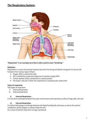
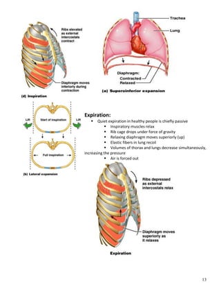
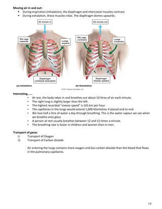
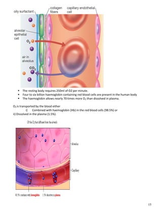
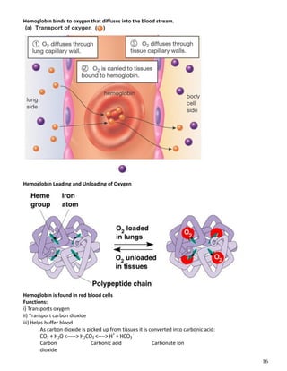
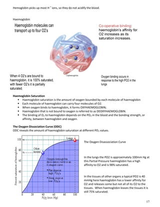
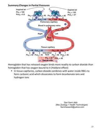
1. 1
The Respiratory System:
“Respiration” is an everyday term that is often used to mean “breathing.”
Definition:
Respiration is a series of chemical reactions by which the living cell obtains energy for its various life
functions from various types of food
Oxygen (O2) is used by the cells
O2 is needed for conversion of glucose to cellular energy (ATP)
Carbon dioxide (CO2) is produced as a waste product
The body’s cells die if either the respiratory or cardiovascular system fails
Types of respiration
Two types of respiration
i) External Respiration
ii) Internal Respiration
I) External Respiration:
It is the direct exchange between the outer environment and respiratory surfaces (lungs, gills, skin etc)
ii) Internal Respiration:
It involves both gaseous exchange between the body fluid (blood) and tissue as well as the cellular
metabolism where Oxygen is utilized inside the cell.
As a result of cellular respiration energy is produced
2. 2
Cellular respiration is the aerobic breakdown of glucose in the mitochondria to make ATP.
Respiration Includes
I) Pulmonary ventilation
II) External respiration
iii) Transport of respiratory gases
iv) Internal respiration
i) Pulmonary ventilation
Air moves in and out of lungs
Continuous replacement of gases in alveoli (air sacs)
ii) External respiration
Gas exchange between blood and air at alveoli
O2 (oxygen) in air diffuses into blood
CO2 (carbon dioxide) in blood diffuses into air
iii) Transport of respiratory gases
Between the lungs and the cells of the body
Performed by the cardiovascular system
Blood is the transporting fluid
iv) Internal respiration
Gas exchange in capillaries between blood and tissue cells
O2 in blood diffuses into tissues
CO2 waste in tissues diffuses into blood
Properties of Respiratory surfaces
- Large Surface area
- Moisturize surfaces
- Thin epithelium
- Ventilation
- Capillary network
Zones of Respiratory Organs
Respiratory organ consists of two zones
1) Conducting zone
2) Respiratory zone
1) Conducting zone:
Respiratory passages that carry air to the site of gas exchange
Filters, humidifies/moisturizes and warms air
2) Respiratory zone:
Site of gaseous exchange
Composed of
Alveolar ducts
Alveolar sacs
3. 3
Respiratory System
“Respiratory systems constitute those organs in animals that exchange gases with the environment.
Respiratory System of MAN:
Components:
Pathway of Inhaled and Exhaled Air:
1) Nasal cavity
2) Pharynx (Throat)
3) Larynx (Voice Box)
4) Trachea (Windpipe)
5) Bronchi
6) Bronchioles
7) Alveoli (Site of gas exchange)
1) Nose
Provides airway for air passage
Moistens and warms air (lined by mucus membrane of ciliated epithelium)
Filters air (Hair and cilia)
Resonating chamber for speech
Olfactory receptors
4. 4
2) The Pharynx (throat)
Length is about 4.5 inches
Parts: three parts
i) Naso-pharynx
ii) Oro-pharynx and
iii) Laryngo-pharynx
Houses tonsils (they respond to inhaled antigens)
Uvula closes off nasopharynx during swallowing so food does not go into nose
Epiglottis posterior to the tongue: keeps food out of airway
Oropharynx and laryngopharynx serve as common passageway for food and air
Lined with stratified squamous epithelium for protection
3) The Larynx (voice box):
Extends from the level of the 4th
to the 6th
cervical vertebrae
Attaches to hyoid bone superiorly
Inferiorly is continuous with trachea (windpipe)
Functions: Perform three functions
1. Produces vocalizations (speech)
2. Provides an open airway (breathing)
3. Switching mechanism to route air and food into proper channels
Closed during swallowing
Open during breathing
5. 5
4) Trachea (the windpipe):
About 5 inches in length and one inch in diameter
Descends into mediastinum
Divides in thorax into two main (primary) bronchi
16-20 C-shaped rings of hyaline cartilage
joined by fibroelastic connective tissue
Flexible for bending stays open despite
pressure changes during breathing
Trachealis muscle can decrease diameter of trachea
Inner lining of trachea is also lined with mucous epithelial ciliated cells – filters, warms and
moistens incoming air
7. 7
Trachea (Windpipe):
Rings of cartilage maintain shape of trachea, to prevent it from closing. Forks into two bronchi.
- Right main bronchus (more susceptible to aspiration)
- Left main bronchus
5) Bronchi (Sing. Bronchus):
Each bronchus leads into a lung and branches into smaller and smaller bronchioles, resembling an inverted
tree. Same cartilageous rings as in Trachea but is irregularly distributed cartilageous plates.
Main (primary) bronchi divide into secondary bronchi, each supplies to one lobe of Lung
3 on the right (as there are 3 lobes in right Lung)
2 on the left (as there are 2 lobes in left Lung)
Secondary (Lobar) bronchi branch into tertiary
(segmental bronchi)
Continues dividing: about 23 times
6) Bronchioles:
Fine tubes (one mm or less in diameter) that allow passage of air.
Smallest (terminal) bronchioles are less than 0.5 mm diameter
Bronchioles totally lack cartilages.
Mainly composed of circular smooth muscle layer which constricts bronchioles.
Epithelium of bronchioles is covered with cilia and mucus.
Mucus traps dust and other particles.
Ciliary Escalator: Cilia beat upwards and remove trapped particles from lower respiratory airways. Rate
about 1 to 3 cm per hour.
Branchioles continously divides and subdivides deep into the Lungs and finally open into a large numbers
of air sacs.
8. 8
Carina
Point where trachea branches (when alive and standing is at T7)
Mucosa highly sensitive to irritants: cough reflex
9. 9
Respiratory Zone:
End-point of respiratory tree
Structures that contain air-exchange chambers are called alveoli
Respiratory bronchioles lead into alveolar ducts
Ducts lead into terminal clusters called alveolar sacs – are microscopic chambers
There are 3 million alveoli!
Gas Exchange:
Air filled alveoli account for most of the lung volume
Very great area for gas exchange (1500 sq ft)
Alveolar wall
Single layer of squamous epithelial cells
0.5um (15 X thinner than tissue paper)
External wall covered by capillaries
This “air-blood barrier” (the respiratory membrane) is where gas exchange occurs
Oxygen diffuses from air in alveolus (singular of alveoli) to blood in capillary
Carbon dioxide diffuses from the blood in the capillary into the air in the alveolus
In the alveolus
The respiratory surface is
made up of the alveoli and
capillary walls.
11. 11
Surfactant:
Surfactant is a detergent-like substance which is secreted in fluid coating alveolar surfaces – it decreases
surface tension so prevent collapsing of alveoli
Without it the walls would stick together during exhalation
Reduces surface tension of the lung allowing the oxygen and carbon dioxide across the membrane.
Premature babies – problem breathing is largely because lack surfactant
Lungs:
Each is cone-shaped with anterior, lateral and posterior surfaces contacting ribs
Superior tip is apex, just deep to clavicle
Concave inferior surface resting on diaphragm is the base
Lungs and Pleura
Around each lung is a flattened sac of membrane called pleura
Parietal pleura – outer layer
Visceral pleura – directly on lung
Pleural cavity – slit-like potential space filled with
pleural fluid
Lungs can slide but separation from pleura
is resisted (like film between 2 plates of glass)
Lungs cling to thoracic wall and are forced
to expand and recoil as volume of thoracic cavity
changes during breathing
12. 12
Lobes of Lungs:
Right lung: 3 lobes
Upper lobe
Middle lobe
Lower lobe
Left lung: 2 lobes
Upper lobe
Lower lobe
Ventilation/Breathing:
Breathing = “pulmonary ventilation”
Pulmonary means related to the lungs
Two phases
Inspiration (inhalation) – air in
Expiration (exhalation) – air out
Mechanical forces cause the movement of air
Gases always flow from higher pressure to lower
For air to enter the thorax, the pressure of the air in it has to be lower than atmospheric
pressure
Making the volume of the thorax larger means the air inside it is under less pressure
(the air has more space for as many gas particles, therefore it is under less pressure)
The diaphragm and intercostal muscles accomplish this
Muscles of Inspiration:
During inspiration, the dome shaped diaphragm flattens as it contracts
This increases the height of the thoracic cavity
The external intercostal muscles contract to raise the ribs
This increases the
circumference of the thoracic cavity
13. 13
Expiration:
Quiet expiration in healthy people is chiefly passive
Inspiratory muscles relax
Rib cage drops under force of gravity
Relaxing diaphragm moves superiorly (up)
Elastic fibers in lung recoil
Volumes of thorax and lungs decrease simultaneously,
increasing the pressure
Air is forced out
14. 14
Moving air in and out:
During inspiration (inhalation), the diaphragm and intercostal muscles contract.
During exhalation, these muscles relax. The diaphragm domes upwards.
Interesting......
• At rest, the body takes in and breathes out about 10 litres of air each minute.
• The right lung is slightly larger than the left.
• The highest recorded "sneeze speed" is 165 km per hour.
• The capillaries in the lungs would extend 1,600 kilometres if placed end to end.
• We lose half a litre of water a day through breathing. This is the water vapour we see when
we breathe onto glass.
• A person at rest usually breathes between 12 and 15 times a minute.
• The breathing rate is faster in children and women than in men.
Transport of gases
i) Transport of Oxygen
ii) Transport of Carbon dioxide
Air entering the lungs contains more oxygen and less carbon dioxide than the blood that flows
in the pulmonary capillaries.
15. 15
The resting body requires 250ml of O2 per minute.
Four to six billion haemoglobin containing red blood cells are present in the human body
The haemoglobin allows nearly 70 times more O2 than dissolved in plasma.
O2 is transported by the blood either
i) Combined with haemoglobin (Hb) in the red blood cells (98.5%) or
ii) Dissolved in the plasma (1.5%).
16. 16
Hemoglobin binds to oxygen that diffuses into the blood stream.
Hemoglobin Loading and Unloading of Oxygen
Hemoglobin is found in red blood cells
Functions:
i) Transports oxygen
ii) Transport carbon dioxide
iii) Helps buffer blood
As carbon dioxide is picked up from tissues it is converted into carbonic acid:
CO2 + H2O <-----> H2CO3 <----> H+
+ HCO3
-
Carbon Carbonic acid Carbonate ion
dioxide
17. 17
Hemoglobin picks up most H +
ions, so they do not acidify the blood.
Haemoglobin
Haemoglobin Saturation
Haemoglobin saturation is the amount of oxygen bounded by each molecule of haemoglobin
Each molecule of haemoglobin can carry four molecules of O2.
When oxygen binds to haemoglobin, it forms OXYHAEMOGLOBIN;
Haemoglobin that is not bound to oxygen is referred to as DEOXYHAEMOGLOBIN.
The binding of O2 to haemoglobin depends on the PO2 in the blood and the bonding strength, or
affinity, between haemoglobin and oxygen.
The Oxygen Dissociation Curve (ODC)
ODC reveals the amount of haemoglobin saturation at different PO2 values.
The Oxygen Disassociation Curve
In the lungs the PO2 is approximately 100mm Hg at
this Partial Pressure haemoglobin has a high
affinity to O2 and is 98% saturated.
In the tissues of other organs a typical PO2 is 40
mmHg here haemoglobin has a lower affinity for
O2 and releases some but not all of its O2 to the
tissues. When haemoglobin leaves the tissues it is
still 75% saturated.
19. 19
Factors Altering Haemoglobin Saturation
Hemoglobin and Oxygen Transport
Oxygen is transported by hemoglobin (98.5%) and is dissolved in plasma (1.5%)
Oxygen-hemoglobin dissociation curve shows that hemoglobin is almost completely saturated
when P02 is 80 mm Hg or above. At lower partial pressures, the hemoglobin releases oxygen.
Factors Affecting Haemoglobin Saturation
Blood temperature…
Blood acidity…(pH of blood)
Carbon Dioxide concentration
Also heavy exercise......
Factors affecting Disassociation
BLOOD TEMPERATURE
increased blood temperature reduces haemoglobin affinity for O2
hence more O2 is delivered to warmed-up tissue
Factors Affecting Haemoglobin Saturation – Blood Acidity If the blood becomes more acidic the
dissociation curve shifts right.
This means that more oxygen is being uploaded from the haemoglobin at tissue level.
See overhead.
Factors Affecting Haemoglobin Saturation – Blood Acidity
The rightward shift of the curve is due to a decline in pH. This is referred to as the BOHR effect.
Factors Affecting Haemoglobin Saturation – Blood Acidity
The pH in the lungs is generally high.
So haemoglobin passing through the lungs has a strong affinity for oxygen, encouraging high saturation.
20. 20
At the tissue level, however the pH is lower, causing oxygen to dissociate from haemoglobin, thereby
supplying oxygen to the tissues.
• Factors Affecting Haemoglobin Saturation – Blood Acidity
• With exercise, the ability to upload oxygen to the muscles increases as the muscle ph decreases.
Factors Affecting Haemoglobin Saturation – Blood Temperature Increased blood temperature shifts the
dissociation curve to the right, indicating that oxygen is uploaded more efficiently. Factors Affecting
Haemoglobin Saturation – Blood Temperature Because of this, the haemoglobin will upload more oxygen
when blood circulates through the metabolically heated active muscles.
In the lungs, where the blood might be a bit cooler, haemoglobin’s affinity for oxygen is increased. This
encourages oxygen binding.
BLOOD PH
lowering of blood pH (making blood more acidic)
caused by presence of H+
ions from lactic acid or carbonic acid
reduces affinity of Hb for O2
and more O2 is delivered to acidic sites which are working harder
CARBON DIOXIDE CONCENTRATION
the higher CO2 concentration in tissue
the less the affinity of Hb for O2
so the harder the tissue is working, the more O2 is released
Key Point
Increased temperature and hydrogen ion (H+
) (pH) concentration in exercising muscle affect the
oxygen dissociation curve, allowing more oxygen to be uploaded to supply the active muscles.
Oxygen Content (2)
CaO 2 = (1.34 x Hb x SaO2) + (0.003 x PaO2)
amount carried by Hb amount dissolved in plasma
CaO2 = (1.34 x 14 x 0.98) + (0.003 x 100)
CaO2 = 18.6 ml/dl
* at 100 % Saturation, 1 g of Hb binds 1.34 ml of Oxygen !
SaO2: means saturation of HB with oxygen, PaO2: means partial pressure of Oxygen, CaO 2 : carrying
capacity of Hb
Carbon dioxide transport
Carbon dioxide is transported from tissues toward lungs in several ways
i) As Carboxy-haemoglobin (23%) (HbNHCOOH)
ii) by other plasma proteins (7%)
iii) As Bicarbonate (HCO3) (70%)
CO2 + H2O <-----> H2CO3 <----> H+
+ HCO3
-
Carbonic acid anhydrase
23. 23
Summary:Changes in Partial Pressures
Hemoglobin that has released oxygen binds more readily to carbon dioxide than
hemoglobin that has oxygen bound to it (Haldane effect)
In tissue capillaries, carbon dioxide combines with water inside RBCs to
form carbonic acid which dissociates to form bicarbonate ions and
hydrogen ions
Qari Sami ullah
(Msc.Zoology + Health Technologist)
Samihaseen8@yahoo.com