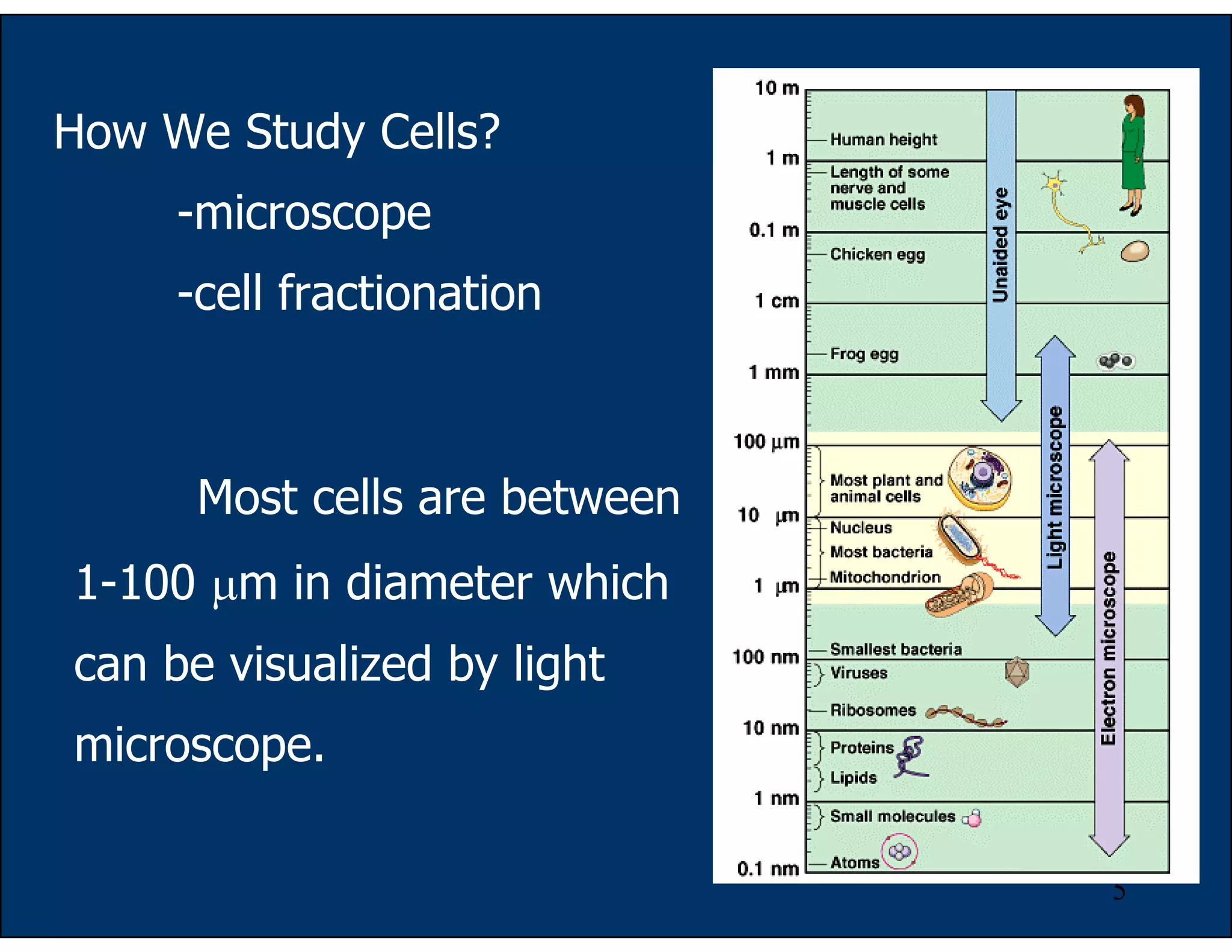
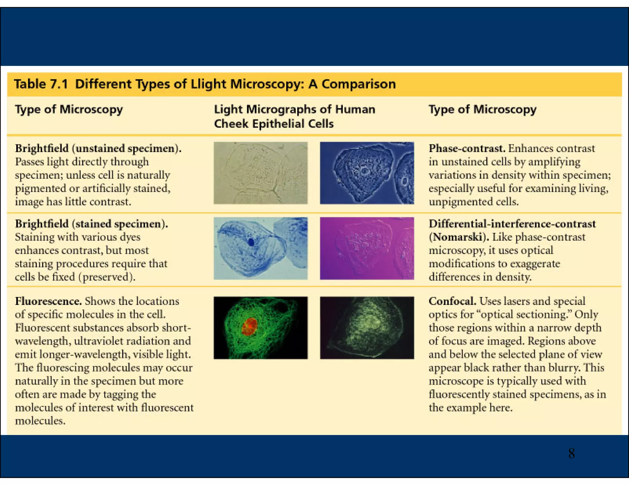
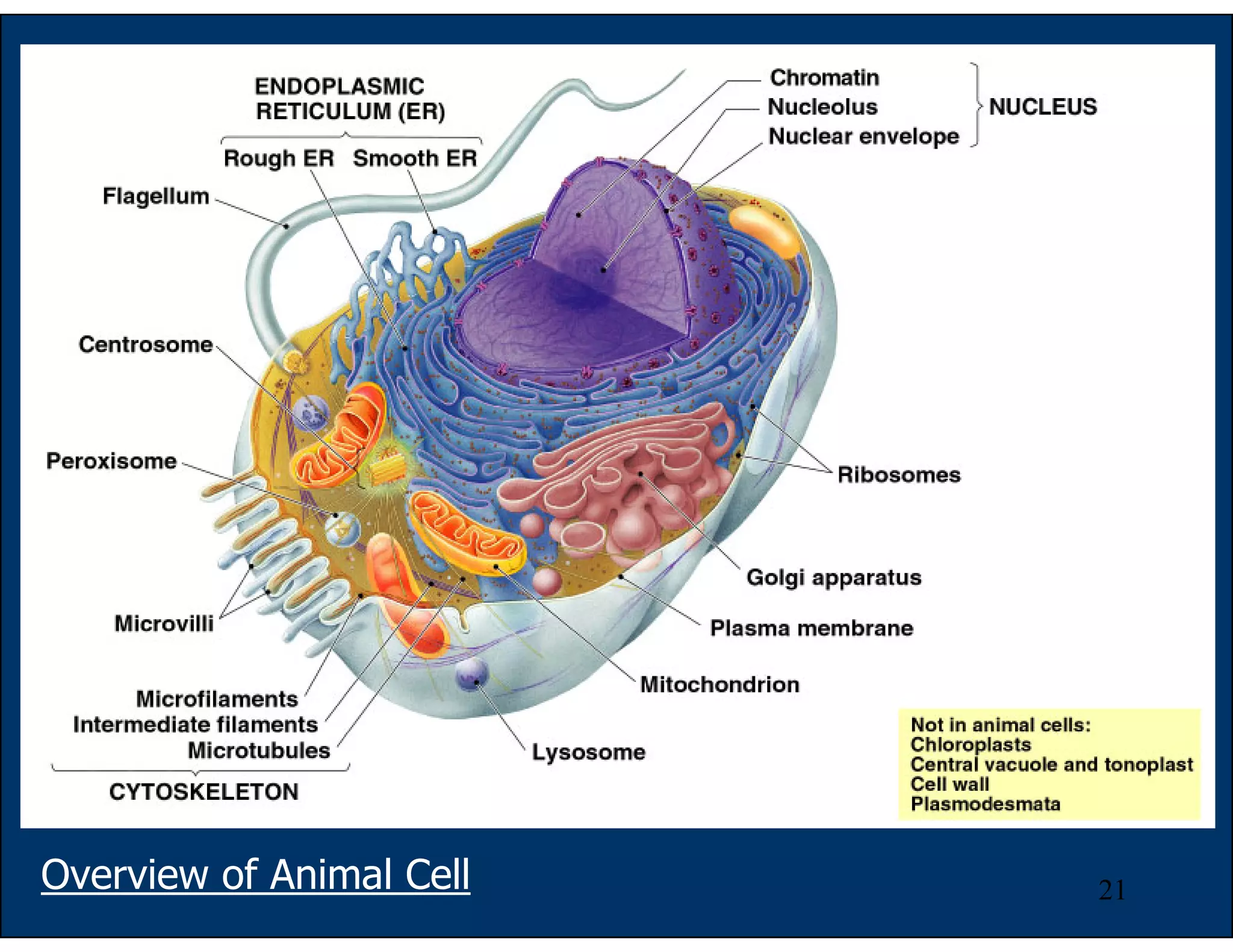
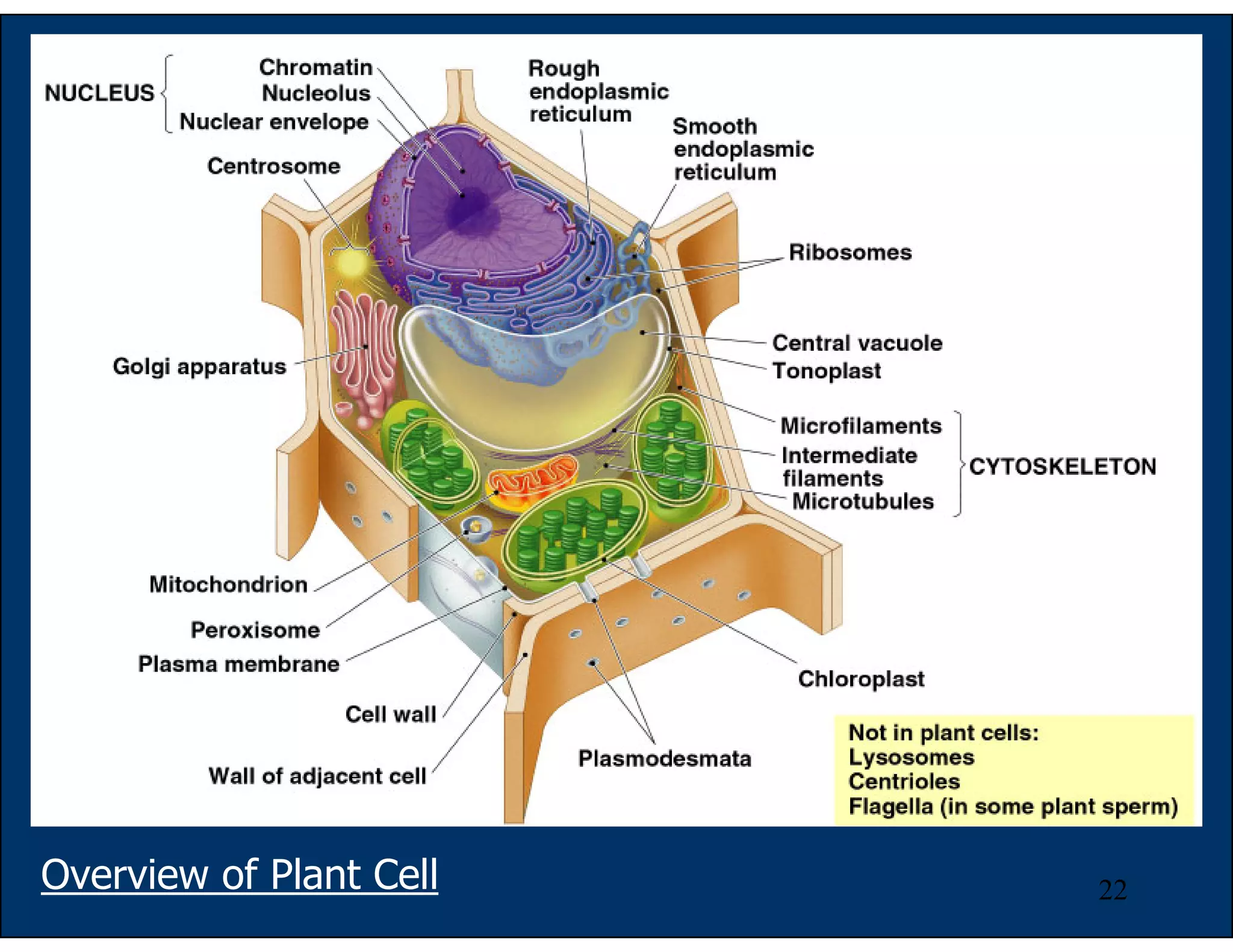
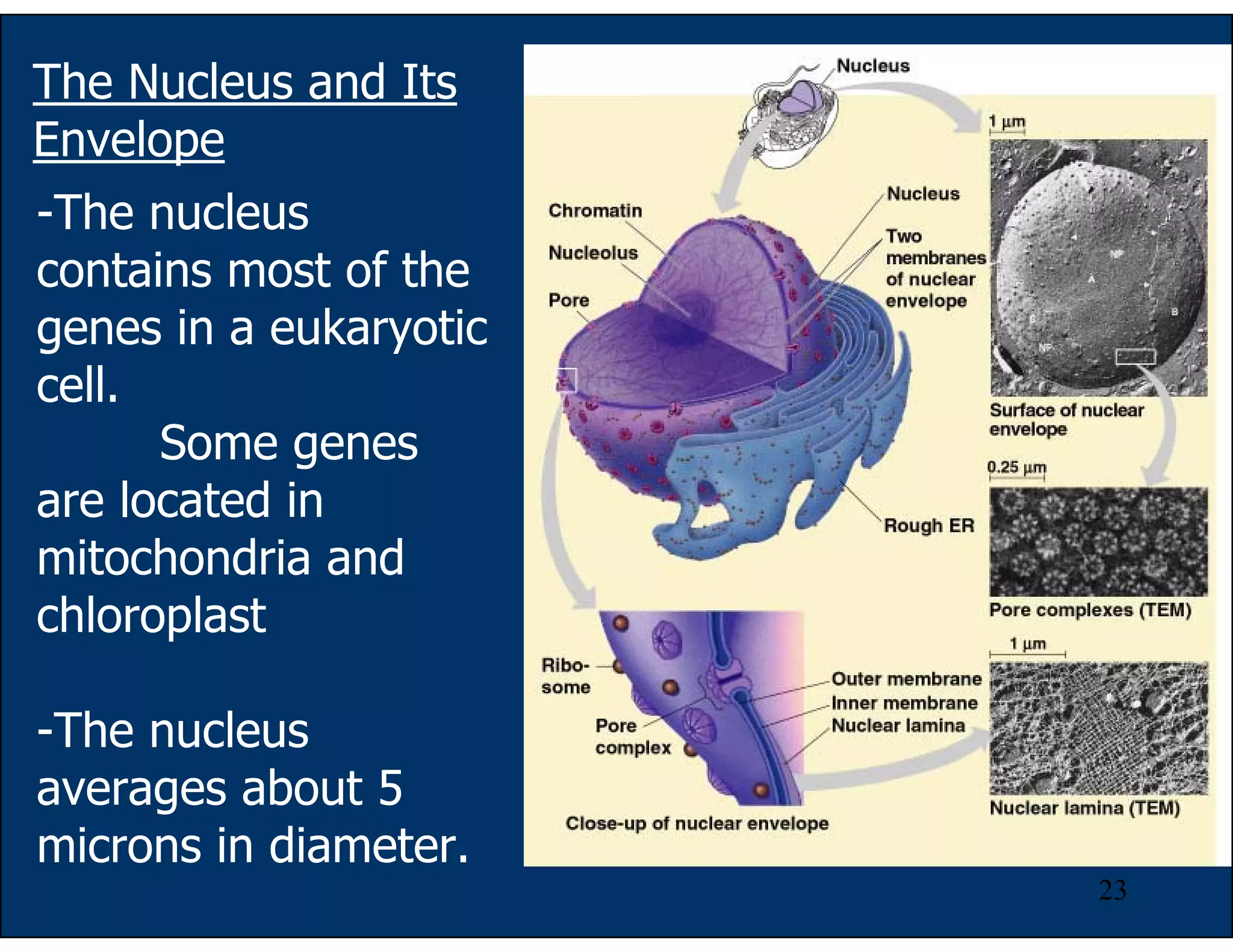
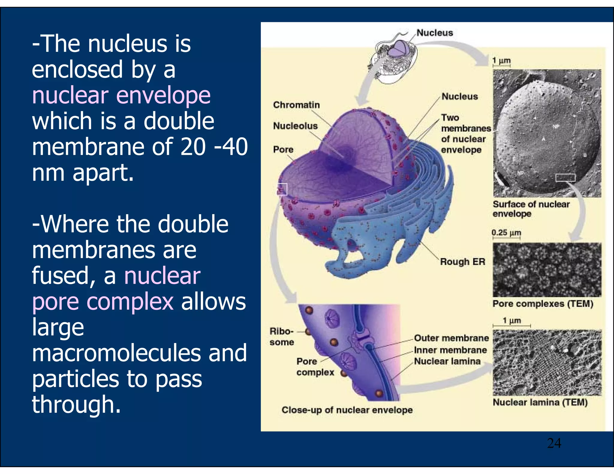
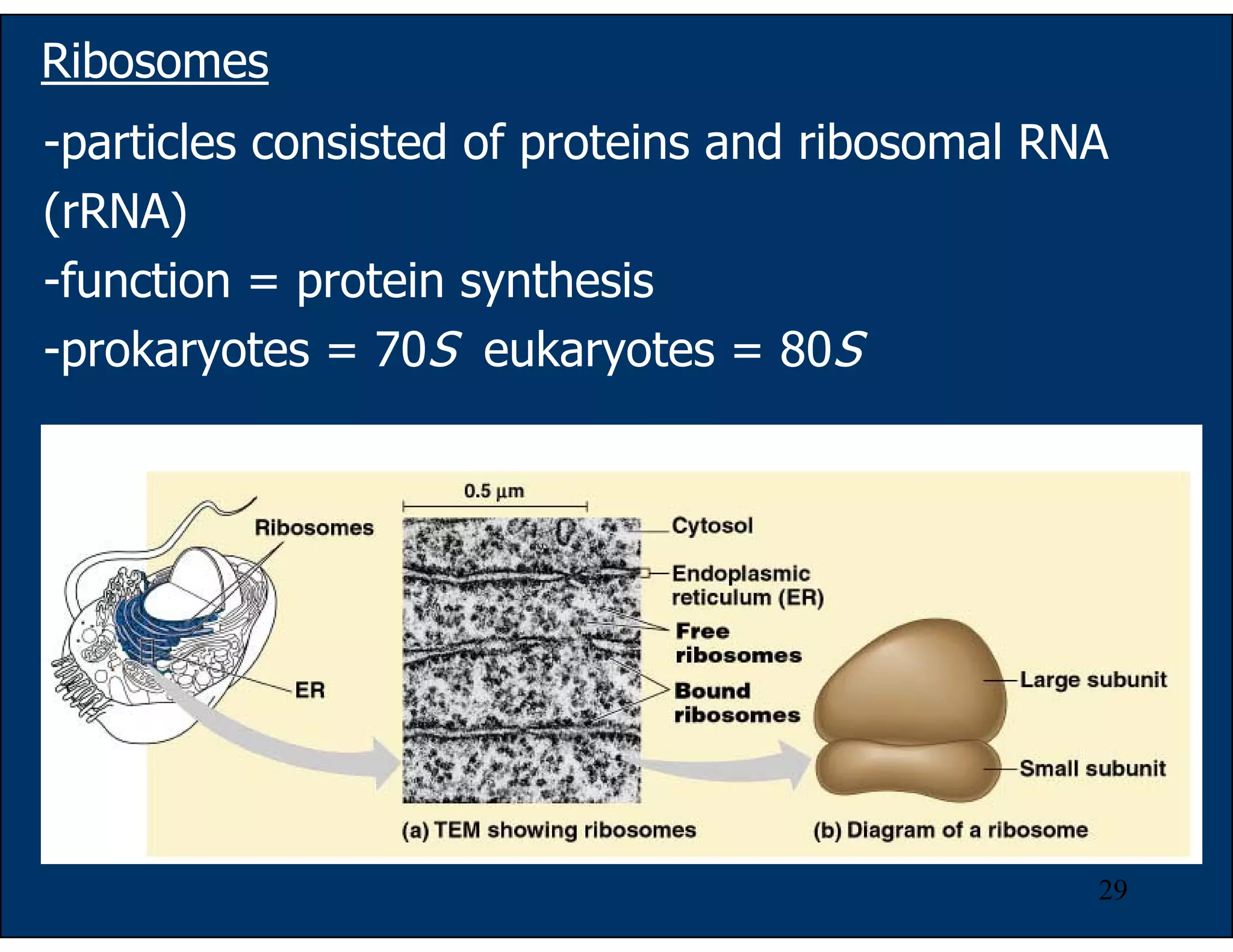
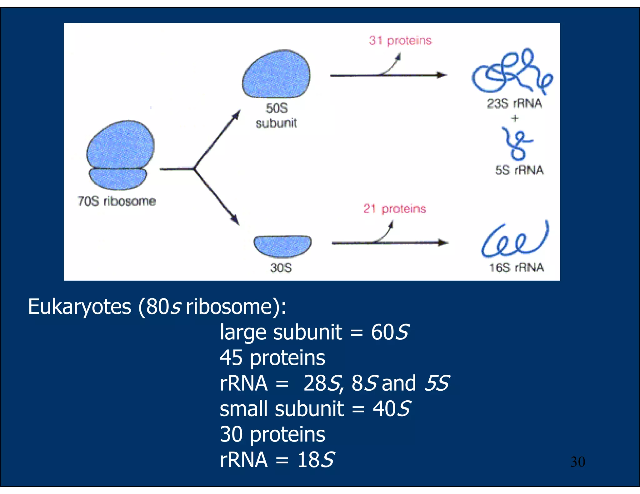
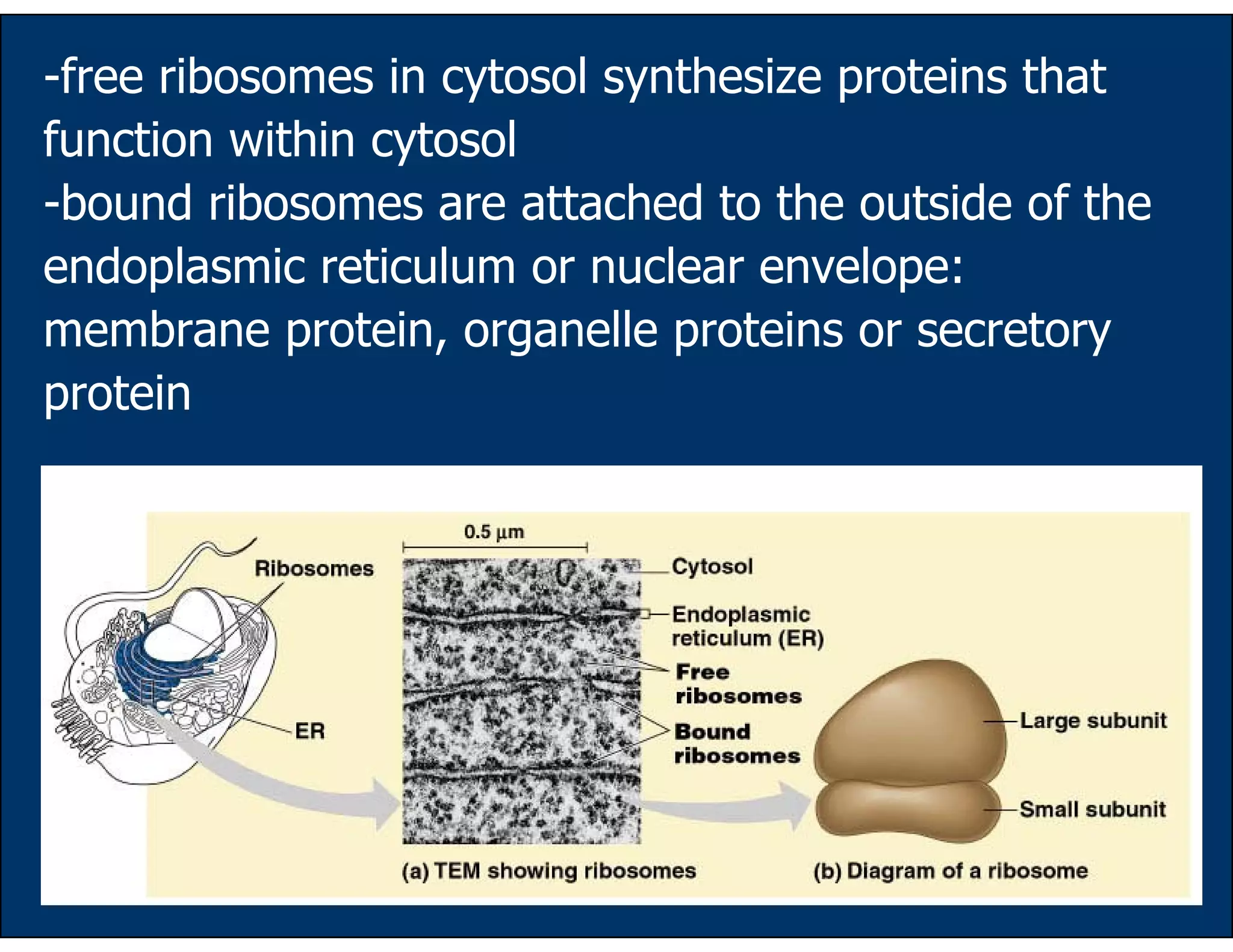
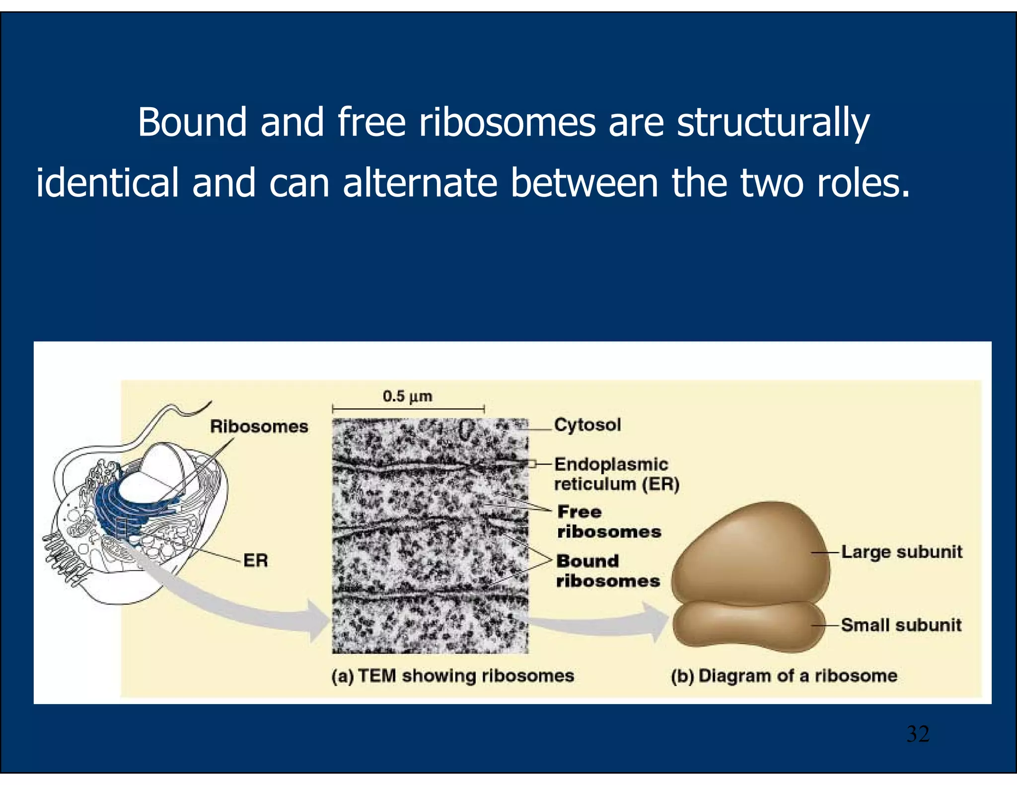
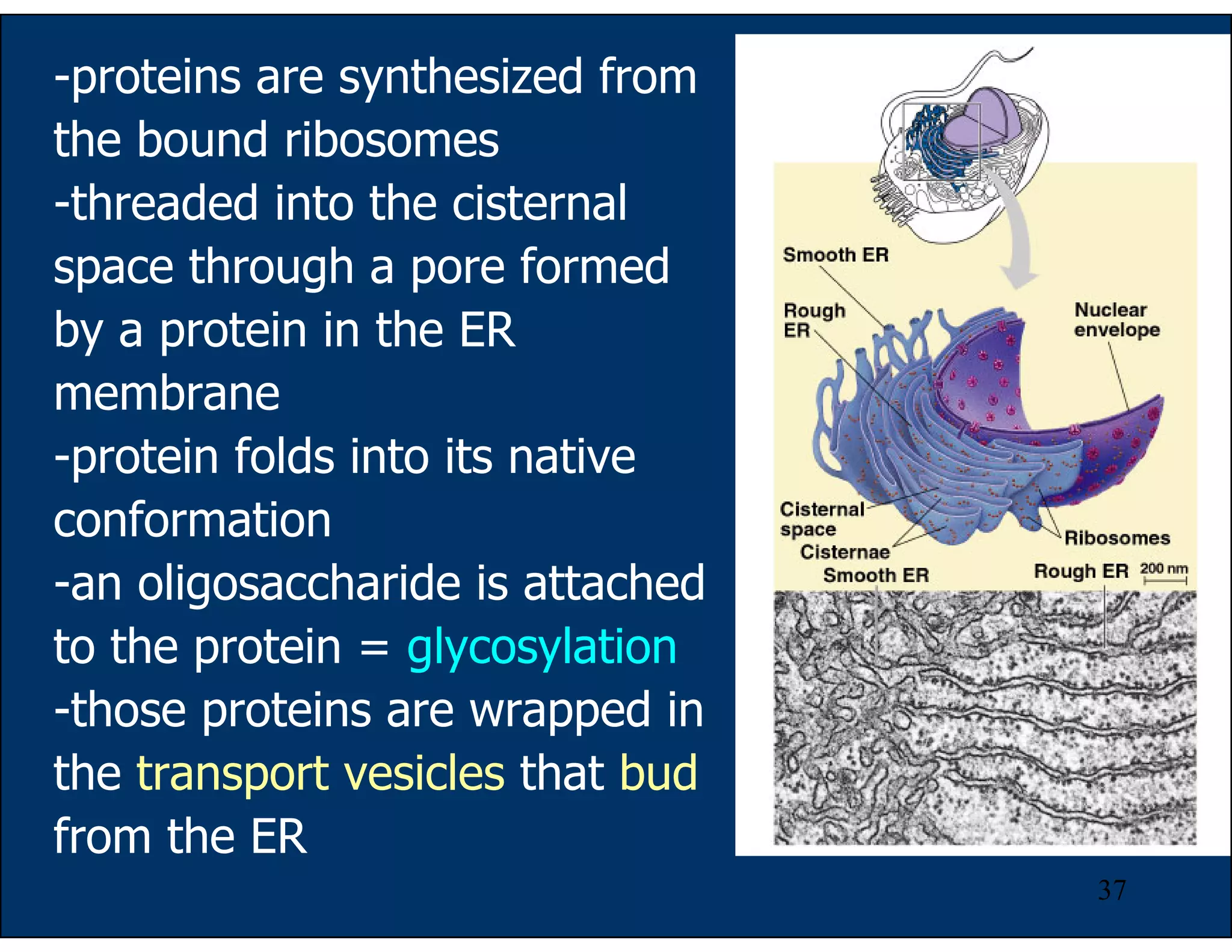
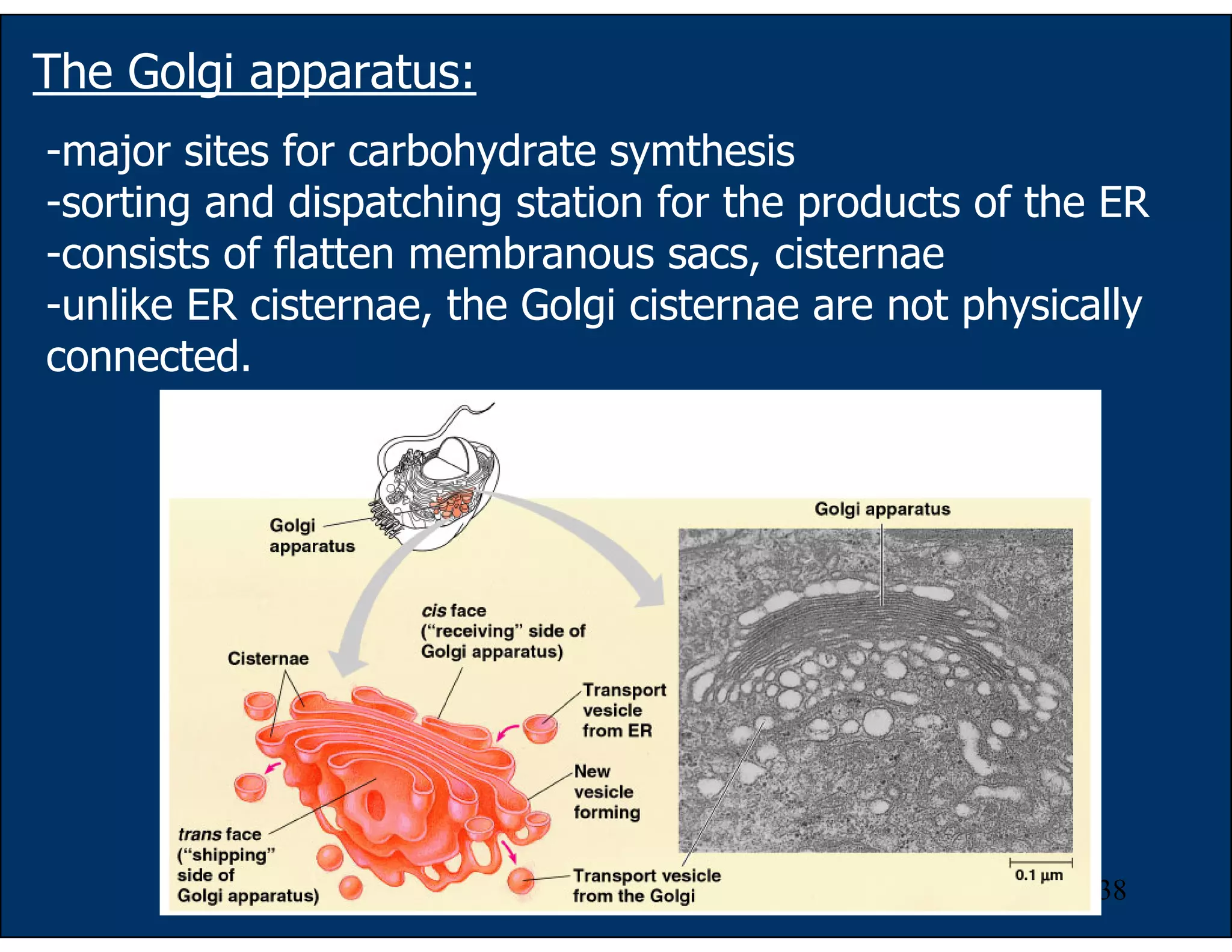
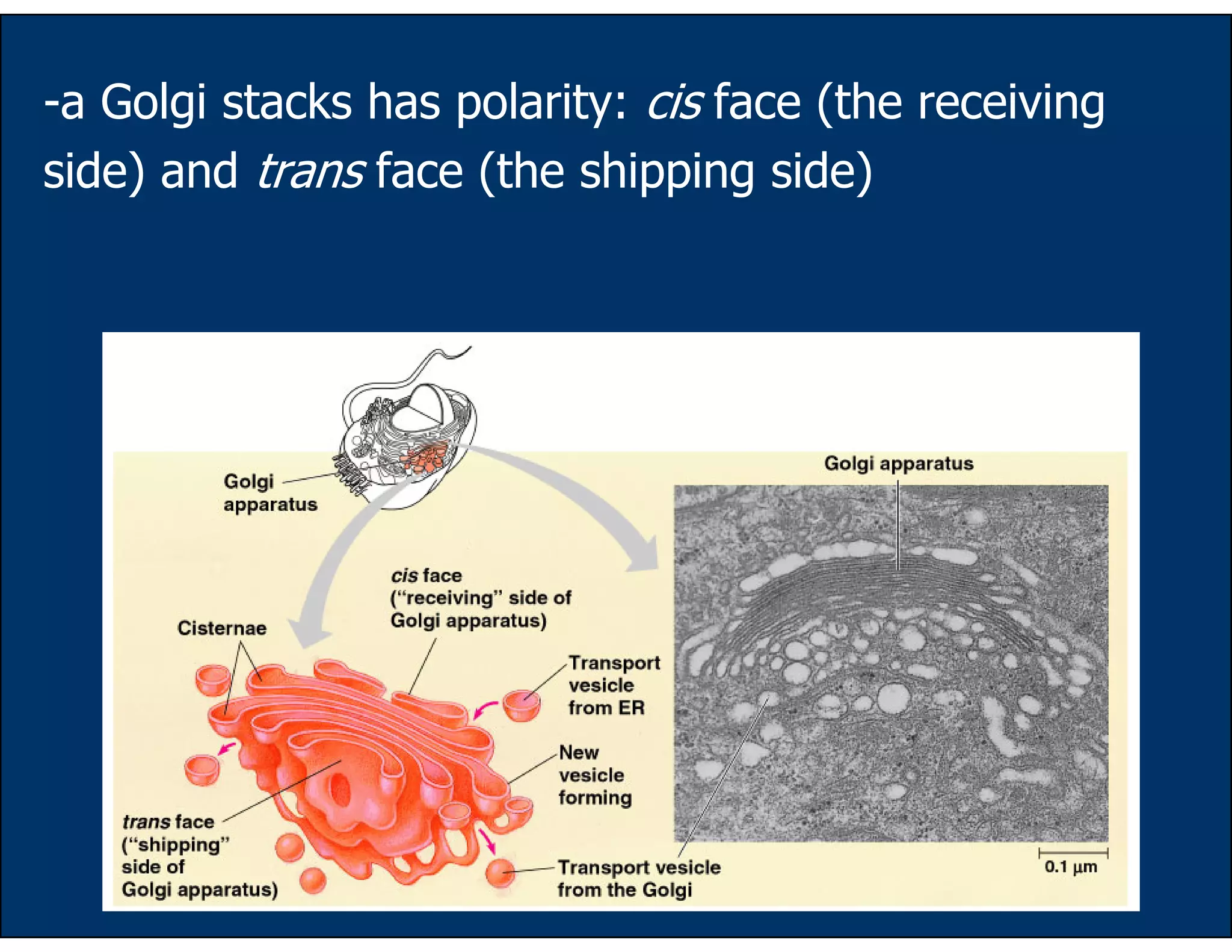
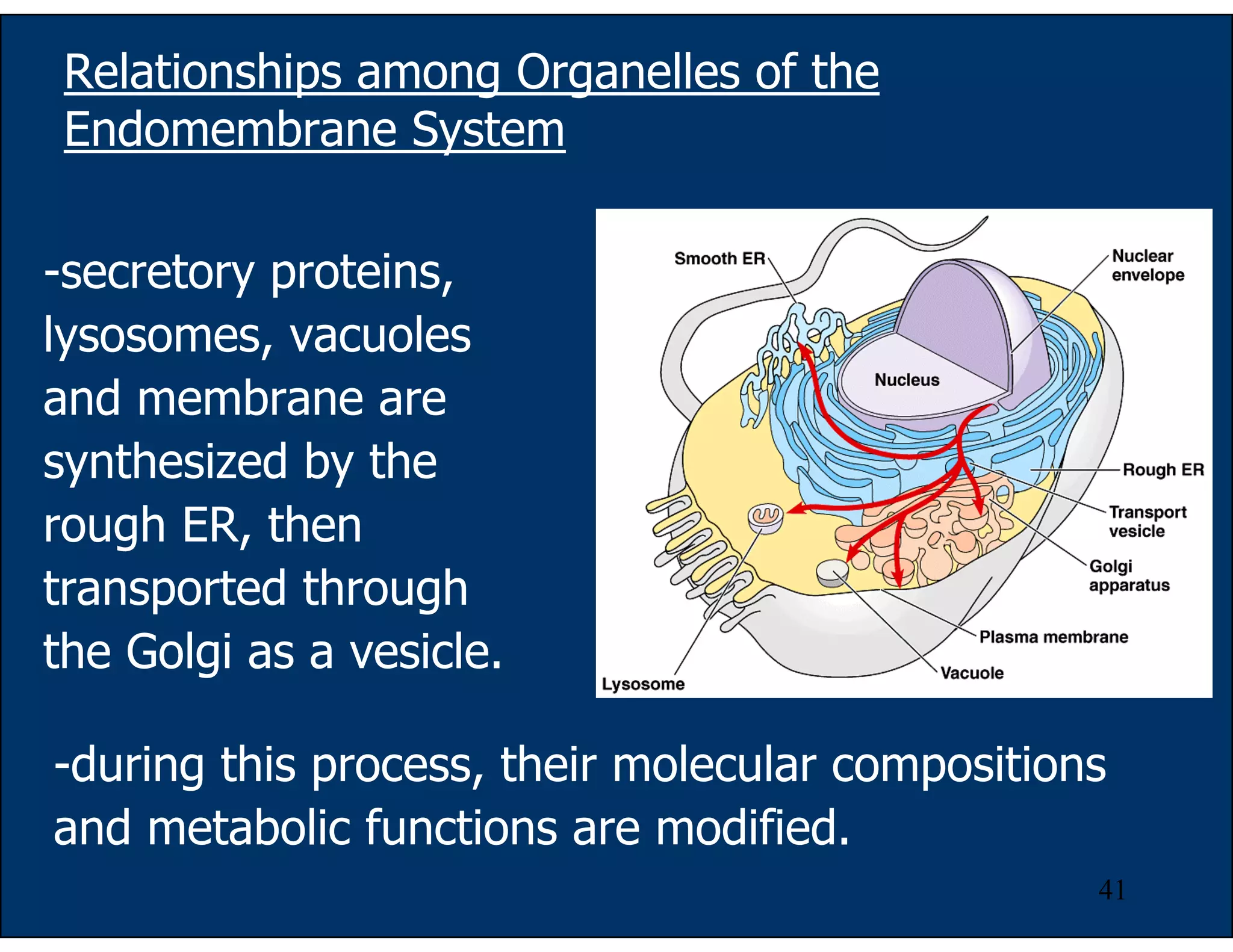
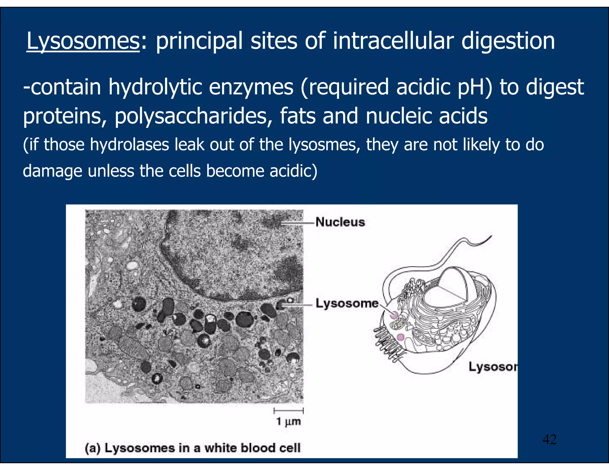
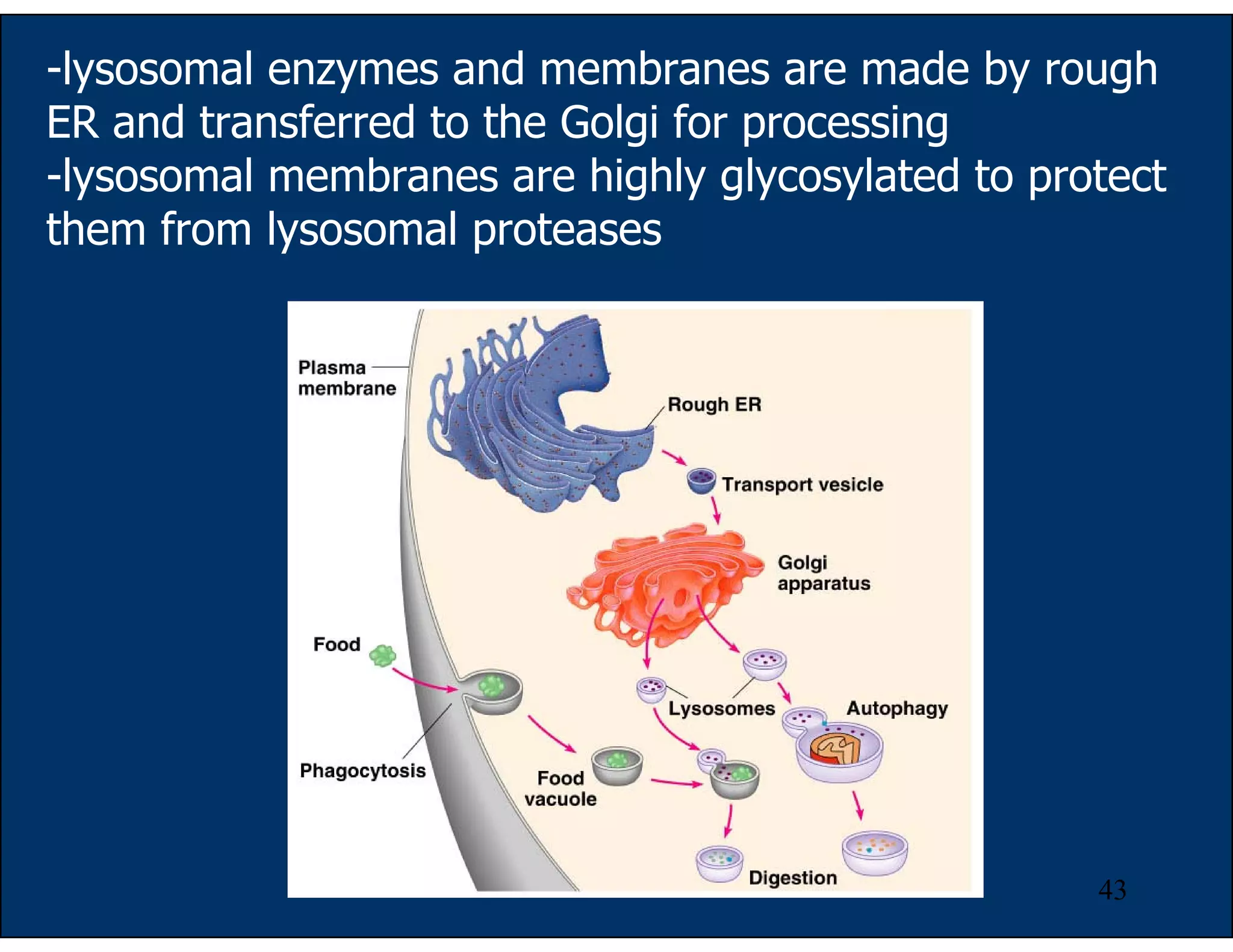
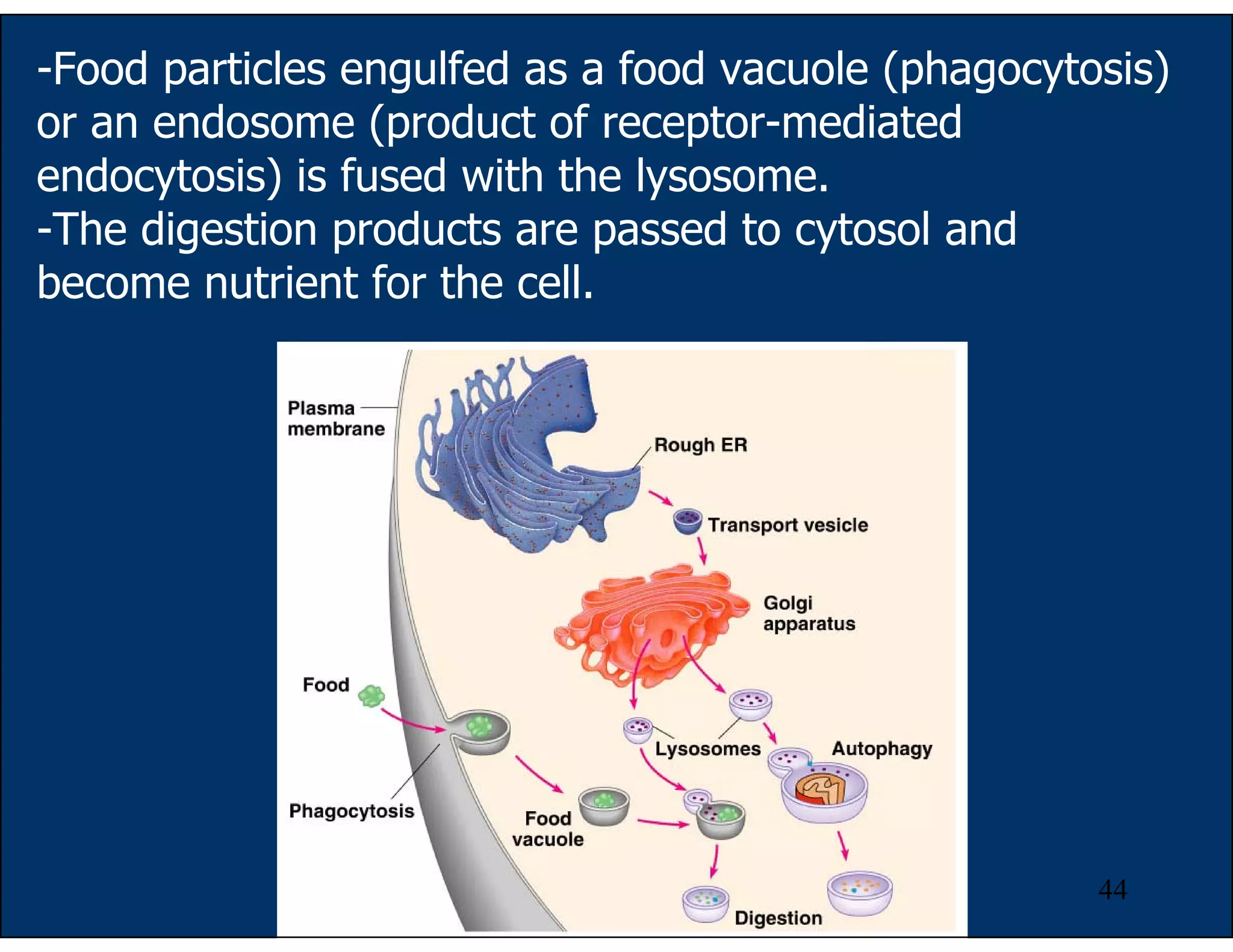
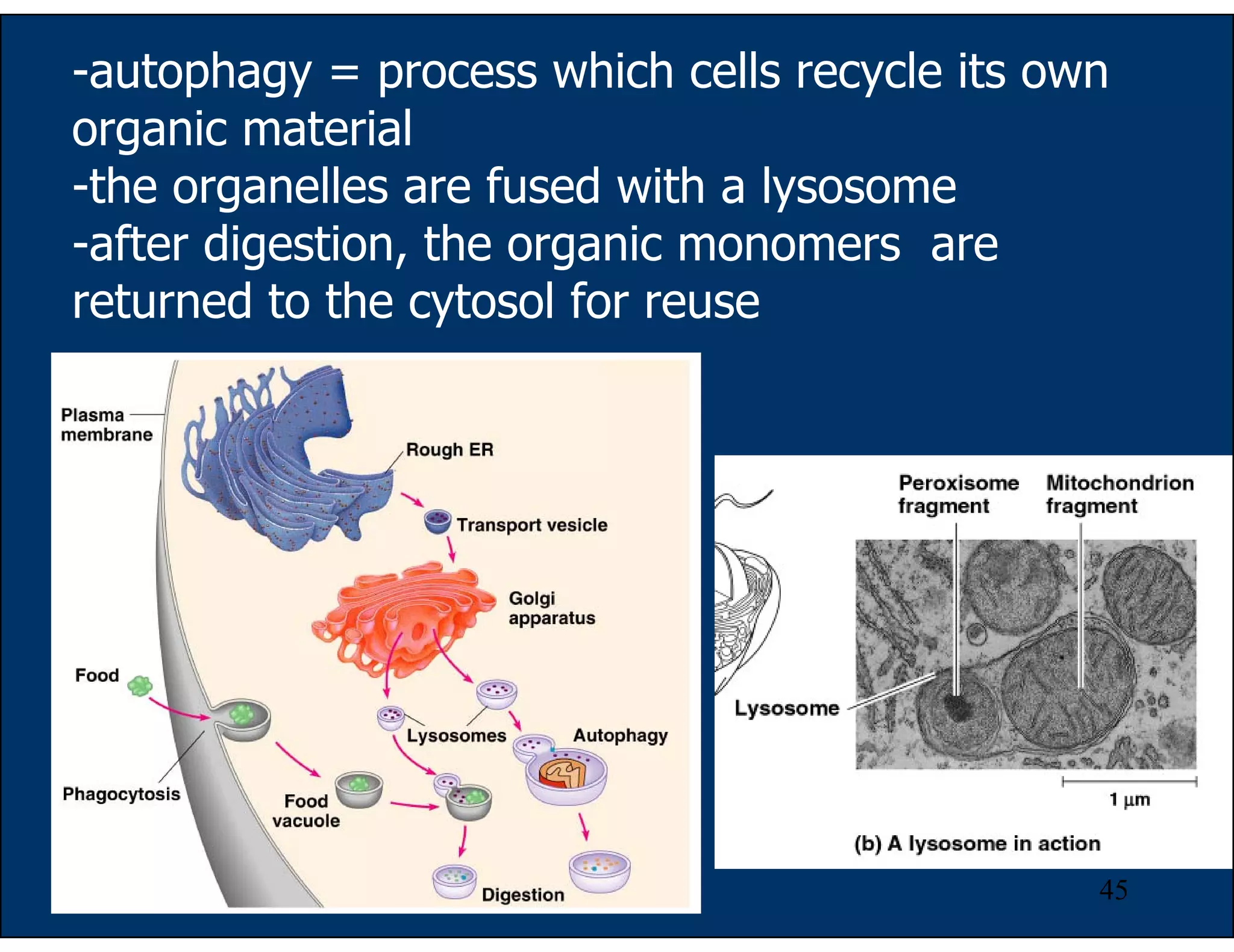
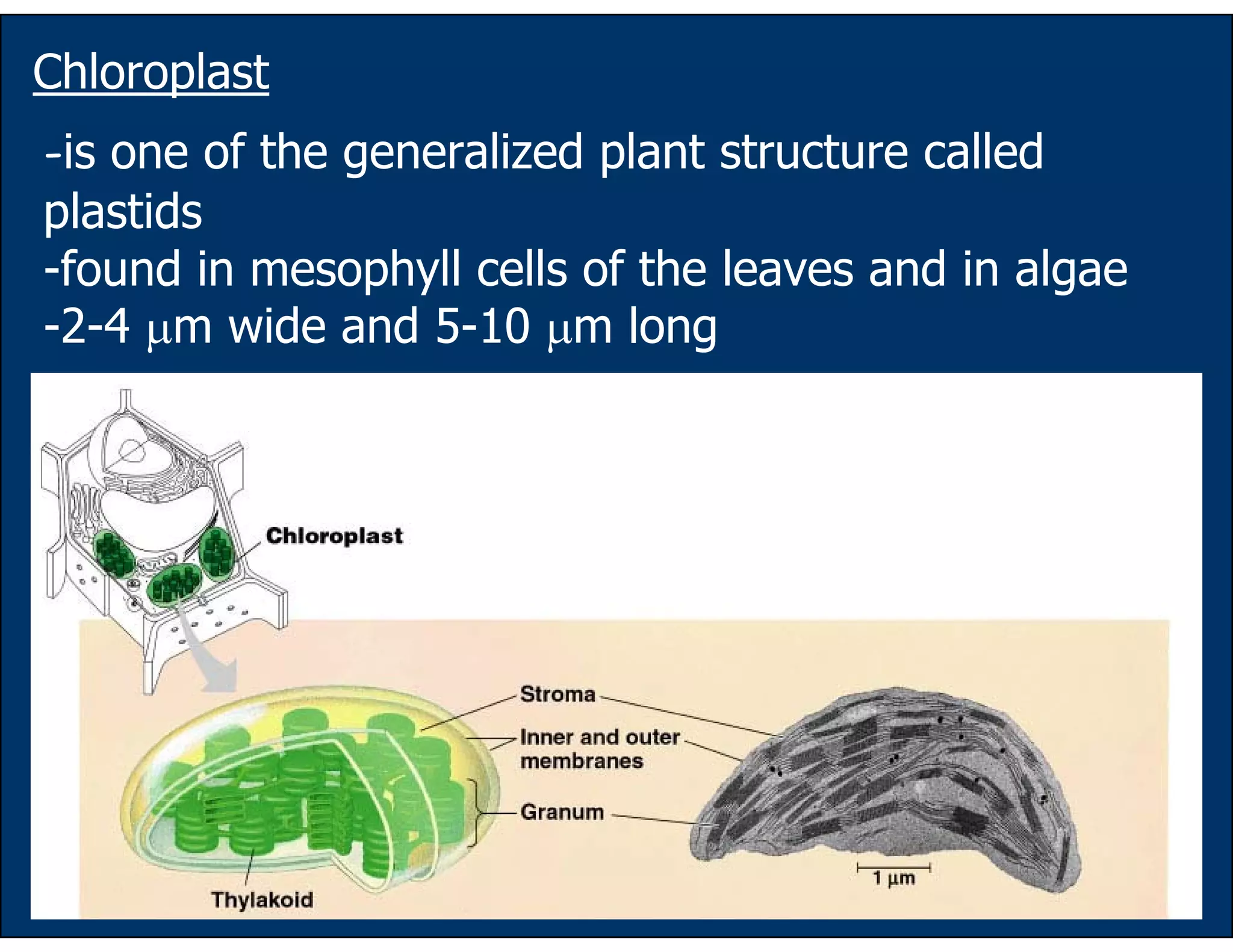
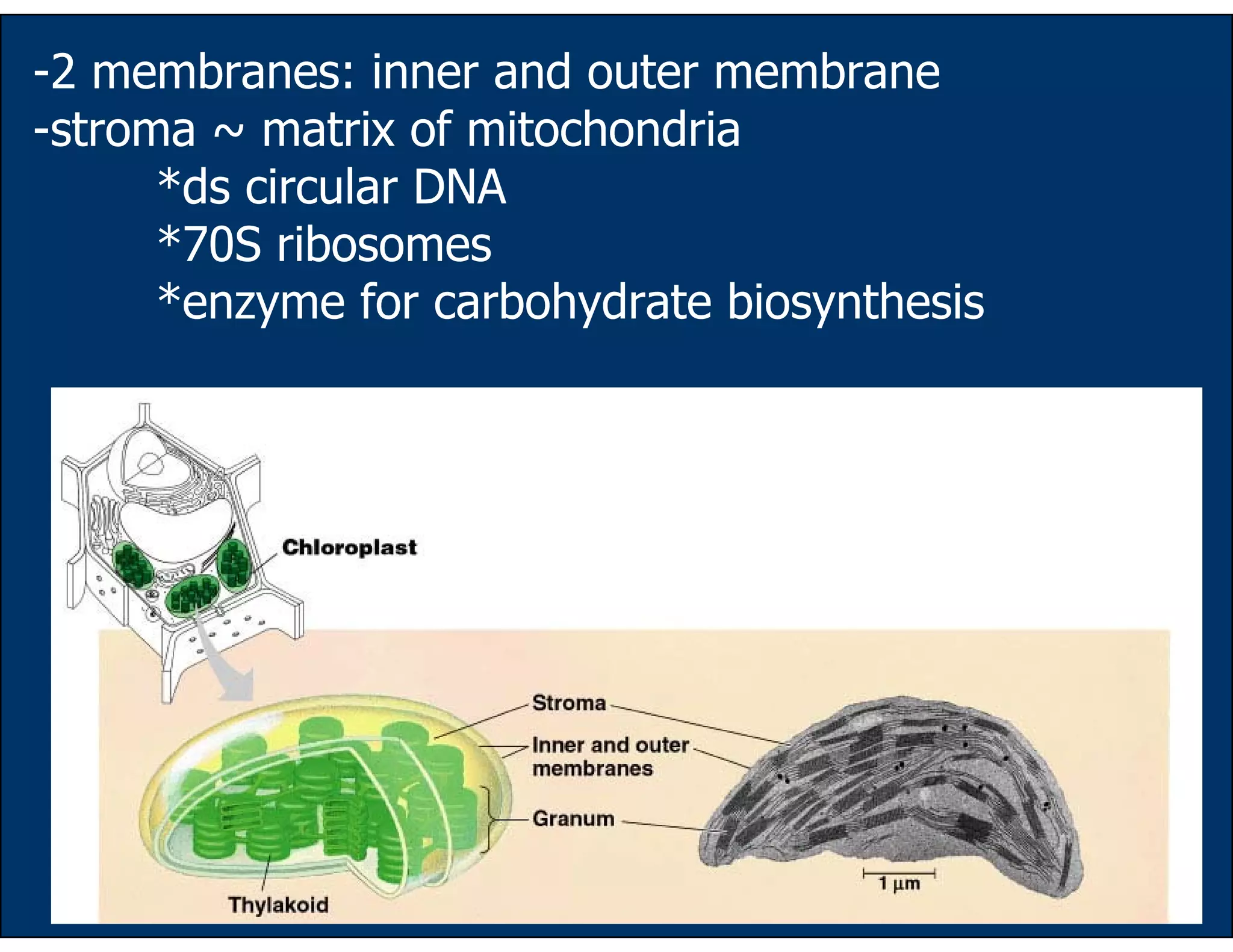
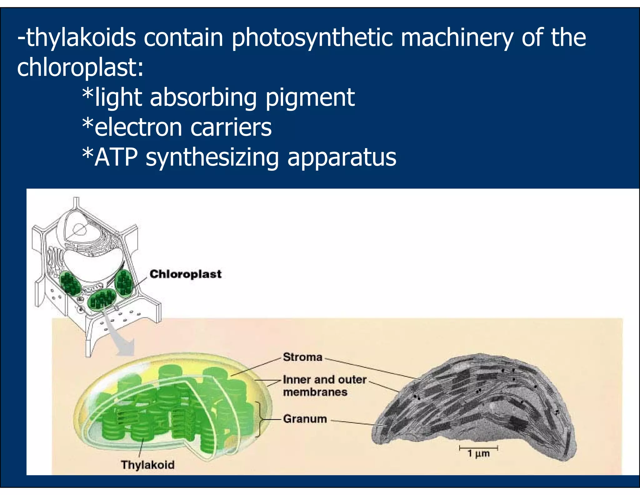
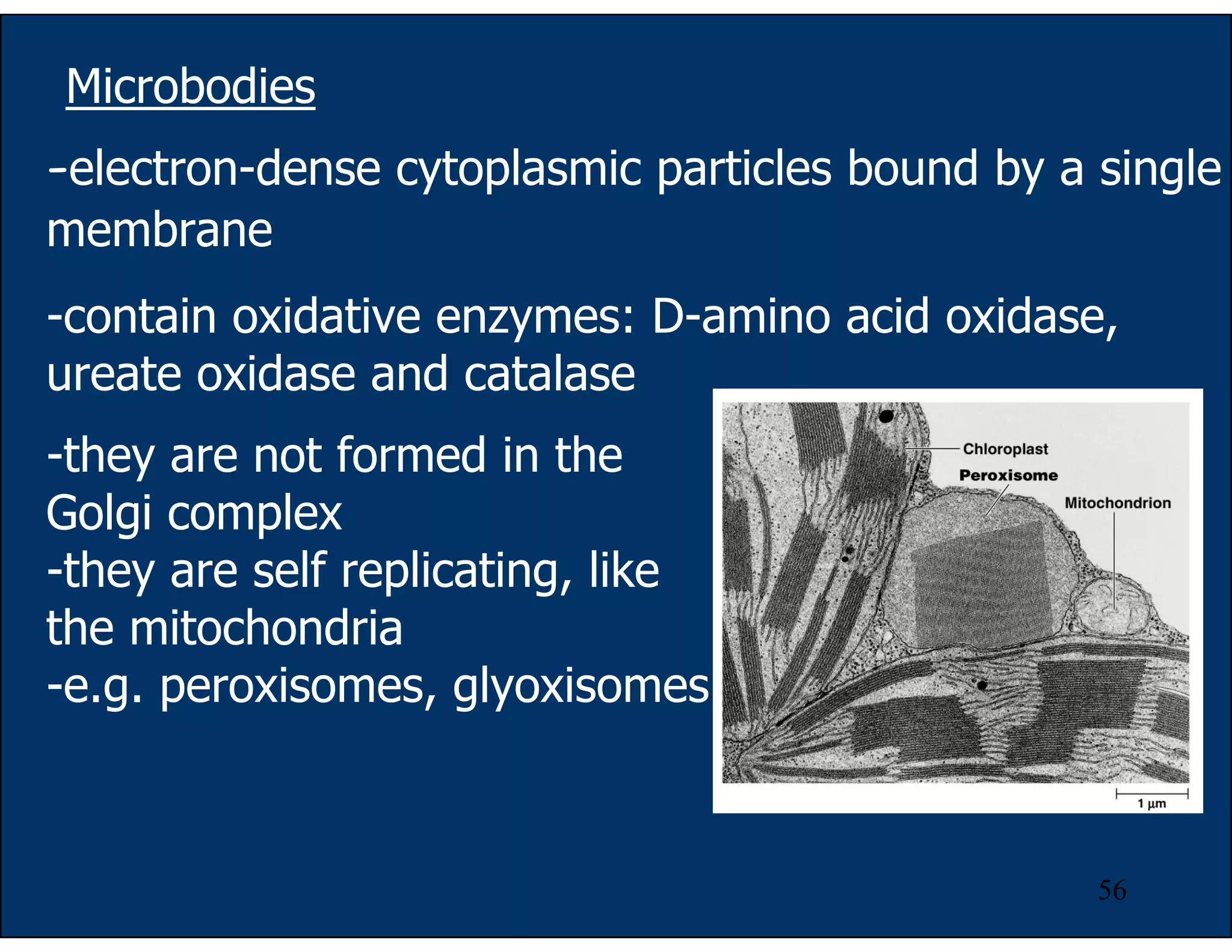
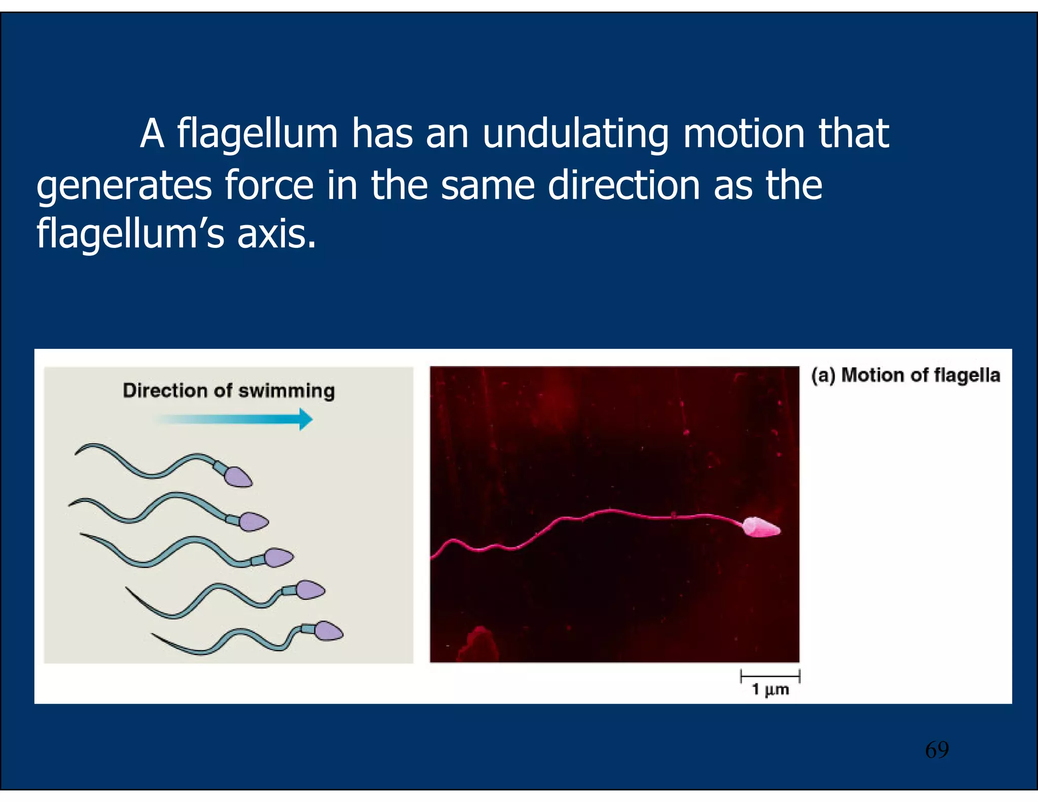
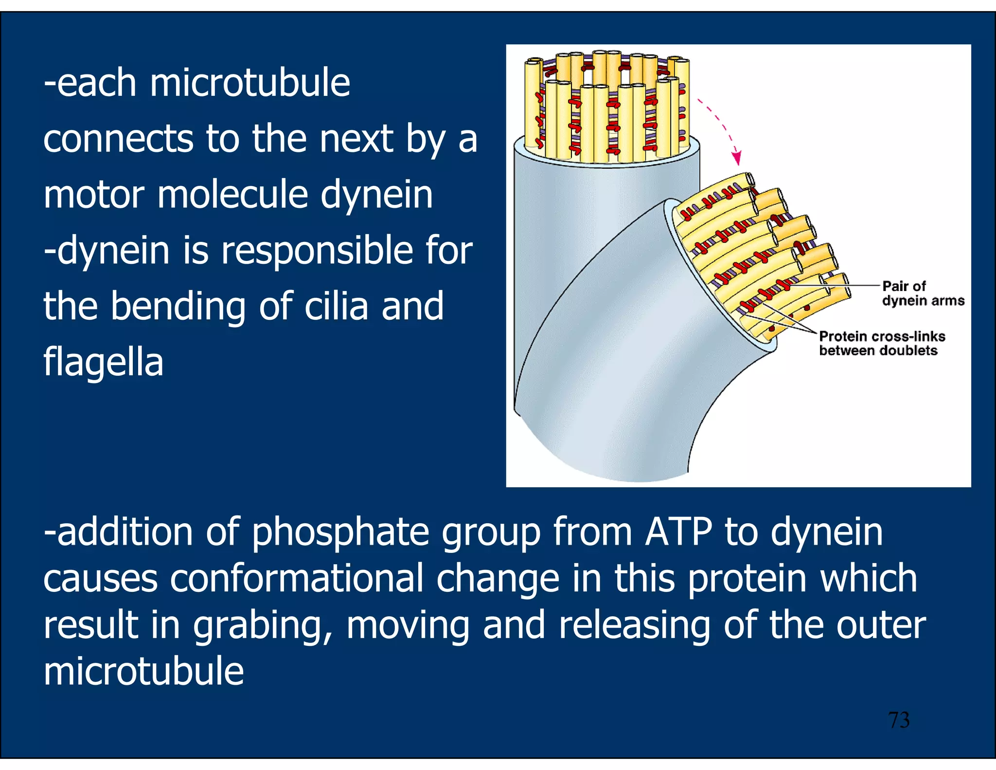
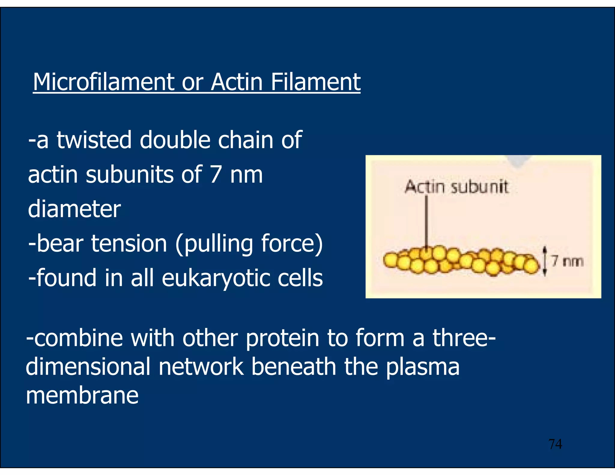
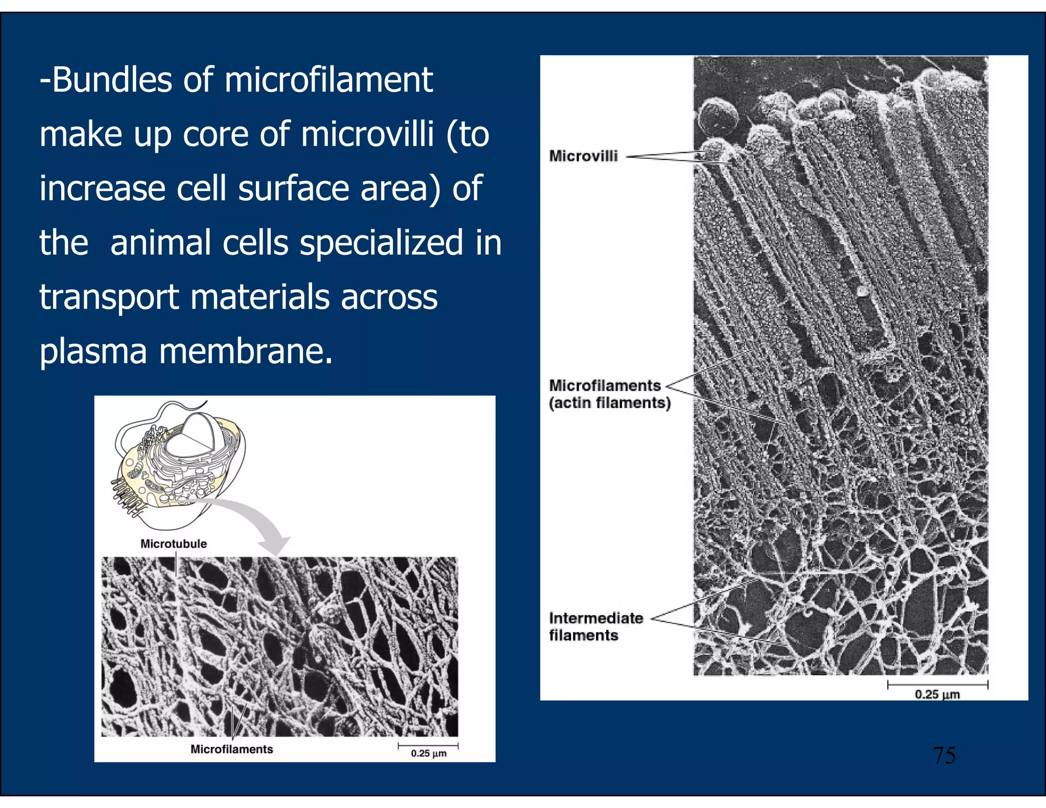
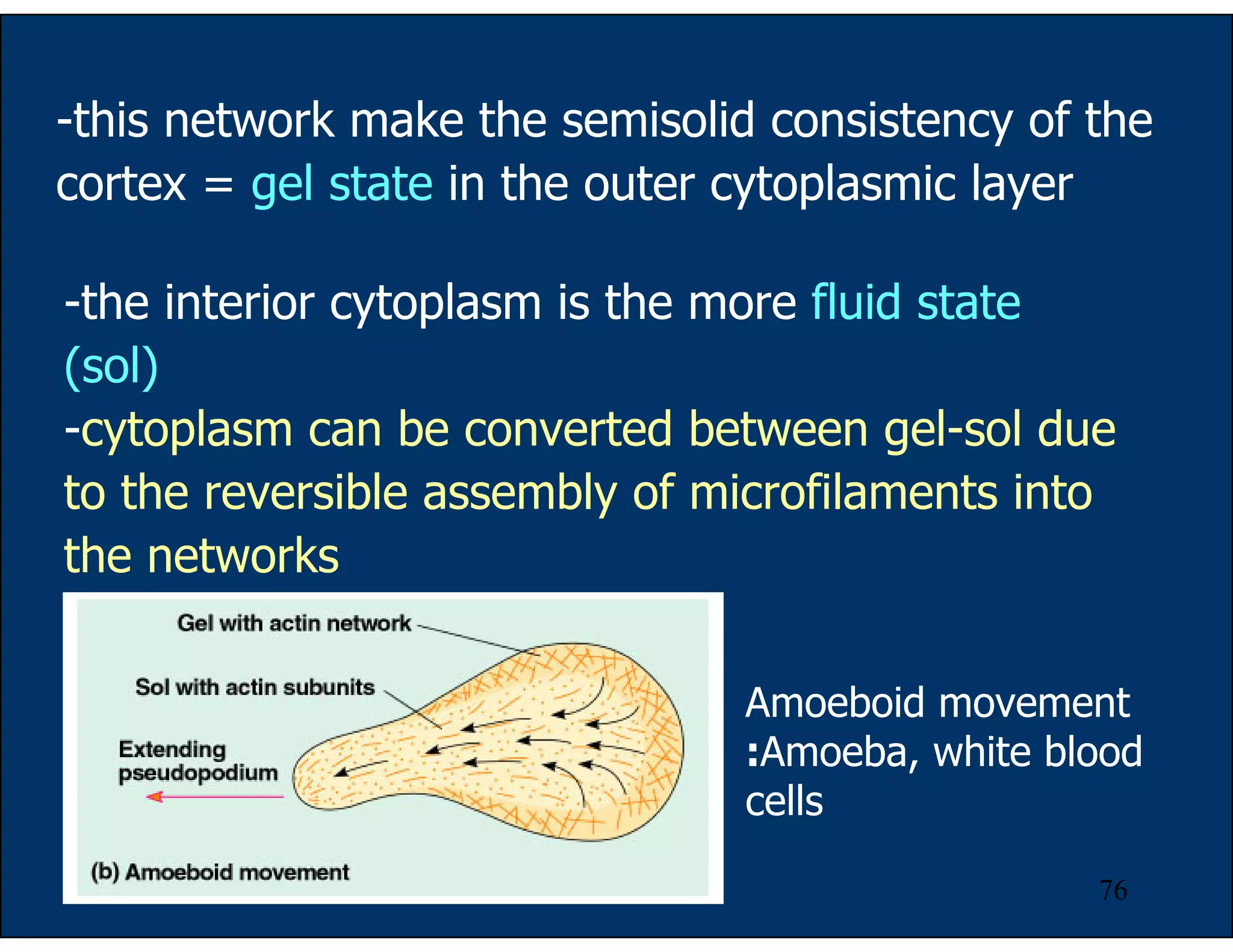
SlideShare a Scribd company logo
1 of 83
Download to read offline
1
The Cell Structure and Function
The cell is the lowest level of structure capable
of performing all the activities of life.
The first cells were observed and named by
Robert Hooke in 1665 from slice of cork.
2
The Cell Thoery
Proposed by Matthais Schleiden and Theodor
Schwann in 1839:
-All living things are made up of cells.
-Cells are the smallest working unit of all
living things.
-All cells come from preexisting cells through
cells division.
3
Some organisms consist of a single cells =
unicellular organism, others are multicellular
aggregates of specialized cells.
4
Whether multicellular or unicellular, all
organisms must accomplish the same functions:
uptake and processing of nutrients
excretion of wastes
response to environmental stimuli
and reproduction among others.
5
How We Study Cells?
-microscope
-cell fractionation
Most cells are between
1-100 µm in diameter which
can be visualized by light
microscope.
6
Light microscope (LM)
In the LM, visible light is passed through the
specimen and then through glass lenses.
The lenses refract light such that the image is
magnified into the eyes or the video screen.
magnification and resolving power
7
Magnification = the ratio of an object’s image to its
real size.
Magnification of LM ~ X1,000
Resolving power = the measure of the image clarity.
It is the minimum distance two points can be
separated and still viewed as two separate points.
Resolution is limited by the shortest wavelength
of the light ~ one half of the wavelength used.
The minimum resolution of a light microscope
is about 2 microns, the size of a small bacterium.
8
9
Electron microscope (EM)
-focuses a beam of electrons through the specimen
or onto its surface
-theoretically, the resolution of a modern EM could
reach 0.1 nanometer (nm), but the practical limit is
closer to about 2 nm
-Transmission and Scanning electron microscope
10
Transmission electron microscope (TEM)
-study internal ultrastructure of cells
-electron beam was aimed through the thin section
of specimen
-the image was focused and magnified by
electromagnet (instead of glass lenses)
-to enhance the image,
the thin section of
preserved cells are
stained with atoms of
heavy metals
-the micrograph is 2
dimensional
11
Scanning electron microscope (SEM)
-study the surface structure of the cells
-the sample surface is covered with the thin film of
gold
-the electron beam excites the electrons on the
sample surface
-the secondary electrons
are collected and focused
on a screen
-the image appeared 3-
dimensional
12
Cell Fractionation
-to separate the organelles of cells for functional
study
-the disrupted cells are centrifuged at different
speed and duration to fractionate components of
different sizes
13
Cell fractionation prepares quantities of
specific cell components/organelles for functional
analysis = biochemical studies.
Microscopes are a major tool in cytology = the
study of cell structures.
Cytology coupled with biochemistry, the study
of molecules and chemical processes in metabolism,
developed modern cell biology.
14
A Panoramic View of the Cell
Basic features of cells:
-All cells are bounded by a plasma membrane.
-The semifluid substance within the membrane is
the cytosol, containing the organelles.
-All cells contain chromosomes which carry genes in
the form of DNA.
-All cells also have ribosomes, tiny organelles that
make proteins using the instructions contained in
genes.
15
-A major difference between prokaryotic and
eukaryotic cells is the location of chromosomes.
-eukaryotic cell, chromosomes are contained in
a membrane-enclosed organelle, the nucleus.
-prokaryotic cell, the DNA is concentrated in
the nucleoid without a membrane separating it from
the rest of the cell.
16
Bacteria: a prokaryotic cell
17
In general, eukaryotic cells are much bigger than prokaryotic
cells:
Most bacteria are 1-10 microns in diameter.
Eukaryotic cells are typically 10-100 microns in
diameter.
-A large cell requires "much more" in terms of the
cellular components.
-Uptake from the environment is also a problem for
large cells: there is less surface area compared to the
volume.
-Distribution of nutrients from one portion of a large
cell to another is also a problem, simply because of the
distance required for the nutrients to travel.
Problems with Cell Size
18
Thus most cells are small: sufficient surface area to
accommodate the volume.
Larger organisms do not generally have larger cells than smaller
organisms, - just simply more cells.
19
The Plasma Membrane
-double layer of phospholipids
-various proteins are attached to it
-carbohydrate side chains are found only on the outer surface
of plasma membrane
20
The Plasma Membrane
- function of plasma membrane = selective barrier that
allows passage of oxygen, nutrients, and wastes for the
whole volume of the cell.
21
Overview of Animal Cell
22
Overview of Plant Cell
23
-The nucleus
contains most of the
genes in a eukaryotic
cell.
Some genes
are located in
mitochondria and
chloroplast
-The nucleus
averages about 5
microns in diameter.
The Nucleus and Its
Envelope
24
-The nucleus is
enclosed by a
nuclear envelope
which is a double
membrane of 20 -40
nm apart.
-Where the double
membranes are
fused, a nuclear
pore complex allows
large
macromolecules and
particles to pass
through.
25
-The nuclear side of
the envelope is lined
by the nuclear
lamina, a network of
intermediate
filaments that
maintain the shape
of the nucleus.
26
Level of Chromatin Packing
In the nucleus,
the DNA and the
associated proteins are
organised into firous
material called
chromatin.
In a normal cell
they appear as diffuse
mass.
27
However, when the
cell prepares to divide, the
chromatin fibers coil up to
be seen as separate
structures, chromosomes.
28
Nucleolus = a region (in
the nucleus) of densely
stained fibers and
granules adjoining
chromatin.
In the nucleolus,
rRNA is synthesized and
assembled with proteins
from the cytoplasm to
form ribosomal subunits.
The subunits pass
from the nuclear pores to
the cytoplasm where
they combine to form
ribosomes.
29
Ribosomes
-particles consisted of proteins and ribosomal RNA
(rRNA)
-function = protein synthesis
-prokaryotes = 70S eukaryotes = 80S
30
Eukaryotes (80s ribosome):
large subunit = 60S
45 proteins
rRNA = 28S, 8S and 5S
small subunit = 40S
30 proteins
rRNA = 18S
31
-free ribosomes in cytosol synthesize proteins that
function within cytosol
-bound ribosomes are attached to the outside of the
endoplasmic reticulum or nuclear envelope:
membrane protein, organelle proteins or secretory
protein
32
Bound and free ribosomes are structurally
identical and can alternate between the two roles.
33
The Endomembrane System
The collection of membranes inside and around
a eukaryotic cell.
These membranes are related either through
direct physical contact or by transfer of vesicles (sac
of membrane).
In spite of these links, these membranes have
diverse functions and structures:
nuclear envelope, endoplasmic reticulum,
Golgi apparatus, lysosomes,
vacuoles, and the plasma membrane.
34
Endoplasmic reticulum (ER)
-ER consists of a network of
membranous tubules and sacs
called cisternae. (cisterna = a
reservoir for a liquid)
-the network are interconnected
- The ER membrane is
continuous with the nuclear
envelope and the cisternal space
of the ER is continuous with the
space between the two
membranes of the nuclear
envelope.
35
-smooth and rough ER (with &
without ribosomes)
-Smooth ER:
synthesis of lipid (oils,
phospholipids, and steroids)
glycogen metabolism in
the liver cells
detoxification of drugs
and poisons
store calcium for muscle
contraction
36
Rough ER: ribosomes are
attached to the outside
-is abundant in cells that
secrete protein
-synthesis secretory
proteins, cell membrane protein
and organelle protein
(proteins are targeted to determined
location according to the sorting signals.
Sorting signals are encoded in a.a
sequences or in the attached
oligosaccharides)
-synthesis of
phospholipids and ER
associated protein
37
-proteins are synthesized from
the bound ribosomes
-threaded into the cisternal
space through a pore formed
by a protein in the ER
membrane
-protein folds into its native
conformation
-an oligosaccharide is attached
to the protein = glycosylation
-those proteins are wrapped in
the transport vesicles that bud
from the ER
38
The Golgi apparatus:
-major sites for carbohydrate symthesis
-sorting and dispatching station for the products of the ER
-consists of flatten membranous sacs, cisternae
-unlike ER cisternae, the Golgi cisternae are not physically
connected.
39
-a Golgi stacks has polarity: cis face (the receiving
side) and trans face (the shipping side)
40
-transport vesicle from ER fuses to the cis face to transfer the
material to the Golgi
-proteins and lipids are altered; glycosylation and
phosphorylation (tagging the sorting signal)
-oligosaccharides portion of the glycoproteins are modified:
Golgi removes some sugar monomers and substitutes others
-some polysaccharides are synthesized in the Golgi e.g pectin
and cellulose of the plant cell wall and most of the
glycosaminoglycans of animal extracellular matrix
-Golgi products (that will be secreted) depart from the trans
face of the Golgi by transport vesicle for the correct docking.
41
Relationships among Organelles of the
Endomembrane System
-secretory proteins,
lysosomes, vacuoles
and membrane are
synthesized by the
rough ER, then
transported through
the Golgi as a vesicle.
-during this process, their molecular compositions
and metabolic functions are modified.
42
Lysosomes: principal sites of intracellular digestion
-contain hydrolytic enzymes (required acidic pH) to digest
proteins, polysaccharides, fats and nucleic acids
(if those hydrolases leak out of the lysosmes, they are not likely to do
damage unless the cells become acidic)
43
-lysosomal enzymes and membranes are made by rough
ER and transferred to the Golgi for processing
-lysosomal membranes are highly glycosylated to protect
them from lysosomal proteases
44
-Food particles engulfed as a food vacuole (phagocytosis)
or an endosome (product of receptor-mediated
endocytosis) is fused with the lysosome.
-The digestion products are passed to cytosol and
become nutrient for the cell.
45
-autophagy = process which cells recycle its own
organic material
-the organelles are fused with a lysosome
-after digestion, the organic monomers are
returned to the cytosol for reuse
46
Vacuoles:
-membrane-bound sacs
-diverse functions in cell maintenance
food vacuoles formed by phagocytosis and
digested by lysosomes
contractile vacuoles (in protists) pump excess
water out of the cells
central vacuole (a versatile compartment in
plants) stores protein and metabolic by-products,
reservoir of inorganic ions, pigments
47
48
Mitochondria and Chloroplast
-both are energy transformers of cells
mitochondria = cellular respiration
chloroplast = photosynthesis
-both are not part of the endomembrane system
-most of their proteins are synthesized by the free
ribosomes in the cytosol
-a few of the proteins are synthesized from their own
ribosomes
49
-both organelles contain small quantity of DNA that
direct the synthesis of polypeptides produced by the
internal ribosomes
-both organelles grow and reproduce as
semiautonomous organelles
50
51
Mitochondria
-1-10 µm long
-some cells contain a single large mitochondrion but
most cells contain several mitochondria
-enclosed by two membrane: outer and inner
membrane with different permeability
-cristae = fold innermembrane to increase the
surface area
-matrix and intermembrane space
52
-matrix contains:
-ds circular DNA
-prokaryote like ribosome (70S)
-enzymes in TCA cycle
-enzymes for β-oxidation of fatty acid
Glucose and fatty acids are catabolized in the
matrix of mitochondria.
-innermembrane of mitochondria contains:
-electron transport chain
-ATP synthase
The energy from catabolism in the matrix is
converted into ATP.
53
Chloroplast
-is one of the generalized plant structure called
plastids
-found in mesophyll cells of the leaves and in algae
-2-4 µm wide and 5-10 µm long
54
-2 membranes: inner and outer membrane
-stroma ~ matrix of mitochondria
*ds circular DNA
*70S ribosomes
*enzyme for carbohydrate biosynthesis
55
-thylakoids contain photosynthetic machinery of the
chloroplast:
*light absorbing pigment
*electron carriers
*ATP synthesizing apparatus
56
-electron-dense cytoplasmic particles bound by a single
membrane
-contain oxidative enzymes: D-amino acid oxidase,
ureate oxidase and catalase
Microbodies
-they are not formed in the
Golgi complex
-they are self replicating, like
the mitochondria
-e.g. peroxisomes, glyoxisomes
57
Peroxisomes
-single membrane
-contain enzymes that transfer
hydrogen from various
substrate to oxygen and
produce H2O2 as intermediate
product
-but peroxisomes contain
another enzyme that convert
H2O2 to H2O
-some peroxisomes break down fatty acids to smaller molecules
that are transported to mitochondria for fuel
Glyoxysomes = specialized peroxisomes (found in fat storing
tissues of plant seeds) convert fatty acid to sugar which can be
used as energy for seedling.
58
The Cytoskeleton
-a network of fibers extending throughout the cytoplasm
-function:
provide mechanical strength to the cell
establish cell shape
locomotion (several types of cell motility)
intracellular transport of organelles
-3 main types of fiber:
1.microtubules: determine the positions of membrane-
enclosed organelles and intracellular transport
2.microfilament: determine the shape of the cell and
necessary for the whole cell locomotion
3.intermediate filament: provide mechanical strength
and resistance to shear stress
59
60
Cytoskeleton and Cell Motility
Cell motility requires interaction of
cytoskeleton with proteins called motor molecules
in ATP dependent manner.
Sliding of
neighboring
microtubules moves
cilia and flagella.
In muscle contraction, motor molecules slide
microfimaments.
61
Motor molecule attached to receptor on
organelles can “walk” the organelles along
microtubules or microfilaments.
Walking of organelles along microtubules
e.g. migration of
neurotransmitters
containing vesicles to
the tips of nerve cell
axon.
62
-are straight, hollow
cylinders
-have a diameter of
about 25 nm
-are variable in length
but can grow 1000 times as
long as they are thick
-are built by the
assembly of dimers of alpha
tubulin and beta tubulin.
-are found in both
animal and plant cells
Microtubules
63
-grow at each end by the
polymerization of tubulin
dimers (powered by the
hydrolysis of GTP)
-shrink at each end by
the release of tubulin dimers
(depolymerization)
-participate in a wide
variety of cell activities. Most
involve motion. The motion is
provided by protein "motors"
that use the energy of ATP to
move along the microtubule.
64
Microtubule motors
There are two major groups of microtubule motors:
kinesins (most of these move toward the plus end
of the microtubules) and
dyneins (which move toward the minus end).
Examples:
The rapid transport of organelles, like vesicles and
mitochondria, along the axons of neurons takes place
along microtubules with their plus ends pointed toward
the end of the axon. The motors are kinesins.
The migration of chromosomes in mitosis and
meiosis takes place on microtubules that make up the
spindle fibers. Both kinesins and dyneins are used as
motors
65
-function of microtubules:
*shape and support the cell
*resist compression
*serve as track along which organelle can
move via motor molecule
*responsible for separation of chromosome
during cell division
*component of centrosome, centrioles, cilia
and flagella
66
Centrosomes and Centrioles
Centrosomes (microtubule-organizing center) = a
region near the nucleus from which microtubules
sprouts.
67
Centrioles
-each centrosomes contain
a pair of centrioles
-found in animal cells
-composed of 9 sets of
triplet microtubules
arranged in a ring
-centrioles replicate before
cell division
-may help organize
microtubule assembly
but centrioles are not essential for this function:
centrosomes of most plants lack centrioles.
68
Cilia and Flagella
-both cilia and flagella are constructed from
microtubules
-both provide either locomotion for the cell or move
fluid pass the cell
-found in prokaryotes and eukaryotes
-cilia and flagella differ in their beating pattern
cilia sweep
mucus carrying
trapped debris
from the lungs.
69
A flagellum has an undulating motion that
generates force in the same direction as the
flagellum’s axis.
70
Cilia works like oars. The alternating power and
recovery strokes generating force in a direction
perpendicular to the cilium’s axis.
71
72
-celia and flagella have the same ultrastructure =
microtubules core sheathed in an extension of
plasma membrane
-“9+2” arrangement of microtubules (9 doublets +
2 single)
-both are anchored in the cell by a basal body,
whose structure is identical to a centriole
73
-each microtubule
connects to the next by a
motor molecule dynein
-dynein is responsible for
the bending of cilia and
flagella
-addition of phosphate group from ATP to dynein
causes conformational change in this protein which
result in grabing, moving and releasing of the outer
microtubule
74
Microfilament or Actin Filament
-a twisted double chain of
actin subunits of 7 nm
diameter
-bear tension (pulling force)
-found in all eukaryotic cells
-combine with other protein to form a three-
dimensional network beneath the plasma
membrane
75
-Bundles of microfilament
make up core of microvilli (to
increase cell surface area) of
the animal cells specialized in
transport materials across
plasma membrane.
76
-this network make the semisolid consistency of the
cortex = gel state in the outer cytoplasmic layer
-the interior cytoplasm is the more fluid state
(sol)
-cytoplasm can be converted between gel-sol due
to the reversible assembly of microfilaments into
the networks
Amoeboid movement
:Amoeba, white blood
cells
77
Cytoplasmic streaming: plant cells
-a circular flow of cytoplasm within cells
-this movement is a result of actin-myosin
interactions and sol-gel transformations
-speed the distribution of materials within the cells
78
Muscle fiber
-made of actin filaments arranged parallel to one
another along the length of a muscle cell
-a motor molecule myosin is interdigitated with actin
filament
-muscle contraction results from the sliding of actin
and myosin
79
-cytoplasmic ropelike fibers with average 10 nm in diameter
(and thus are "intermediate" in size between actin filaments
(8 nm) and microtubules (25 nm)
-There are several types of intermediate filament, each
constructed from one or more proteins characteristic of it.
keratins are found in epithelial cells and also form hair
and nails (over 20 different kinds of keratins have been found)
nuclear lamins form a meshwork that stabilizes the
inner membrane of the nuclear envelope
neurofilaments strengthen the long axons of neurons;
vimentins provide mechanical strength to muscle (and
other) cells.
Intermediate filament
80
Despite their chemical diversity,
intermediate filament play similar
roles in the cells: providing a
supporting framework within the
cells
81
-other intermediate filaments make up the nuclear
lamina that lines the interior of the nuclear envelope
82
The vimentin intermediate filament network (red) extends into
the lamella where tetramers colocalize with the actin-bundling
protein, fimbrin (green). These fluorescent foci are specialized
focal adhesions found in osteoclasts and macrophages called
podosomes
83
-intermediate filaments strengthen the long extension
(axon) of nerve cells that transmit impulse

More Related Content

What's hot (20)

4. Cell Structure and Function
4. Cell Structure and Function4. Cell Structure and Function
4. Cell Structure and Function
 
Cell biology
Cell biologyCell biology
Cell biology
 
Cell structure
Cell structureCell structure
Cell structure
 
Lesson 1 introduction to human anat and cell structure
Lesson 1 introduction to human anat and cell structureLesson 1 introduction to human anat and cell structure
Lesson 1 introduction to human anat and cell structure
 
Structure of cell
Structure of cell  Structure of cell
Structure of cell
 
Ultrastructure
UltrastructureUltrastructure
Ultrastructure
 
Additional Notes: Cell theory
Additional Notes: Cell theoryAdditional Notes: Cell theory
Additional Notes: Cell theory
 
HUMAN CELL ANATOMY
HUMAN CELL ANATOMYHUMAN CELL ANATOMY
HUMAN CELL ANATOMY
 
Copy of cell introduction
Copy of cell introductionCopy of cell introduction
Copy of cell introduction
 
Cells
CellsCells
Cells
 
Cell
CellCell
Cell
 
module 2: ultra structure of cell
module 2: ultra structure of cellmodule 2: ultra structure of cell
module 2: ultra structure of cell
 
Introduction to the cell
Introduction to the cellIntroduction to the cell
Introduction to the cell
 
CELL ANATOMY
CELL ANATOMYCELL ANATOMY
CELL ANATOMY
 
Cell membrane (Plasma Membrane) & Cellular Junctions
Cell membrane (Plasma Membrane) & Cellular JunctionsCell membrane (Plasma Membrane) & Cellular Junctions
Cell membrane (Plasma Membrane) & Cellular Junctions
 
Cytoplasm
CytoplasmCytoplasm
Cytoplasm
 
Cell Structure
Cell StructureCell Structure
Cell Structure
 
Cell - structure and functions
Cell - structure and functionsCell - structure and functions
Cell - structure and functions
 
HUMAN CELL AND TISSUE ( cytology )
HUMAN CELL AND TISSUE ( cytology )HUMAN CELL AND TISSUE ( cytology )
HUMAN CELL AND TISSUE ( cytology )
 
Anatomy & Physiology Lecture Notes - Ch. 3 cells - part 1
Anatomy & Physiology Lecture Notes - Ch. 3 cells - part 1Anatomy & Physiology Lecture Notes - Ch. 3 cells - part 1
Anatomy & Physiology Lecture Notes - Ch. 3 cells - part 1
 

Similar to Cell structure

Lecture 1 Cell.ppt
Lecture 1 Cell.pptLecture 1 Cell.ppt
Lecture 1 Cell.pptNevrusBalliu
 
RavenCh4Lecture.ppt
RavenCh4Lecture.pptRavenCh4Lecture.ppt
RavenCh4Lecture.pptLoveMaribao
 
cell and cell theory ppt.pdf
cell and cell theory ppt.pdfcell and cell theory ppt.pdf
cell and cell theory ppt.pdfAlrizGMemis
 
Cell structures, cell theory, biological diversity
Cell structures, cell theory, biological diversityCell structures, cell theory, biological diversity
Cell structures, cell theory, biological diversityjun de la Ceruz
 
Ch. 7 life is cellular By Hamdy Karim.
Ch. 7 life is cellular By Hamdy Karim.Ch. 7 life is cellular By Hamdy Karim.
Ch. 7 life is cellular By Hamdy Karim.Hamdy Karim
 
Cells (Eukaryote & Prokaryote, Parts & Functions)
Cells (Eukaryote & Prokaryote, Parts & Functions)Cells (Eukaryote & Prokaryote, Parts & Functions)
Cells (Eukaryote & Prokaryote, Parts & Functions)Dorothy Claire
 
cytoplasm and its organelles
cytoplasm and its organellescytoplasm and its organelles
cytoplasm and its organellesFatimaSundus1
 
Pyur Life - Cell Structure & Function
Pyur Life -  Cell Structure  & FunctionPyur Life -  Cell Structure  & Function
Pyur Life - Cell Structure & FunctionPYUR Life
 
3. eukaryotes, their structure & em
3. eukaryotes, their structure & em3. eukaryotes, their structure & em
3. eukaryotes, their structure & emsophiespyrou
 
Cell structure and function
Cell structure and functionCell structure and function
Cell structure and functionpugazhkurianc
 
Introduction to physiology lecture
Introduction to physiology lectureIntroduction to physiology lecture
Introduction to physiology lectureLawrence James
 
Cellular organization.ppt
Cellular organization.pptCellular organization.ppt
Cellular organization.pptSomyaJain603714
 

Similar to Cell structure (20)

UNIT - 1_1 to 80.pptx
UNIT - 1_1 to 80.pptxUNIT - 1_1 to 80.pptx
UNIT - 1_1 to 80.pptx
 
Lecture 1 Cell.ppt
Lecture 1 Cell.pptLecture 1 Cell.ppt
Lecture 1 Cell.ppt
 
RavenCh4Lecture.ppt
RavenCh4Lecture.pptRavenCh4Lecture.ppt
RavenCh4Lecture.ppt
 
cell and cell theory ppt.pdf
cell and cell theory ppt.pdfcell and cell theory ppt.pdf
cell and cell theory ppt.pdf
 
Cell structures, cell theory, biological diversity
Cell structures, cell theory, biological diversityCell structures, cell theory, biological diversity
Cell structures, cell theory, biological diversity
 
Ch. 7 life is cellular By Hamdy Karim.
Ch. 7 life is cellular By Hamdy Karim.Ch. 7 life is cellular By Hamdy Karim.
Ch. 7 life is cellular By Hamdy Karim.
 
Cells
CellsCells
Cells
 
Cells (Eukaryote & Prokaryote, Parts & Functions)
Cells (Eukaryote & Prokaryote, Parts & Functions)Cells (Eukaryote & Prokaryote, Parts & Functions)
Cells (Eukaryote & Prokaryote, Parts & Functions)
 
Tour of the Cell
Tour of the CellTour of the Cell
Tour of the Cell
 
Introduction to cell
Introduction to cellIntroduction to cell
Introduction to cell
 
Genetics
GeneticsGenetics
Genetics
 
cytoplasm and its organelles
cytoplasm and its organellescytoplasm and its organelles
cytoplasm and its organelles
 
The Cell
The CellThe Cell
The Cell
 
Pyur Life - Cell Structure & Function
Pyur Life -  Cell Structure  & FunctionPyur Life -  Cell Structure  & Function
Pyur Life - Cell Structure & Function
 
3. eukaryotes, their structure & em
3. eukaryotes, their structure & em3. eukaryotes, their structure & em
3. eukaryotes, their structure & em
 
Cell structure and function
Cell structure and functionCell structure and function
Cell structure and function
 
Cell structure
Cell structureCell structure
Cell structure
 
Introduction to physiology lecture
Introduction to physiology lectureIntroduction to physiology lecture
Introduction to physiology lecture
 
Cellular organization.ppt
Cellular organization.pptCellular organization.ppt
Cellular organization.ppt
 
Cell biology
Cell biologyCell biology
Cell biology
 

More from maricrisnebiar

More from maricrisnebiar (9)

12a belk extended self in a digital world
12a belk extended self in a digital world12a belk extended self in a digital world
12a belk extended self in a digital world
 
2 nursing process
2 nursing process2 nursing process
2 nursing process
 
107 ncm-checklists all
107 ncm-checklists all107 ncm-checklists all
107 ncm-checklists all
 
Activity 1
Activity 1Activity 1
Activity 1
 
11.3 tfn
11.3 tfn11.3 tfn
11.3 tfn
 
Anaphy learning-activity-4.3-
Anaphy learning-activity-4.3-Anaphy learning-activity-4.3-
Anaphy learning-activity-4.3-
 
Anaphy la 3.2
Anaphy la 3.2Anaphy la 3.2
Anaphy la 3.2
 
Activity 1 c1
Activity 1 c1Activity 1 c1
Activity 1 c1
 
Analysis activity-7
Analysis activity-7Analysis activity-7
Analysis activity-7
 

Recently uploaded

Hi,Fi Call Girl In Marathahalli - 7001305949 with real photos and phone numbers
Hi,Fi Call Girl In Marathahalli - 7001305949 with real photos and phone numbersHi,Fi Call Girl In Marathahalli - 7001305949 with real photos and phone numbers
Hi,Fi Call Girl In Marathahalli - 7001305949 with real photos and phone numbersnarwatsonia7
 
Call Girls Gurgaon Parul 9711199012 Independent Escort Service Gurgaon
Call Girls Gurgaon Parul 9711199012 Independent Escort Service GurgaonCall Girls Gurgaon Parul 9711199012 Independent Escort Service Gurgaon
Call Girls Gurgaon Parul 9711199012 Independent Escort Service GurgaonCall Girls Service Gurgaon
 
Hi,Fi Call Girl In Whitefield - [ Cash on Delivery ] Contact 7001305949 Escor...
Hi,Fi Call Girl In Whitefield - [ Cash on Delivery ] Contact 7001305949 Escor...Hi,Fi Call Girl In Whitefield - [ Cash on Delivery ] Contact 7001305949 Escor...
Hi,Fi Call Girl In Whitefield - [ Cash on Delivery ] Contact 7001305949 Escor...narwatsonia7
 
VIP Call Girls Hyderabad Megha 9907093804 Independent Escort Service Hyderabad
VIP Call Girls Hyderabad Megha 9907093804 Independent Escort Service HyderabadVIP Call Girls Hyderabad Megha 9907093804 Independent Escort Service Hyderabad
VIP Call Girls Hyderabad Megha 9907093804 Independent Escort Service Hyderabaddelhimodelshub1
 
Call Girl Hyderabad Madhuri 9907093804 Independent Escort Service Hyderabad
Call Girl Hyderabad Madhuri 9907093804 Independent Escort Service HyderabadCall Girl Hyderabad Madhuri 9907093804 Independent Escort Service Hyderabad
Call Girl Hyderabad Madhuri 9907093804 Independent Escort Service Hyderabaddelhimodelshub1
 
Call Girls Service Chandigarh Grishma ❤️🍑 9907093804 👄🫦 Independent Escort Se...
Call Girls Service Chandigarh Grishma ❤️🍑 9907093804 👄🫦 Independent Escort Se...Call Girls Service Chandigarh Grishma ❤️🍑 9907093804 👄🫦 Independent Escort Se...
Call Girls Service Chandigarh Grishma ❤️🍑 9907093804 👄🫦 Independent Escort Se...High Profile Call Girls Chandigarh Aarushi
 
Russian Call Girls Hyderabad Saloni 9907093804 Independent Escort Service Hyd...
Russian Call Girls Hyderabad Saloni 9907093804 Independent Escort Service Hyd...Russian Call Girls Hyderabad Saloni 9907093804 Independent Escort Service Hyd...
Russian Call Girls Hyderabad Saloni 9907093804 Independent Escort Service Hyd...delhimodelshub1
 
Leading transformational change: inner and outer skills
Leading transformational change: inner and outer skillsLeading transformational change: inner and outer skills
Leading transformational change: inner and outer skillsHelenBevan4
 
Gurgaon iffco chowk 🔝 Call Girls Service 🔝 ( 8264348440 ) unlimited hard sex ...
Gurgaon iffco chowk 🔝 Call Girls Service 🔝 ( 8264348440 ) unlimited hard sex ...Gurgaon iffco chowk 🔝 Call Girls Service 🔝 ( 8264348440 ) unlimited hard sex ...
Gurgaon iffco chowk 🔝 Call Girls Service 🔝 ( 8264348440 ) unlimited hard sex ...soniya singh
 
Models Call Girls Electronic City | 7001305949 At Low Cost Cash Payment Booking
Models Call Girls Electronic City | 7001305949 At Low Cost Cash Payment BookingModels Call Girls Electronic City | 7001305949 At Low Cost Cash Payment Booking
Models Call Girls Electronic City | 7001305949 At Low Cost Cash Payment Bookingnarwatsonia7
 
College Call Girls Hyderabad Sakshi 9907093804 Independent Escort Service Hyd...
College Call Girls Hyderabad Sakshi 9907093804 Independent Escort Service Hyd...College Call Girls Hyderabad Sakshi 9907093804 Independent Escort Service Hyd...
College Call Girls Hyderabad Sakshi 9907093804 Independent Escort Service Hyd...delhimodelshub1
 
Call Girl Service ITPL - [ Cash on Delivery ] Contact 7001305949 Escorts Service
Call Girl Service ITPL - [ Cash on Delivery ] Contact 7001305949 Escorts ServiceCall Girl Service ITPL - [ Cash on Delivery ] Contact 7001305949 Escorts Service
Call Girl Service ITPL - [ Cash on Delivery ] Contact 7001305949 Escorts Servicenarwatsonia7
 
Call Girl Chandigarh Mallika ❤️🍑 9907093804 👄🫦 Independent Escort Service Cha...
Call Girl Chandigarh Mallika ❤️🍑 9907093804 👄🫦 Independent Escort Service Cha...Call Girl Chandigarh Mallika ❤️🍑 9907093804 👄🫦 Independent Escort Service Cha...
Call Girl Chandigarh Mallika ❤️🍑 9907093804 👄🫦 Independent Escort Service Cha...High Profile Call Girls Chandigarh Aarushi
 
Call Girl Gurgaon Saloni 9711199012 Independent Escort Service Gurgaon
Call Girl Gurgaon Saloni 9711199012 Independent Escort Service GurgaonCall Girl Gurgaon Saloni 9711199012 Independent Escort Service Gurgaon
Call Girl Gurgaon Saloni 9711199012 Independent Escort Service GurgaonCall Girls Service Gurgaon
 
No Advance 9053900678 Chandigarh Call Girls , Indian Call Girls For Full Ni...
No Advance 9053900678 Chandigarh  Call Girls , Indian Call Girls  For Full Ni...No Advance 9053900678 Chandigarh  Call Girls , Indian Call Girls  For Full Ni...
No Advance 9053900678 Chandigarh Call Girls , Indian Call Girls For Full Ni...Vip call girls In Chandigarh
 
Low Rate Call Girls In Bommanahalli Just Call 7001305949
Low Rate Call Girls In Bommanahalli Just Call 7001305949Low Rate Call Girls In Bommanahalli Just Call 7001305949
Low Rate Call Girls In Bommanahalli Just Call 7001305949ps5894268
 
Call Girls LB Nagar 7001305949 all area service COD available Any Time
Call Girls LB Nagar 7001305949 all area service COD available Any TimeCall Girls LB Nagar 7001305949 all area service COD available Any Time
Call Girls LB Nagar 7001305949 all area service COD available Any Timedelhimodelshub1
 

Recently uploaded (20)

Call Girls in Lucknow Esha 🔝 8923113531 🔝 🎶 Independent Escort Service Lucknow
Call Girls in Lucknow Esha 🔝 8923113531  🔝 🎶 Independent Escort Service LucknowCall Girls in Lucknow Esha 🔝 8923113531  🔝 🎶 Independent Escort Service Lucknow
Call Girls in Lucknow Esha 🔝 8923113531 🔝 🎶 Independent Escort Service Lucknow
 
Hi,Fi Call Girl In Marathahalli - 7001305949 with real photos and phone numbers
Hi,Fi Call Girl In Marathahalli - 7001305949 with real photos and phone numbersHi,Fi Call Girl In Marathahalli - 7001305949 with real photos and phone numbers
Hi,Fi Call Girl In Marathahalli - 7001305949 with real photos and phone numbers
 
Call Girls Gurgaon Parul 9711199012 Independent Escort Service Gurgaon
Call Girls Gurgaon Parul 9711199012 Independent Escort Service GurgaonCall Girls Gurgaon Parul 9711199012 Independent Escort Service Gurgaon
Call Girls Gurgaon Parul 9711199012 Independent Escort Service Gurgaon
 
Hi,Fi Call Girl In Whitefield - [ Cash on Delivery ] Contact 7001305949 Escor...
Hi,Fi Call Girl In Whitefield - [ Cash on Delivery ] Contact 7001305949 Escor...Hi,Fi Call Girl In Whitefield - [ Cash on Delivery ] Contact 7001305949 Escor...
Hi,Fi Call Girl In Whitefield - [ Cash on Delivery ] Contact 7001305949 Escor...
 
VIP Call Girls Hyderabad Megha 9907093804 Independent Escort Service Hyderabad
VIP Call Girls Hyderabad Megha 9907093804 Independent Escort Service HyderabadVIP Call Girls Hyderabad Megha 9907093804 Independent Escort Service Hyderabad
VIP Call Girls Hyderabad Megha 9907093804 Independent Escort Service Hyderabad
 
Call Girl Hyderabad Madhuri 9907093804 Independent Escort Service Hyderabad
Call Girl Hyderabad Madhuri 9907093804 Independent Escort Service HyderabadCall Girl Hyderabad Madhuri 9907093804 Independent Escort Service Hyderabad
Call Girl Hyderabad Madhuri 9907093804 Independent Escort Service Hyderabad
 
Call Girls Service Chandigarh Grishma ❤️🍑 9907093804 👄🫦 Independent Escort Se...
Call Girls Service Chandigarh Grishma ❤️🍑 9907093804 👄🫦 Independent Escort Se...Call Girls Service Chandigarh Grishma ❤️🍑 9907093804 👄🫦 Independent Escort Se...
Call Girls Service Chandigarh Grishma ❤️🍑 9907093804 👄🫦 Independent Escort Se...
 
Call Girl Guwahati Aashi 👉 7001305949 👈 🔝 Independent Escort Service Guwahati
Call Girl Guwahati Aashi 👉 7001305949 👈 🔝 Independent Escort Service GuwahatiCall Girl Guwahati Aashi 👉 7001305949 👈 🔝 Independent Escort Service Guwahati
Call Girl Guwahati Aashi 👉 7001305949 👈 🔝 Independent Escort Service Guwahati
 
Russian Call Girls Hyderabad Saloni 9907093804 Independent Escort Service Hyd...
Russian Call Girls Hyderabad Saloni 9907093804 Independent Escort Service Hyd...Russian Call Girls Hyderabad Saloni 9907093804 Independent Escort Service Hyd...
Russian Call Girls Hyderabad Saloni 9907093804 Independent Escort Service Hyd...
 
Leading transformational change: inner and outer skills
Leading transformational change: inner and outer skillsLeading transformational change: inner and outer skills
Leading transformational change: inner and outer skills
 
Gurgaon iffco chowk 🔝 Call Girls Service 🔝 ( 8264348440 ) unlimited hard sex ...
Gurgaon iffco chowk 🔝 Call Girls Service 🔝 ( 8264348440 ) unlimited hard sex ...Gurgaon iffco chowk 🔝 Call Girls Service 🔝 ( 8264348440 ) unlimited hard sex ...
Gurgaon iffco chowk 🔝 Call Girls Service 🔝 ( 8264348440 ) unlimited hard sex ...
 
Models Call Girls Electronic City | 7001305949 At Low Cost Cash Payment Booking
Models Call Girls Electronic City | 7001305949 At Low Cost Cash Payment BookingModels Call Girls Electronic City | 7001305949 At Low Cost Cash Payment Booking
Models Call Girls Electronic City | 7001305949 At Low Cost Cash Payment Booking
 
Call Girls Guwahati Aaradhya 👉 7001305949👈 🎶 Independent Escort Service Guwahati
Call Girls Guwahati Aaradhya 👉 7001305949👈 🎶 Independent Escort Service GuwahatiCall Girls Guwahati Aaradhya 👉 7001305949👈 🎶 Independent Escort Service Guwahati
Call Girls Guwahati Aaradhya 👉 7001305949👈 🎶 Independent Escort Service Guwahati
 
College Call Girls Hyderabad Sakshi 9907093804 Independent Escort Service Hyd...
College Call Girls Hyderabad Sakshi 9907093804 Independent Escort Service Hyd...College Call Girls Hyderabad Sakshi 9907093804 Independent Escort Service Hyd...
College Call Girls Hyderabad Sakshi 9907093804 Independent Escort Service Hyd...
 
Call Girl Service ITPL - [ Cash on Delivery ] Contact 7001305949 Escorts Service
Call Girl Service ITPL - [ Cash on Delivery ] Contact 7001305949 Escorts ServiceCall Girl Service ITPL - [ Cash on Delivery ] Contact 7001305949 Escorts Service
Call Girl Service ITPL - [ Cash on Delivery ] Contact 7001305949 Escorts Service
 
Call Girl Chandigarh Mallika ❤️🍑 9907093804 👄🫦 Independent Escort Service Cha...
Call Girl Chandigarh Mallika ❤️🍑 9907093804 👄🫦 Independent Escort Service Cha...Call Girl Chandigarh Mallika ❤️🍑 9907093804 👄🫦 Independent Escort Service Cha...
Call Girl Chandigarh Mallika ❤️🍑 9907093804 👄🫦 Independent Escort Service Cha...
 
Call Girl Gurgaon Saloni 9711199012 Independent Escort Service Gurgaon
Call Girl Gurgaon Saloni 9711199012 Independent Escort Service GurgaonCall Girl Gurgaon Saloni 9711199012 Independent Escort Service Gurgaon
Call Girl Gurgaon Saloni 9711199012 Independent Escort Service Gurgaon
 
No Advance 9053900678 Chandigarh Call Girls , Indian Call Girls For Full Ni...
No Advance 9053900678 Chandigarh  Call Girls , Indian Call Girls  For Full Ni...No Advance 9053900678 Chandigarh  Call Girls , Indian Call Girls  For Full Ni...
No Advance 9053900678 Chandigarh Call Girls , Indian Call Girls For Full Ni...
 
Low Rate Call Girls In Bommanahalli Just Call 7001305949
Low Rate Call Girls In Bommanahalli Just Call 7001305949Low Rate Call Girls In Bommanahalli Just Call 7001305949
Low Rate Call Girls In Bommanahalli Just Call 7001305949
 
Call Girls LB Nagar 7001305949 all area service COD available Any Time
Call Girls LB Nagar 7001305949 all area service COD available Any TimeCall Girls LB Nagar 7001305949 all area service COD available Any Time
Call Girls LB Nagar 7001305949 all area service COD available Any Time
 

Cell structure

  • 1. 1 The Cell Structure and Function The cell is the lowest level of structure capable of performing all the activities of life. The first cells were observed and named by Robert Hooke in 1665 from slice of cork.
  • 2. 2 The Cell Thoery Proposed by Matthais Schleiden and Theodor Schwann in 1839: -All living things are made up of cells. -Cells are the smallest working unit of all living things. -All cells come from preexisting cells through cells division.
  • 3. 3 Some organisms consist of a single cells = unicellular organism, others are multicellular aggregates of specialized cells.
  • 4. 4 Whether multicellular or unicellular, all organisms must accomplish the same functions: uptake and processing of nutrients excretion of wastes response to environmental stimuli and reproduction among others.
  • 5. 5 How We Study Cells? -microscope -cell fractionation Most cells are between 1-100 µm in diameter which can be visualized by light microscope.
  • 6. 6 Light microscope (LM) In the LM, visible light is passed through the specimen and then through glass lenses. The lenses refract light such that the image is magnified into the eyes or the video screen. magnification and resolving power
  • 7. 7 Magnification = the ratio of an object’s image to its real size. Magnification of LM ~ X1,000 Resolving power = the measure of the image clarity. It is the minimum distance two points can be separated and still viewed as two separate points. Resolution is limited by the shortest wavelength of the light ~ one half of the wavelength used. The minimum resolution of a light microscope is about 2 microns, the size of a small bacterium.
  • 8. 8
  • 9. 9 Electron microscope (EM) -focuses a beam of electrons through the specimen or onto its surface -theoretically, the resolution of a modern EM could reach 0.1 nanometer (nm), but the practical limit is closer to about 2 nm -Transmission and Scanning electron microscope
  • 10. 10 Transmission electron microscope (TEM) -study internal ultrastructure of cells -electron beam was aimed through the thin section of specimen -the image was focused and magnified by electromagnet (instead of glass lenses) -to enhance the image, the thin section of preserved cells are stained with atoms of heavy metals -the micrograph is 2 dimensional
  • 11. 11 Scanning electron microscope (SEM) -study the surface structure of the cells -the sample surface is covered with the thin film of gold -the electron beam excites the electrons on the sample surface -the secondary electrons are collected and focused on a screen -the image appeared 3- dimensional
  • 12. 12 Cell Fractionation -to separate the organelles of cells for functional study -the disrupted cells are centrifuged at different speed and duration to fractionate components of different sizes
  • 13. 13 Cell fractionation prepares quantities of specific cell components/organelles for functional analysis = biochemical studies. Microscopes are a major tool in cytology = the study of cell structures. Cytology coupled with biochemistry, the study of molecules and chemical processes in metabolism, developed modern cell biology.
  • 14. 14 A Panoramic View of the Cell Basic features of cells: -All cells are bounded by a plasma membrane. -The semifluid substance within the membrane is the cytosol, containing the organelles. -All cells contain chromosomes which carry genes in the form of DNA. -All cells also have ribosomes, tiny organelles that make proteins using the instructions contained in genes.
  • 15. 15 -A major difference between prokaryotic and eukaryotic cells is the location of chromosomes. -eukaryotic cell, chromosomes are contained in a membrane-enclosed organelle, the nucleus. -prokaryotic cell, the DNA is concentrated in the nucleoid without a membrane separating it from the rest of the cell.
  • 17. 17 In general, eukaryotic cells are much bigger than prokaryotic cells: Most bacteria are 1-10 microns in diameter. Eukaryotic cells are typically 10-100 microns in diameter. -A large cell requires "much more" in terms of the cellular components. -Uptake from the environment is also a problem for large cells: there is less surface area compared to the volume. -Distribution of nutrients from one portion of a large cell to another is also a problem, simply because of the distance required for the nutrients to travel. Problems with Cell Size
  • 18. 18 Thus most cells are small: sufficient surface area to accommodate the volume. Larger organisms do not generally have larger cells than smaller organisms, - just simply more cells.
  • 19. 19 The Plasma Membrane -double layer of phospholipids -various proteins are attached to it -carbohydrate side chains are found only on the outer surface of plasma membrane
  • 20. 20 The Plasma Membrane - function of plasma membrane = selective barrier that allows passage of oxygen, nutrients, and wastes for the whole volume of the cell.
  • 23. 23 -The nucleus contains most of the genes in a eukaryotic cell. Some genes are located in mitochondria and chloroplast -The nucleus averages about 5 microns in diameter. The Nucleus and Its Envelope
  • 24. 24 -The nucleus is enclosed by a nuclear envelope which is a double membrane of 20 -40 nm apart. -Where the double membranes are fused, a nuclear pore complex allows large macromolecules and particles to pass through.
  • 25. 25 -The nuclear side of the envelope is lined by the nuclear lamina, a network of intermediate filaments that maintain the shape of the nucleus.
  • 26. 26 Level of Chromatin Packing In the nucleus, the DNA and the associated proteins are organised into firous material called chromatin. In a normal cell they appear as diffuse mass.
  • 27. 27 However, when the cell prepares to divide, the chromatin fibers coil up to be seen as separate structures, chromosomes.
  • 28. 28 Nucleolus = a region (in the nucleus) of densely stained fibers and granules adjoining chromatin. In the nucleolus, rRNA is synthesized and assembled with proteins from the cytoplasm to form ribosomal subunits. The subunits pass from the nuclear pores to the cytoplasm where they combine to form ribosomes.
  • 29. 29 Ribosomes -particles consisted of proteins and ribosomal RNA (rRNA) -function = protein synthesis -prokaryotes = 70S eukaryotes = 80S
  • 30. 30 Eukaryotes (80s ribosome): large subunit = 60S 45 proteins rRNA = 28S, 8S and 5S small subunit = 40S 30 proteins rRNA = 18S
  • 31. 31 -free ribosomes in cytosol synthesize proteins that function within cytosol -bound ribosomes are attached to the outside of the endoplasmic reticulum or nuclear envelope: membrane protein, organelle proteins or secretory protein
  • 32. 32 Bound and free ribosomes are structurally identical and can alternate between the two roles.
  • 33. 33 The Endomembrane System The collection of membranes inside and around a eukaryotic cell. These membranes are related either through direct physical contact or by transfer of vesicles (sac of membrane). In spite of these links, these membranes have diverse functions and structures: nuclear envelope, endoplasmic reticulum, Golgi apparatus, lysosomes, vacuoles, and the plasma membrane.
  • 34. 34 Endoplasmic reticulum (ER) -ER consists of a network of membranous tubules and sacs called cisternae. (cisterna = a reservoir for a liquid) -the network are interconnected - The ER membrane is continuous with the nuclear envelope and the cisternal space of the ER is continuous with the space between the two membranes of the nuclear envelope.
  • 35. 35 -smooth and rough ER (with & without ribosomes) -Smooth ER: synthesis of lipid (oils, phospholipids, and steroids) glycogen metabolism in the liver cells detoxification of drugs and poisons store calcium for muscle contraction
  • 36. 36 Rough ER: ribosomes are attached to the outside -is abundant in cells that secrete protein -synthesis secretory proteins, cell membrane protein and organelle protein (proteins are targeted to determined location according to the sorting signals. Sorting signals are encoded in a.a sequences or in the attached oligosaccharides) -synthesis of phospholipids and ER associated protein
  • 37. 37 -proteins are synthesized from the bound ribosomes -threaded into the cisternal space through a pore formed by a protein in the ER membrane -protein folds into its native conformation -an oligosaccharide is attached to the protein = glycosylation -those proteins are wrapped in the transport vesicles that bud from the ER
  • 38. 38 The Golgi apparatus: -major sites for carbohydrate symthesis -sorting and dispatching station for the products of the ER -consists of flatten membranous sacs, cisternae -unlike ER cisternae, the Golgi cisternae are not physically connected.
  • 39. 39 -a Golgi stacks has polarity: cis face (the receiving side) and trans face (the shipping side)
  • 40. 40 -transport vesicle from ER fuses to the cis face to transfer the material to the Golgi -proteins and lipids are altered; glycosylation and phosphorylation (tagging the sorting signal) -oligosaccharides portion of the glycoproteins are modified: Golgi removes some sugar monomers and substitutes others -some polysaccharides are synthesized in the Golgi e.g pectin and cellulose of the plant cell wall and most of the glycosaminoglycans of animal extracellular matrix -Golgi products (that will be secreted) depart from the trans face of the Golgi by transport vesicle for the correct docking.
  • 41. 41 Relationships among Organelles of the Endomembrane System -secretory proteins, lysosomes, vacuoles and membrane are synthesized by the rough ER, then transported through the Golgi as a vesicle. -during this process, their molecular compositions and metabolic functions are modified.
  • 42. 42 Lysosomes: principal sites of intracellular digestion -contain hydrolytic enzymes (required acidic pH) to digest proteins, polysaccharides, fats and nucleic acids (if those hydrolases leak out of the lysosmes, they are not likely to do damage unless the cells become acidic)
  • 43. 43 -lysosomal enzymes and membranes are made by rough ER and transferred to the Golgi for processing -lysosomal membranes are highly glycosylated to protect them from lysosomal proteases
  • 44. 44 -Food particles engulfed as a food vacuole (phagocytosis) or an endosome (product of receptor-mediated endocytosis) is fused with the lysosome. -The digestion products are passed to cytosol and become nutrient for the cell.
  • 45. 45 -autophagy = process which cells recycle its own organic material -the organelles are fused with a lysosome -after digestion, the organic monomers are returned to the cytosol for reuse
  • 46. 46 Vacuoles: -membrane-bound sacs -diverse functions in cell maintenance food vacuoles formed by phagocytosis and digested by lysosomes contractile vacuoles (in protists) pump excess water out of the cells central vacuole (a versatile compartment in plants) stores protein and metabolic by-products, reservoir of inorganic ions, pigments
  • 47. 47
  • 48. 48 Mitochondria and Chloroplast -both are energy transformers of cells mitochondria = cellular respiration chloroplast = photosynthesis -both are not part of the endomembrane system -most of their proteins are synthesized by the free ribosomes in the cytosol -a few of the proteins are synthesized from their own ribosomes
  • 49. 49 -both organelles contain small quantity of DNA that direct the synthesis of polypeptides produced by the internal ribosomes -both organelles grow and reproduce as semiautonomous organelles
  • 50. 50
  • 51. 51 Mitochondria -1-10 µm long -some cells contain a single large mitochondrion but most cells contain several mitochondria -enclosed by two membrane: outer and inner membrane with different permeability -cristae = fold innermembrane to increase the surface area -matrix and intermembrane space
  • 52. 52 -matrix contains: -ds circular DNA -prokaryote like ribosome (70S) -enzymes in TCA cycle -enzymes for β-oxidation of fatty acid Glucose and fatty acids are catabolized in the matrix of mitochondria. -innermembrane of mitochondria contains: -electron transport chain -ATP synthase The energy from catabolism in the matrix is converted into ATP.
  • 53. 53 Chloroplast -is one of the generalized plant structure called plastids -found in mesophyll cells of the leaves and in algae -2-4 µm wide and 5-10 µm long
  • 54. 54 -2 membranes: inner and outer membrane -stroma ~ matrix of mitochondria *ds circular DNA *70S ribosomes *enzyme for carbohydrate biosynthesis
  • 55. 55 -thylakoids contain photosynthetic machinery of the chloroplast: *light absorbing pigment *electron carriers *ATP synthesizing apparatus
  • 56. 56 -electron-dense cytoplasmic particles bound by a single membrane -contain oxidative enzymes: D-amino acid oxidase, ureate oxidase and catalase Microbodies -they are not formed in the Golgi complex -they are self replicating, like the mitochondria -e.g. peroxisomes, glyoxisomes
  • 57. 57 Peroxisomes -single membrane -contain enzymes that transfer hydrogen from various substrate to oxygen and produce H2O2 as intermediate product -but peroxisomes contain another enzyme that convert H2O2 to H2O -some peroxisomes break down fatty acids to smaller molecules that are transported to mitochondria for fuel Glyoxysomes = specialized peroxisomes (found in fat storing tissues of plant seeds) convert fatty acid to sugar which can be used as energy for seedling.
  • 58. 58 The Cytoskeleton -a network of fibers extending throughout the cytoplasm -function: provide mechanical strength to the cell establish cell shape locomotion (several types of cell motility) intracellular transport of organelles -3 main types of fiber: 1.microtubules: determine the positions of membrane- enclosed organelles and intracellular transport 2.microfilament: determine the shape of the cell and necessary for the whole cell locomotion 3.intermediate filament: provide mechanical strength and resistance to shear stress
  • 59. 59
  • 60. 60 Cytoskeleton and Cell Motility Cell motility requires interaction of cytoskeleton with proteins called motor molecules in ATP dependent manner. Sliding of neighboring microtubules moves cilia and flagella. In muscle contraction, motor molecules slide microfimaments.
  • 61. 61 Motor molecule attached to receptor on organelles can “walk” the organelles along microtubules or microfilaments. Walking of organelles along microtubules e.g. migration of neurotransmitters containing vesicles to the tips of nerve cell axon.
  • 62. 62 -are straight, hollow cylinders -have a diameter of about 25 nm -are variable in length but can grow 1000 times as long as they are thick -are built by the assembly of dimers of alpha tubulin and beta tubulin. -are found in both animal and plant cells Microtubules
  • 63. 63 -grow at each end by the polymerization of tubulin dimers (powered by the hydrolysis of GTP) -shrink at each end by the release of tubulin dimers (depolymerization) -participate in a wide variety of cell activities. Most involve motion. The motion is provided by protein "motors" that use the energy of ATP to move along the microtubule.
  • 64. 64 Microtubule motors There are two major groups of microtubule motors: kinesins (most of these move toward the plus end of the microtubules) and dyneins (which move toward the minus end). Examples: The rapid transport of organelles, like vesicles and mitochondria, along the axons of neurons takes place along microtubules with their plus ends pointed toward the end of the axon. The motors are kinesins. The migration of chromosomes in mitosis and meiosis takes place on microtubules that make up the spindle fibers. Both kinesins and dyneins are used as motors
  • 65. 65 -function of microtubules: *shape and support the cell *resist compression *serve as track along which organelle can move via motor molecule *responsible for separation of chromosome during cell division *component of centrosome, centrioles, cilia and flagella
  • 66. 66 Centrosomes and Centrioles Centrosomes (microtubule-organizing center) = a region near the nucleus from which microtubules sprouts.
  • 67. 67 Centrioles -each centrosomes contain a pair of centrioles -found in animal cells -composed of 9 sets of triplet microtubules arranged in a ring -centrioles replicate before cell division -may help organize microtubule assembly but centrioles are not essential for this function: centrosomes of most plants lack centrioles.
  • 68. 68 Cilia and Flagella -both cilia and flagella are constructed from microtubules -both provide either locomotion for the cell or move fluid pass the cell -found in prokaryotes and eukaryotes -cilia and flagella differ in their beating pattern cilia sweep mucus carrying trapped debris from the lungs.
  • 69. 69 A flagellum has an undulating motion that generates force in the same direction as the flagellum’s axis.
  • 70. 70 Cilia works like oars. The alternating power and recovery strokes generating force in a direction perpendicular to the cilium’s axis.
  • 71. 71
  • 72. 72 -celia and flagella have the same ultrastructure = microtubules core sheathed in an extension of plasma membrane -“9+2” arrangement of microtubules (9 doublets + 2 single) -both are anchored in the cell by a basal body, whose structure is identical to a centriole
  • 73. 73 -each microtubule connects to the next by a motor molecule dynein -dynein is responsible for the bending of cilia and flagella -addition of phosphate group from ATP to dynein causes conformational change in this protein which result in grabing, moving and releasing of the outer microtubule
  • 74. 74 Microfilament or Actin Filament -a twisted double chain of actin subunits of 7 nm diameter -bear tension (pulling force) -found in all eukaryotic cells -combine with other protein to form a three- dimensional network beneath the plasma membrane
  • 75. 75 -Bundles of microfilament make up core of microvilli (to increase cell surface area) of the animal cells specialized in transport materials across plasma membrane.
  • 76. 76 -this network make the semisolid consistency of the cortex = gel state in the outer cytoplasmic layer -the interior cytoplasm is the more fluid state (sol) -cytoplasm can be converted between gel-sol due to the reversible assembly of microfilaments into the networks Amoeboid movement :Amoeba, white blood cells
  • 77. 77 Cytoplasmic streaming: plant cells -a circular flow of cytoplasm within cells -this movement is a result of actin-myosin interactions and sol-gel transformations -speed the distribution of materials within the cells
  • 78. 78 Muscle fiber -made of actin filaments arranged parallel to one another along the length of a muscle cell -a motor molecule myosin is interdigitated with actin filament -muscle contraction results from the sliding of actin and myosin
  • 79. 79 -cytoplasmic ropelike fibers with average 10 nm in diameter (and thus are "intermediate" in size between actin filaments (8 nm) and microtubules (25 nm) -There are several types of intermediate filament, each constructed from one or more proteins characteristic of it. keratins are found in epithelial cells and also form hair and nails (over 20 different kinds of keratins have been found) nuclear lamins form a meshwork that stabilizes the inner membrane of the nuclear envelope neurofilaments strengthen the long axons of neurons; vimentins provide mechanical strength to muscle (and other) cells. Intermediate filament
  • 80. 80 Despite their chemical diversity, intermediate filament play similar roles in the cells: providing a supporting framework within the cells
  • 81. 81 -other intermediate filaments make up the nuclear lamina that lines the interior of the nuclear envelope
  • 82. 82 The vimentin intermediate filament network (red) extends into the lamella where tetramers colocalize with the actin-bundling protein, fimbrin (green). These fluorescent foci are specialized focal adhesions found in osteoclasts and macrophages called podosomes
  • 83. 83 -intermediate filaments strengthen the long extension (axon) of nerve cells that transmit impulse