SlideShare a Scribd company logo
1 of 80
Tour of the Cell
Overview: The Fundamental Units of Life
• All organisms are made of cells
• The cell is the simplest collection of matter
that can be alive
• Cell structure is correlated to cellular function
• All cells are related by their descent from earlier
cells
© 2011 Pearson Education, Inc.
Concept 6.1: Biologists use microscopes and
the tools of biochemistry to study cells
• In a light microscope (LM), visible light is
passed through a specimen and then through
glass lenses
• Most subcellular structures, including
organelles (membrane-enclosed
compartments), are too small to be resolved by
an LM
© 2011 Pearson Education, Inc.
• Three important parameters of microscopy
– Magnification, the ratio of an object’s image size
to its real size
– Resolution, the measure of the clarity of the
image, or the minimum distance of two
distinguishable points
– Contrast, visible differences in parts of the
sample
© 2011 Pearson Education, Inc.
Figure 6.2 10 m
1 m
0.1 m
1 cm
1 mm
100 m
10 m
1 m
100 nm
10 nm
1 nm
0.1 nm Atoms
Small molecules
Lipids
Proteins
Ribosomes
Viruses
Smallest bacteria
Mitochondrion
Most bacteria
Nucleus
Most plant and
animal cells
Human egg
Frog egg
Chicken egg
Length of some
nerve and
muscle cells
Human height
Unaided
eye
Light
microscopy
Electron
microscopy
Super-
resolution
microscopy
Brightfield
(unstained specimen)
Brightfield
(stained specimen)
50
m
Confocal
Differential-interference-
contrast (Nomarski)
Fluorescence
10 m
Deconvolution
Super-resolution
Scanning electron
microscopy (SEM)
Transmission electron
microscopy (TEM)
Cross section
of cilium
Longitudinal section
of cilium
Cilia
Electron Microscopy (EM)
1
m
10
m
50
m
2 m
2 m
Light Microscopy (LM)
Phase-contrast
Figure 6.3
• Scanning electron microscopes (SEMs) focus
a beam of electrons onto the surface of a
specimen, providing images that look 3-D
• Transmission electron microscopes (TEMs)
focus a beam of electrons through a specimen
• TEMs are used mainly to study the internal
structure of cells
© 2011 Pearson Education, Inc.
Cell Fractionation
• Cell fractionation takes cells apart and
separates the major organelles from one
another
• Centrifuges fractionate cells into their
component parts
• Cell fractionation enables scientists to determine
the functions of organelles
• Biochemistry and cytology help correlate cell
function with structure
© 2011 Pearson Education, Inc.
Figure 6.4 TECHNIQUE
Homogenization
Tissue
cells
Homogenate
Centrifugation
Differential
centrifugation
Centrifuged at
1,000 g
(1,000 times the
force of gravity)
for 10 min Supernatant
poured into
next tube
20,000 g
20 min
80,000 g
60 min
Pellet rich in
nuclei and
cellular debris
150,000 g
3 hr
Pellet rich in
mitochondria
(and chloro-
plasts if cells
are from a plant)
Pellet rich in
“microsomes”
(pieces of plasma
membranes and
cells’ internal
membranes) Pellet rich in
ribosomes
Concept 6.2: Eukaryotic cells have internal
membranes that compartmentalize their
functions
• The basic structural and functional unit of every
organism is one of two types of cells: prokaryotic
or eukaryotic
• Only organisms of the domains Bacteria and
Archaea consist of prokaryotic cells
• Protists, fungi, animals, and plants all consist of
eukaryotic cells
© 2011 Pearson Education, Inc.
Comparing Prokaryotic and Eukaryotic
Cells
• Basic features of all cells
– Plasma membrane
– Semifluid substance called cytosol
– Chromosomes (carry genes)
– Ribosomes (make proteins)
© 2011 Pearson Education, Inc.
• Prokaryotic cells are characterized by having
– No nucleus
– DNA in an unbound region called the nucleoid
– No membrane-bound organelles
– Cytoplasm bound by the plasma membrane
© 2011 Pearson Education, Inc.
Fimbriae
Bacterial
chromosome
A typical
rod-shaped
bacterium
(a)
Nucleoid
Ribosomes
Plasma
membrane
Cell wall
Capsule
Flagella A thin section
through the
bacterium Bacillus
coagulans (TEM)
(b)
0.5 m
Figure 6.5
• Eukaryotic cells are characterized by having
– DNA in a nucleus that is bounded by a
membranous nuclear envelope
– Membrane-bound organelles
– Cytoplasm in the region between the plasma
membrane and nucleus
• Eukaryotic cells are generally much larger than
prokaryotic cells
© 2011 Pearson Education, Inc.
• The plasma membrane is a selective barrier
that allows sufficient passage of oxygen,
nutrients, and waste to service the volume of
every cell
• The general structure of a biological membrane
is a double layer of phospholipids
© 2011 Pearson Education, Inc.
Figure 6.6
Outside of cell
Inside of cell
0.1 m
(a) TEM of a plasma
membrane
Hydrophilic
region
Hydrophobic
region
Hydrophilic
region
Carbohydrate side chains
Proteins
Phospholipid
(b) Structure of the plasma membrane
• Metabolic requirements set upper limits on the
size of cells
• The surface area to volume ratio of a cell is
critical
• As the surface area increases by a factor of n2,
the volume increases by a factor of n3
• Small cells have a greater surface area relative
to volume
© 2011 Pearson Education, Inc.
A Panoramic View of the Eukaryotic Cell
• A eukaryotic cell has internal membranes that
partition the cell into organelles
• Plant and animal cells have most of the same
organelles
© 2011 Pearson Education, Inc.
Figure 6.8a
ENDOPLASMIC RETICULUM (ER)
Rough
ER
Smooth
ER
Nuclear
envelope
Nucleolus
Chromatin
Plasma
membrane
Ribosomes
Golgi apparatus
Lysosome
Mitochondrion
Peroxisome
Microvilli
Microtubules
Intermediate filaments
Microfilaments
Centrosome
CYTOSKELETON:
Flagellum NUCLEUS
NUCLEUS
Nuclear
envelope
Nucleolus
Chromatin
Golgi
apparatus
Mitochondrion
Peroxisome
Plasma membrane
Cell wall
Wall of adjacent cell
Plasmodesmata
Chloroplast
Microtubules
Intermediate
filaments
Microfilaments
CYTOSKELETON
Central vacuole
Ribosomes
Smooth
endoplasmic
reticulum
Rough
endoplasmic
reticulum
Figure 6.8c
Concept 6.3: The eukaryotic cell’s genetic
instructions are housed in the nucleus and
carried out by the ribosomes
• The nucleus contains most of the DNA in a
eukaryotic cell
• Ribosomes use the information from the DNA to
make proteins
© 2011 Pearson Education, Inc.
The Nucleus: Information Central
• The nucleus contains most of the cell’s genes
and is usually the most conspicuous organelle
• The nuclear envelope encloses the nucleus,
separating it from the cytoplasm
• The nuclear membrane is a double membrane;
each membrane consists of a lipid bilayer
© 2011 Pearson Education, Inc.
Nucleus
Rough ER
Nucleolus
Chromatin
Nuclear envelope:
Inner membrane
Outer membrane
Nuclear pore
Chromatin
Ribosome
Pore
complex
Close-up
of nuclear
envelope
Figure 6.9a
• Pores regulate the entry and exit of molecules
from the nucleus
• The shape of the nucleus is maintained by the
nuclear lamina, which is composed of protein
© 2011 Pearson Education, Inc.
• In the nucleus, DNA is organized into discrete
units called chromosomes
• Each chromosome is composed of a single DNA
molecule associated with proteins
• The DNA and proteins of chromosomes are
together called chromatin
• The nucleolus is located within the nucleus and
is the site of ribosomal RNA (rRNA) synthesis
© 2011 Pearson Education, Inc.
Ribosomes: Protein Factories
• Ribosomes are particles made of ribosomal
RNA and protein
• Ribosomes carry out protein synthesis in two
locations
– In the cytosol (free ribosomes)
– On the outside of the endoplasmic reticulum or
the nuclear envelope (bound ribosomes)
© 2011 Pearson Education, Inc.
Figure 6.10
0.25 m
Free ribosomes in cytosol
Endoplasmic reticulum (ER)
Ribosomes bound to ER
Large
subunit
Small
subunit
Diagram of a ribosome
TEM showing ER and
ribosomes
Concept 6.4: The endomembrane system
regulates protein traffic and performs
metabolic functions in the cell
• Components of the endomembrane system
– Nuclear envelope
– Endoplasmic reticulum
– Golgi apparatus
– Lysosomes
– Vacuoles
– Plasma membrane
• These components are either continuous or
connected via transfer by vesicles
© 2011 Pearson Education, Inc.
The Endoplasmic Reticulum: Biosynthetic
Factory
• The endoplasmic reticulum (ER) accounts for
more than half of the total membrane in many
eukaryotic cells
• The ER membrane is continuous with the
nuclear envelope
• There are two distinct regions of ER
– Smooth ER, which lacks ribosomes
– Rough ER, surface is studded with ribosomes
© 2011 Pearson Education, Inc.
Figure 6.11a
Smooth ER
Rough ER
Cisternae
Ribosomes
Transport vesicle
Transitional ER
Nuclear
envelope
ER lumen
Functions of Smooth ER
• The smooth ER
– Synthesizes lipids
– Metabolizes carbohydrates
– Detoxifies drugs and poisons
– Stores calcium ions
© 2011 Pearson Education, Inc.
Functions of Rough ER
• The rough ER
– Has bound ribosomes, which secrete
glycoproteins (proteins covalently bonded to
carbohydrates)
– Distributes transport vesicles, proteins
surrounded by membranes
– Is a membrane factory for the cell
© 2011 Pearson Education, Inc.
• The Golgi apparatus consists of flattened
membranous sacs called cisternae
• Functions of the Golgi apparatus
– Modifies products of the ER
– Manufactures certain macromolecules
– Sorts and packages materials into transport
vesicles
The Golgi Apparatus: Shipping and
Receiving Center
© 2011 Pearson Education, Inc.
Figure 6.12
cis face
(“receiving” side of
Golgi apparatus)
trans face
(“shipping” side of
Golgi apparatus)
0.1 m
TEM of Golgi apparatus
Cisternae
Lysosomes: Digestive Compartments
• A lysosome is a membranous sac of
hydrolytic enzymes that can digest
macromolecules
• Lysosomal enzymes can hydrolyze proteins,
fats, polysaccharides, and nucleic acids
• Lysosomal enzymes work best in the acidic
environment inside the lysosome
© 2011 Pearson Education, Inc.
• Some types of cell can engulf another cell by
phagocytosis; this forms a food vacuole
• A lysosome fuses with the food vacuole and
digests the molecules
• Lysosomes also use enzymes to recycle the
cell’s own organelles and macromolecules, a
process called autophagy
© 2011 Pearson Education, Inc.
Figure 6.13b
Vesicle containing
two damaged
organelles
1 m
Mitochondrion
fragment
Peroxisome
fragment
Peroxisome
Vesicle
Mitochondrion
Lysosome
Digestion
(b) Autophagy
Figure 6.13bb
Vesicle containing
two damaged
organelles
1 m
Mitochondrion
fragment
Peroxisome
fragment
Vacuoles: Diverse Maintenance
Compartments
• A plant cell or fungal cell may have one or
several vacuoles, derived from endoplasmic
reticulum and Golgi apparatus
© 2011 Pearson Education, Inc.
• Food vacuoles are formed by phagocytosis
• Contractile vacuoles, found in many freshwater
protists, pump excess water out of cells
• Central vacuoles, found in many mature plant
cells, hold organic compounds and water
© 2011 Pearson Education, Inc.
Figure 6.14
Central vacuole
Cytosol
Nucleus
Cell wall
Chloroplast
Central
vacuole
5 m
Figure 6.15-3
Smooth ER
Nucleus
Rough ER
Plasma
membrane
cis Golgi
trans Golgi
Concept 6.5: Mitochondria and chloroplasts
change energy from one form to another
• Mitochondria are the sites of cellular respiration,
a metabolic process that uses oxygen to
generate ATP
• Chloroplasts, found in plants and algae, are the
sites of photosynthesis
© 2011 Pearson Education, Inc.
• Mitochondria and chloroplasts have similarities
with bacteria
– Enveloped by a double membrane
– Contain free ribosomes and circular DNA
molecules
– Grow and reproduce somewhat independently
in cells
© 2011 Pearson Education, Inc.
The Evolutionary Origins of Mitochondria
and Chloroplasts
• The Endosymbiont theory
– An early ancestor of eukaryotic cells engulfed
a nonphotosynthetic prokaryotic cell, which
formed an endosymbiont relationship with its
host
– The host cell and endosymbiont merged into
a single organism, a eukaryotic cell with a
mitochondrion
– At least one of these cells may have taken up
a photosynthetic prokaryote, becoming the
ancestor of cells that contain chloroplasts
© 2011 Pearson Education, Inc.
Figure 6.16
Nucleus
Endoplasmic
reticulum
Nuclear
envelope
Ancestor of
eukaryotic cells
(host cell)
Engulfing of oxygen-
using nonphotosynthetic
prokaryote, which
becomes a mitochondrion
Mitochondrion
Nonphotosynthetic
eukaryote
Mitochondrion
At least
one cell
Photosynthetic eukaryote
Engulfing of
photosynthetic
prokaryote
Chloroplast
Mitochondria: Chemical Energy Conversion
• Mitochondria are in nearly all eukaryotic cells
• They have a smooth outer membrane and an
inner membrane folded into cristae
• The inner membrane creates two compartments:
intermembrane space and mitochondrial matrix
• Some metabolic steps of cellular respiration are
catalyzed in the mitochondrial matrix
• Cristae present a large surface area for enzymes
that synthesize ATP
© 2011 Pearson Education, Inc.
Figure 6.17a
Intermembrane space
Outer
DNA
Inner
membrane
Cristae
Matrix
Free
ribosomes
in the
mitochondrial
matrix
(a) Diagram and TEM of mitochondrion
0.1 m
membrane
Chloroplasts: Capture of Light Energy
• Chloroplasts contain the green pigment
chlorophyll, as well as enzymes and other
molecules that function in photosynthesis
• Chloroplasts are found in leaves and other
green organs of plants and in algae
© 2011 Pearson Education, Inc.
• Chloroplast structure includes
– Thylakoids, membranous sacs, stacked to
form a granum
– Stroma, the internal fluid
• The chloroplast is one of a group of plant
organelles, called plastids
© 2011 Pearson Education, Inc.
Figure 6.18a
Ribosomes
Stroma
Inner and outer
membranes
Granum
1 m
Intermembrane space
Thylakoid
(a) Diagram and TEM of chloroplast
DNA
Peroxisomes: Oxidation
• Peroxisomes are specialized metabolic
compartments bounded by a single membrane
• Peroxisomes produce hydrogen peroxide and
convert it to water
• Peroxisomes perform reactions with many
different functions
© 2011 Pearson Education, Inc.
Concept 6.6: The cytoskeleton is a network
of fibers that organizes structures and
activities in the cell
• The cytoskeleton is a network of fibers
extending throughout the cytoplasm
• It organizes the cell’s structures and activities,
anchoring many organelles
• It is composed of three types of molecular
structures
– Microtubules
– Microfilaments
– Intermediate filaments
© 2011 Pearson Education, Inc.
Roles of the Cytoskeleton:
Support and Motility
• The cytoskeleton helps to support the cell and
maintain its shape
• It interacts with motor proteins to produce
motility
• Inside the cell, vesicles can travel along
“monorails” provided by the cytoskeleton
• Recent evidence suggests that the cytoskeleton
may help regulate biochemical activities
© 2011 Pearson Education, Inc.
Figure 6.21
ATP
Vesicle
(a)
Motor protein
(ATP powered)
Microtubule
of cytoskeleton
Receptor for
motor protein
0.25 m
Vesicles
Microtubule
(b)
Components of the Cytoskeleton
• Three main types of fibers make up the
cytoskeleton
– Microtubules are the thickest of the three
components of the cytoskeleton
– Microfilaments, also called actin filaments, are
the thinnest components
– Intermediate filaments are fibers with
diameters in a middle range
© 2011 Pearson Education, Inc.
Tubulin dimer
25 nm
 
Column of tubulin dimers
10 m
Table 6.1a
10 m
Actin subunit
7 nm
Table 6.1b
5 m
Keratin proteins
Fibrous subunit (keratins
coiled together)
812 nm
Table 6.1c
Centrosomes and Centrioles
• In many cells, microtubules grow out from a
centrosome near the nucleus
• The centrosome is a “microtubule-organizing
center”
• In animal cells, the centrosome has a pair of
centrioles, each with nine triplets of
microtubules arranged in a ring
© 2011 Pearson Education, Inc.
Centrosome
Longitudinal
section of
one centriole
Centrioles
Microtubule
0.25 m
Microtubules Cross section
of the other centriole
Figure 6.22
Cilia and Flagella
• Microtubules control the beating of cilia and
flagella, locomotor appendages of some cells
• Cilia and flagella differ in their beating patterns
© 2011 Pearson Education, Inc.
Direction of swimming
(b) Motion of cilia
Direction of organism’s movement
Power stroke Recovery stroke
(a) Motion of flagella
5 m
15 m
Figure 6.23
• Cilia and flagella share a common structure
– A core of microtubules sheathed by the plasma
membrane
– A basal body that anchors the cilium or
flagellum
– A motor protein called dynein, which drives the
bending movements of a cilium or flagellum
© 2011 Pearson Education, Inc.
Figure 6.24b
0.1 m
(b) Cross section of
motile cilium
Outer microtubule
doublet
Dynein proteins
Central
microtubule
Radial
spoke
Cross-linking
proteins between
outer doublets
Plasma membrane
Figure 6.24c
0.1 m
Triplet
(c) Cross section of
basal body
• How dynein “walking” moves flagella and cilia
− Dynein arms alternately grab, move, and release
the outer microtubules
– Protein cross-links limit sliding
– Forces exerted by dynein arms cause doublets to
curve, bending the cilium or flagellum
© 2011 Pearson Education, Inc.
Figure 6.25 Microtubule
doublets
Dynein protein
ATP
(a) Effect of unrestrained dynein movement
Cross-linking proteins
between outer doublets
ATP
Anchorage
in cell
(b) Effect of cross-linking proteins
(c) Wavelike motion
1
2
3
Concept 6.7: Extracellular components and
connections between cells help coordinate
cellular activities
• Most cells synthesize and secrete materials that
are external to the plasma membrane
• These extracellular structures include
– Cell walls of plants
– The extracellular matrix (ECM) of animal cells
– Intercellular junctions
© 2011 Pearson Education, Inc.
Cell Walls of Plants
• The cell wall is an extracellular structure that
distinguishes plant cells from animal cells
• Prokaryotes, fungi, and some protists also have
cell walls
• The cell wall protects the plant cell, maintains its
shape, and prevents excessive uptake of water
• Plant cell walls are made of cellulose fibers
embedded in other polysaccharides and protein
© 2011 Pearson Education, Inc.
• Plant cell walls may have multiple layers
– Primary cell wall: relatively thin and flexible
– Middle lamella: thin layer between primary walls
of adjacent cells
– Secondary cell wall (in some cells): added
between the plasma membrane and the primary
cell wall
• Plasmodesmata are channels between adjacent
plant cells
© 2011 Pearson Education, Inc.
Secondary
cell wall
Primary
cell wall
Middle
lamella
Central vacuole
Cytosol
Plasma membrane
Plant cell walls
Plasmodesmata
1 m
Figure 6.28
The Extracellular Matrix (ECM) of Animal
Cells
• Animal cells lack cell walls but are covered by an
elaborate extracellular matrix (ECM)
• The ECM is made up of glycoproteins such as
collagen, proteoglycans, and fibronectin
• ECM proteins bind to receptor proteins in the
plasma membrane called integrins
© 2011 Pearson Education, Inc.
Figure 6.30a
EXTRACELLULAR FLUID
Collagen
Fibronectin
Plasma
membrane
Micro-
filaments
CYTOPLASM
Integrins
Proteoglycan
complex
• Functions of the ECM
– Support
– Adhesion
– Movement
– Regulation
© 2011 Pearson Education, Inc.
Cell Junctions
• Neighboring cells in tissues, organs, or organ
systems often adhere, interact, and
communicate through direct physical contact
• Intercellular junctions facilitate this contact
• There are several types of intercellular junctions
– Plasmodesmata
– Tight junctions
– Desmosomes
– Gap junctions
© 2011 Pearson Education, Inc.
Plasmodesmata in Plant Cells
• Plasmodesmata are channels that perforate
plant cell walls
• Through plasmodesmata, water and small
solutes (and sometimes proteins and RNA) can
pass from cell to cell
© 2011 Pearson Education, Inc.
Tight Junctions, Desmosomes, and Gap
Junctions in Animal Cells
• At tight junctions, membranes of neighboring
cells are pressed together, preventing leakage of
extracellular fluid- skin cells
• Desmosomes (anchoring junctions) fasten cells
together into strong sheets- muscle cells
• Gap junctions (communicating junctions) provide
cytoplasmic channels between adjacent cells-heart
muscle
© 2011 Pearson Education, Inc.
Tight junctions prevent
fluid from moving
across a layer of cells
Extracellular
matrix
Plasma membranes
of adjacent cells
Space
between cells
Ions or small
molecules
Desmosome
Intermediate
filaments
Tight junction
Gap
junction
Figure 6.32a
Who Am I???

More Related Content

Similar to Chapter :Tour of cell ,structure and function of parts

04 lecture presentation
04 lecture presentation04 lecture presentation
04 lecture presentationGian Romano
 
Cell - Fundemental Unit of Life - MBBS.pptx
Cell - Fundemental Unit of Life  - MBBS.pptxCell - Fundemental Unit of Life  - MBBS.pptx
Cell - Fundemental Unit of Life - MBBS.pptxMathew Joseph
 
Chapter 6 - Lecture.pdf
Chapter 6 - Lecture.pdfChapter 6 - Lecture.pdf
Chapter 6 - Lecture.pdfbuggFirst
 
Human cell anatomy
Human cell anatomyHuman cell anatomy
Human cell anatomysaifullah366
 
Sel Campbelll 8 untuk olimpiade SMA .ppt
Sel Campbelll 8 untuk olimpiade SMA .pptSel Campbelll 8 untuk olimpiade SMA .ppt
Sel Campbelll 8 untuk olimpiade SMA .pptMuhammadSamudraIlham1
 
1._Cell_structure_function_1.ppt
1._Cell_structure_function_1.ppt1._Cell_structure_function_1.ppt
1._Cell_structure_function_1.pptAliMu4
 
Cell Structure and Function
Cell Structure and Function Cell Structure and Function
Cell Structure and Function Fasama H. Kollie
 
L1 Introduction to cells.pptx
L1 Introduction to cells.pptxL1 Introduction to cells.pptx
L1 Introduction to cells.pptxAbdulkarim803288
 
06lecturepresentation 111105014946-phpapp01
06lecturepresentation 111105014946-phpapp0106lecturepresentation 111105014946-phpapp01
06lecturepresentation 111105014946-phpapp01Cleophas Rwemera
 
Biochem-Cells.pptx
Biochem-Cells.pptxBiochem-Cells.pptx
Biochem-Cells.pptxpaultembo7
 
Dr. B Ch 02_lecture_presentation
Dr. B Ch 02_lecture_presentationDr. B Ch 02_lecture_presentation
Dr. B Ch 02_lecture_presentationTheSlaps
 
Ch. 4 Cell Structures
Ch. 4 Cell StructuresCh. 4 Cell Structures
Ch. 4 Cell Structureswatsonma12
 
Chapter6 Ap Bionotes A Tour Of The Cell
Chapter6 Ap Bionotes A Tour Of The CellChapter6 Ap Bionotes A Tour Of The Cell
Chapter6 Ap Bionotes A Tour Of The CellHyde Park
 
Chapter4 sections+1 7
Chapter4 sections+1 7Chapter4 sections+1 7
Chapter4 sections+1 7bigdanny
 
Chapter4 sections+1 7
Chapter4 sections+1 7Chapter4 sections+1 7
Chapter4 sections+1 7bigdanny
 

Similar to Chapter :Tour of cell ,structure and function of parts (20)

Cell Notes
Cell NotesCell Notes
Cell Notes
 
04 lecture presentation
04 lecture presentation04 lecture presentation
04 lecture presentation
 
Cell - Fundemental Unit of Life - MBBS.pptx
Cell - Fundemental Unit of Life  - MBBS.pptxCell - Fundemental Unit of Life  - MBBS.pptx
Cell - Fundemental Unit of Life - MBBS.pptx
 
A tour of the cell
A tour of the cellA tour of the cell
A tour of the cell
 
Chapter 4 Notes
Chapter 4 NotesChapter 4 Notes
Chapter 4 Notes
 
Chapter 6 - Lecture.pdf
Chapter 6 - Lecture.pdfChapter 6 - Lecture.pdf
Chapter 6 - Lecture.pdf
 
Human cell anatomy
Human cell anatomyHuman cell anatomy
Human cell anatomy
 
Sel Campbelll 8 untuk olimpiade SMA .ppt
Sel Campbelll 8 untuk olimpiade SMA .pptSel Campbelll 8 untuk olimpiade SMA .ppt
Sel Campbelll 8 untuk olimpiade SMA .ppt
 
1._Cell_structure_function_1.ppt
1._Cell_structure_function_1.ppt1._Cell_structure_function_1.ppt
1._Cell_structure_function_1.ppt
 
Cell Structure and Function
Cell Structure and Function Cell Structure and Function
Cell Structure and Function
 
L1 Introduction to cells.pptx
L1 Introduction to cells.pptxL1 Introduction to cells.pptx
L1 Introduction to cells.pptx
 
06lecturepresentation 111105014946-phpapp01
06lecturepresentation 111105014946-phpapp0106lecturepresentation 111105014946-phpapp01
06lecturepresentation 111105014946-phpapp01
 
Biochem-Cells.pptx
Biochem-Cells.pptxBiochem-Cells.pptx
Biochem-Cells.pptx
 
Biology Theory Lec. 3
Biology Theory Lec. 3Biology Theory Lec. 3
Biology Theory Lec. 3
 
Dr. B Ch 02_lecture_presentation
Dr. B Ch 02_lecture_presentationDr. B Ch 02_lecture_presentation
Dr. B Ch 02_lecture_presentation
 
Ch. 4 Cell Structures
Ch. 4 Cell StructuresCh. 4 Cell Structures
Ch. 4 Cell Structures
 
Chapter6 Ap Bionotes A Tour Of The Cell
Chapter6 Ap Bionotes A Tour Of The CellChapter6 Ap Bionotes A Tour Of The Cell
Chapter6 Ap Bionotes A Tour Of The Cell
 
Chapter4 sections+1 7
Chapter4 sections+1 7Chapter4 sections+1 7
Chapter4 sections+1 7
 
Chapter4 sections+1 7
Chapter4 sections+1 7Chapter4 sections+1 7
Chapter4 sections+1 7
 
Cell
CellCell
Cell
 

More from nowsheranss185151

Chagas- New world Trypanosomiasis, epidemiology.ppt
Chagas- New world Trypanosomiasis, epidemiology.pptChagas- New world Trypanosomiasis, epidemiology.ppt
Chagas- New world Trypanosomiasis, epidemiology.pptnowsheranss185151
 
Chaga’s-disease-cause , effect and prevalence and control measure
Chaga’s-disease-cause , effect and prevalence and control measureChaga’s-disease-cause , effect and prevalence and control measure
Chaga’s-disease-cause , effect and prevalence and control measurenowsheranss185151
 
Cell-Structure and types -Eukaryotic and Prokaryotic .ppt
Cell-Structure and types -Eukaryotic and Prokaryotic .pptCell-Structure and types -Eukaryotic and Prokaryotic .ppt
Cell-Structure and types -Eukaryotic and Prokaryotic .pptnowsheranss185151
 
Eagleman Ch 3. Neurons and Synapses2.pptx
Eagleman Ch 3. Neurons and Synapses2.pptxEagleman Ch 3. Neurons and Synapses2.pptx
Eagleman Ch 3. Neurons and Synapses2.pptxnowsheranss185151
 
Lec3DiseaseTransmission(Revised07).ppt
Lec3DiseaseTransmission(Revised07).pptLec3DiseaseTransmission(Revised07).ppt
Lec3DiseaseTransmission(Revised07).pptnowsheranss185151
 

More from nowsheranss185151 (8)

Chagas- New world Trypanosomiasis, epidemiology.ppt
Chagas- New world Trypanosomiasis, epidemiology.pptChagas- New world Trypanosomiasis, epidemiology.ppt
Chagas- New world Trypanosomiasis, epidemiology.ppt
 
Chaga’s-disease-cause , effect and prevalence and control measure
Chaga’s-disease-cause , effect and prevalence and control measureChaga’s-disease-cause , effect and prevalence and control measure
Chaga’s-disease-cause , effect and prevalence and control measure
 
Cell-Structure and types -Eukaryotic and Prokaryotic .ppt
Cell-Structure and types -Eukaryotic and Prokaryotic .pptCell-Structure and types -Eukaryotic and Prokaryotic .ppt
Cell-Structure and types -Eukaryotic and Prokaryotic .ppt
 
nerve_impulse-0.ppt
nerve_impulse-0.pptnerve_impulse-0.ppt
nerve_impulse-0.ppt
 
Eagleman Ch 3. Neurons and Synapses2.pptx
Eagleman Ch 3. Neurons and Synapses2.pptxEagleman Ch 3. Neurons and Synapses2.pptx
Eagleman Ch 3. Neurons and Synapses2.pptx
 
Criteria 5-2-1.pptx
Criteria 5-2-1.pptxCriteria 5-2-1.pptx
Criteria 5-2-1.pptx
 
Lec3DiseaseTransmission(Revised07).ppt
Lec3DiseaseTransmission(Revised07).pptLec3DiseaseTransmission(Revised07).ppt
Lec3DiseaseTransmission(Revised07).ppt
 
insect head.pptx
insect head.pptxinsect head.pptx
insect head.pptx
 

Recently uploaded

A Critique of the Proposed National Education Policy Reform
A Critique of the Proposed National Education Policy ReformA Critique of the Proposed National Education Policy Reform
A Critique of the Proposed National Education Policy ReformChameera Dedduwage
 
Presiding Officer Training module 2024 lok sabha elections
Presiding Officer Training module 2024 lok sabha electionsPresiding Officer Training module 2024 lok sabha elections
Presiding Officer Training module 2024 lok sabha electionsanshu789521
 
Introduction to AI in Higher Education_draft.pptx
Introduction to AI in Higher Education_draft.pptxIntroduction to AI in Higher Education_draft.pptx
Introduction to AI in Higher Education_draft.pptxpboyjonauth
 
POINT- BIOCHEMISTRY SEM 2 ENZYMES UNIT 5.pptx
POINT- BIOCHEMISTRY SEM 2 ENZYMES UNIT 5.pptxPOINT- BIOCHEMISTRY SEM 2 ENZYMES UNIT 5.pptx
POINT- BIOCHEMISTRY SEM 2 ENZYMES UNIT 5.pptxSayali Powar
 
microwave assisted reaction. General introduction
microwave assisted reaction. General introductionmicrowave assisted reaction. General introduction
microwave assisted reaction. General introductionMaksud Ahmed
 
Arihant handbook biology for class 11 .pdf
Arihant handbook biology for class 11 .pdfArihant handbook biology for class 11 .pdf
Arihant handbook biology for class 11 .pdfchloefrazer622
 
Science 7 - LAND and SEA BREEZE and its Characteristics
Science 7 - LAND and SEA BREEZE and its CharacteristicsScience 7 - LAND and SEA BREEZE and its Characteristics
Science 7 - LAND and SEA BREEZE and its CharacteristicsKarinaGenton
 
Call Girls in Dwarka Mor Delhi Contact Us 9654467111
Call Girls in Dwarka Mor Delhi Contact Us 9654467111Call Girls in Dwarka Mor Delhi Contact Us 9654467111
Call Girls in Dwarka Mor Delhi Contact Us 9654467111Sapana Sha
 
URLs and Routing in the Odoo 17 Website App
URLs and Routing in the Odoo 17 Website AppURLs and Routing in the Odoo 17 Website App
URLs and Routing in the Odoo 17 Website AppCeline George
 
Q4-W6-Restating Informational Text Grade 3
Q4-W6-Restating Informational Text Grade 3Q4-W6-Restating Informational Text Grade 3
Q4-W6-Restating Informational Text Grade 3JemimahLaneBuaron
 
_Math 4-Q4 Week 5.pptx Steps in Collecting Data
_Math 4-Q4 Week 5.pptx Steps in Collecting Data_Math 4-Q4 Week 5.pptx Steps in Collecting Data
_Math 4-Q4 Week 5.pptx Steps in Collecting DataJhengPantaleon
 
Introduction to ArtificiaI Intelligence in Higher Education
Introduction to ArtificiaI Intelligence in Higher EducationIntroduction to ArtificiaI Intelligence in Higher Education
Introduction to ArtificiaI Intelligence in Higher Educationpboyjonauth
 
APM Welcome, APM North West Network Conference, Synergies Across Sectors
APM Welcome, APM North West Network Conference, Synergies Across SectorsAPM Welcome, APM North West Network Conference, Synergies Across Sectors
APM Welcome, APM North West Network Conference, Synergies Across SectorsAssociation for Project Management
 
18-04-UA_REPORT_MEDIALITERAСY_INDEX-DM_23-1-final-eng.pdf
18-04-UA_REPORT_MEDIALITERAСY_INDEX-DM_23-1-final-eng.pdf18-04-UA_REPORT_MEDIALITERAСY_INDEX-DM_23-1-final-eng.pdf
18-04-UA_REPORT_MEDIALITERAСY_INDEX-DM_23-1-final-eng.pdfssuser54595a
 
Solving Puzzles Benefits Everyone (English).pptx
Solving Puzzles Benefits Everyone (English).pptxSolving Puzzles Benefits Everyone (English).pptx
Solving Puzzles Benefits Everyone (English).pptxOH TEIK BIN
 
The Most Excellent Way | 1 Corinthians 13
The Most Excellent Way | 1 Corinthians 13The Most Excellent Way | 1 Corinthians 13
The Most Excellent Way | 1 Corinthians 13Steve Thomason
 
Grant Readiness 101 TechSoup and Remy Consulting
Grant Readiness 101 TechSoup and Remy ConsultingGrant Readiness 101 TechSoup and Remy Consulting
Grant Readiness 101 TechSoup and Remy ConsultingTechSoup
 
Employee wellbeing at the workplace.pptx
Employee wellbeing at the workplace.pptxEmployee wellbeing at the workplace.pptx
Employee wellbeing at the workplace.pptxNirmalaLoungPoorunde1
 
MENTAL STATUS EXAMINATION format.docx
MENTAL     STATUS EXAMINATION format.docxMENTAL     STATUS EXAMINATION format.docx
MENTAL STATUS EXAMINATION format.docxPoojaSen20
 

Recently uploaded (20)

A Critique of the Proposed National Education Policy Reform
A Critique of the Proposed National Education Policy ReformA Critique of the Proposed National Education Policy Reform
A Critique of the Proposed National Education Policy Reform
 
Presiding Officer Training module 2024 lok sabha elections
Presiding Officer Training module 2024 lok sabha electionsPresiding Officer Training module 2024 lok sabha elections
Presiding Officer Training module 2024 lok sabha elections
 
Introduction to AI in Higher Education_draft.pptx
Introduction to AI in Higher Education_draft.pptxIntroduction to AI in Higher Education_draft.pptx
Introduction to AI in Higher Education_draft.pptx
 
POINT- BIOCHEMISTRY SEM 2 ENZYMES UNIT 5.pptx
POINT- BIOCHEMISTRY SEM 2 ENZYMES UNIT 5.pptxPOINT- BIOCHEMISTRY SEM 2 ENZYMES UNIT 5.pptx
POINT- BIOCHEMISTRY SEM 2 ENZYMES UNIT 5.pptx
 
microwave assisted reaction. General introduction
microwave assisted reaction. General introductionmicrowave assisted reaction. General introduction
microwave assisted reaction. General introduction
 
Arihant handbook biology for class 11 .pdf
Arihant handbook biology for class 11 .pdfArihant handbook biology for class 11 .pdf
Arihant handbook biology for class 11 .pdf
 
Science 7 - LAND and SEA BREEZE and its Characteristics
Science 7 - LAND and SEA BREEZE and its CharacteristicsScience 7 - LAND and SEA BREEZE and its Characteristics
Science 7 - LAND and SEA BREEZE and its Characteristics
 
Call Girls in Dwarka Mor Delhi Contact Us 9654467111
Call Girls in Dwarka Mor Delhi Contact Us 9654467111Call Girls in Dwarka Mor Delhi Contact Us 9654467111
Call Girls in Dwarka Mor Delhi Contact Us 9654467111
 
URLs and Routing in the Odoo 17 Website App
URLs and Routing in the Odoo 17 Website AppURLs and Routing in the Odoo 17 Website App
URLs and Routing in the Odoo 17 Website App
 
Q4-W6-Restating Informational Text Grade 3
Q4-W6-Restating Informational Text Grade 3Q4-W6-Restating Informational Text Grade 3
Q4-W6-Restating Informational Text Grade 3
 
_Math 4-Q4 Week 5.pptx Steps in Collecting Data
_Math 4-Q4 Week 5.pptx Steps in Collecting Data_Math 4-Q4 Week 5.pptx Steps in Collecting Data
_Math 4-Q4 Week 5.pptx Steps in Collecting Data
 
Introduction to ArtificiaI Intelligence in Higher Education
Introduction to ArtificiaI Intelligence in Higher EducationIntroduction to ArtificiaI Intelligence in Higher Education
Introduction to ArtificiaI Intelligence in Higher Education
 
APM Welcome, APM North West Network Conference, Synergies Across Sectors
APM Welcome, APM North West Network Conference, Synergies Across SectorsAPM Welcome, APM North West Network Conference, Synergies Across Sectors
APM Welcome, APM North West Network Conference, Synergies Across Sectors
 
18-04-UA_REPORT_MEDIALITERAСY_INDEX-DM_23-1-final-eng.pdf
18-04-UA_REPORT_MEDIALITERAСY_INDEX-DM_23-1-final-eng.pdf18-04-UA_REPORT_MEDIALITERAСY_INDEX-DM_23-1-final-eng.pdf
18-04-UA_REPORT_MEDIALITERAСY_INDEX-DM_23-1-final-eng.pdf
 
TataKelola dan KamSiber Kecerdasan Buatan v022.pdf
TataKelola dan KamSiber Kecerdasan Buatan v022.pdfTataKelola dan KamSiber Kecerdasan Buatan v022.pdf
TataKelola dan KamSiber Kecerdasan Buatan v022.pdf
 
Solving Puzzles Benefits Everyone (English).pptx
Solving Puzzles Benefits Everyone (English).pptxSolving Puzzles Benefits Everyone (English).pptx
Solving Puzzles Benefits Everyone (English).pptx
 
The Most Excellent Way | 1 Corinthians 13
The Most Excellent Way | 1 Corinthians 13The Most Excellent Way | 1 Corinthians 13
The Most Excellent Way | 1 Corinthians 13
 
Grant Readiness 101 TechSoup and Remy Consulting
Grant Readiness 101 TechSoup and Remy ConsultingGrant Readiness 101 TechSoup and Remy Consulting
Grant Readiness 101 TechSoup and Remy Consulting
 
Employee wellbeing at the workplace.pptx
Employee wellbeing at the workplace.pptxEmployee wellbeing at the workplace.pptx
Employee wellbeing at the workplace.pptx
 
MENTAL STATUS EXAMINATION format.docx
MENTAL     STATUS EXAMINATION format.docxMENTAL     STATUS EXAMINATION format.docx
MENTAL STATUS EXAMINATION format.docx
 

Chapter :Tour of cell ,structure and function of parts

  • 1. Tour of the Cell
  • 2. Overview: The Fundamental Units of Life • All organisms are made of cells • The cell is the simplest collection of matter that can be alive • Cell structure is correlated to cellular function • All cells are related by their descent from earlier cells © 2011 Pearson Education, Inc.
  • 3. Concept 6.1: Biologists use microscopes and the tools of biochemistry to study cells • In a light microscope (LM), visible light is passed through a specimen and then through glass lenses • Most subcellular structures, including organelles (membrane-enclosed compartments), are too small to be resolved by an LM © 2011 Pearson Education, Inc.
  • 4. • Three important parameters of microscopy – Magnification, the ratio of an object’s image size to its real size – Resolution, the measure of the clarity of the image, or the minimum distance of two distinguishable points – Contrast, visible differences in parts of the sample © 2011 Pearson Education, Inc.
  • 5. Figure 6.2 10 m 1 m 0.1 m 1 cm 1 mm 100 m 10 m 1 m 100 nm 10 nm 1 nm 0.1 nm Atoms Small molecules Lipids Proteins Ribosomes Viruses Smallest bacteria Mitochondrion Most bacteria Nucleus Most plant and animal cells Human egg Frog egg Chicken egg Length of some nerve and muscle cells Human height Unaided eye Light microscopy Electron microscopy Super- resolution microscopy
  • 6. Brightfield (unstained specimen) Brightfield (stained specimen) 50 m Confocal Differential-interference- contrast (Nomarski) Fluorescence 10 m Deconvolution Super-resolution Scanning electron microscopy (SEM) Transmission electron microscopy (TEM) Cross section of cilium Longitudinal section of cilium Cilia Electron Microscopy (EM) 1 m 10 m 50 m 2 m 2 m Light Microscopy (LM) Phase-contrast Figure 6.3
  • 7. • Scanning electron microscopes (SEMs) focus a beam of electrons onto the surface of a specimen, providing images that look 3-D • Transmission electron microscopes (TEMs) focus a beam of electrons through a specimen • TEMs are used mainly to study the internal structure of cells © 2011 Pearson Education, Inc.
  • 8. Cell Fractionation • Cell fractionation takes cells apart and separates the major organelles from one another • Centrifuges fractionate cells into their component parts • Cell fractionation enables scientists to determine the functions of organelles • Biochemistry and cytology help correlate cell function with structure © 2011 Pearson Education, Inc.
  • 9. Figure 6.4 TECHNIQUE Homogenization Tissue cells Homogenate Centrifugation Differential centrifugation Centrifuged at 1,000 g (1,000 times the force of gravity) for 10 min Supernatant poured into next tube 20,000 g 20 min 80,000 g 60 min Pellet rich in nuclei and cellular debris 150,000 g 3 hr Pellet rich in mitochondria (and chloro- plasts if cells are from a plant) Pellet rich in “microsomes” (pieces of plasma membranes and cells’ internal membranes) Pellet rich in ribosomes
  • 10. Concept 6.2: Eukaryotic cells have internal membranes that compartmentalize their functions • The basic structural and functional unit of every organism is one of two types of cells: prokaryotic or eukaryotic • Only organisms of the domains Bacteria and Archaea consist of prokaryotic cells • Protists, fungi, animals, and plants all consist of eukaryotic cells © 2011 Pearson Education, Inc.
  • 11. Comparing Prokaryotic and Eukaryotic Cells • Basic features of all cells – Plasma membrane – Semifluid substance called cytosol – Chromosomes (carry genes) – Ribosomes (make proteins) © 2011 Pearson Education, Inc.
  • 12. • Prokaryotic cells are characterized by having – No nucleus – DNA in an unbound region called the nucleoid – No membrane-bound organelles – Cytoplasm bound by the plasma membrane © 2011 Pearson Education, Inc.
  • 13. Fimbriae Bacterial chromosome A typical rod-shaped bacterium (a) Nucleoid Ribosomes Plasma membrane Cell wall Capsule Flagella A thin section through the bacterium Bacillus coagulans (TEM) (b) 0.5 m Figure 6.5
  • 14. • Eukaryotic cells are characterized by having – DNA in a nucleus that is bounded by a membranous nuclear envelope – Membrane-bound organelles – Cytoplasm in the region between the plasma membrane and nucleus • Eukaryotic cells are generally much larger than prokaryotic cells © 2011 Pearson Education, Inc.
  • 15. • The plasma membrane is a selective barrier that allows sufficient passage of oxygen, nutrients, and waste to service the volume of every cell • The general structure of a biological membrane is a double layer of phospholipids © 2011 Pearson Education, Inc.
  • 16. Figure 6.6 Outside of cell Inside of cell 0.1 m (a) TEM of a plasma membrane Hydrophilic region Hydrophobic region Hydrophilic region Carbohydrate side chains Proteins Phospholipid (b) Structure of the plasma membrane
  • 17. • Metabolic requirements set upper limits on the size of cells • The surface area to volume ratio of a cell is critical • As the surface area increases by a factor of n2, the volume increases by a factor of n3 • Small cells have a greater surface area relative to volume © 2011 Pearson Education, Inc.
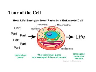
  • 18. A Panoramic View of the Eukaryotic Cell • A eukaryotic cell has internal membranes that partition the cell into organelles • Plant and animal cells have most of the same organelles © 2011 Pearson Education, Inc.
  • 19. Figure 6.8a ENDOPLASMIC RETICULUM (ER) Rough ER Smooth ER Nuclear envelope Nucleolus Chromatin Plasma membrane Ribosomes Golgi apparatus Lysosome Mitochondrion Peroxisome Microvilli Microtubules Intermediate filaments Microfilaments Centrosome CYTOSKELETON: Flagellum NUCLEUS
  • 20. NUCLEUS Nuclear envelope Nucleolus Chromatin Golgi apparatus Mitochondrion Peroxisome Plasma membrane Cell wall Wall of adjacent cell Plasmodesmata Chloroplast Microtubules Intermediate filaments Microfilaments CYTOSKELETON Central vacuole Ribosomes Smooth endoplasmic reticulum Rough endoplasmic reticulum Figure 6.8c
  • 21. Concept 6.3: The eukaryotic cell’s genetic instructions are housed in the nucleus and carried out by the ribosomes • The nucleus contains most of the DNA in a eukaryotic cell • Ribosomes use the information from the DNA to make proteins © 2011 Pearson Education, Inc.
  • 22. The Nucleus: Information Central • The nucleus contains most of the cell’s genes and is usually the most conspicuous organelle • The nuclear envelope encloses the nucleus, separating it from the cytoplasm • The nuclear membrane is a double membrane; each membrane consists of a lipid bilayer © 2011 Pearson Education, Inc.
  • 23. Nucleus Rough ER Nucleolus Chromatin Nuclear envelope: Inner membrane Outer membrane Nuclear pore Chromatin Ribosome Pore complex Close-up of nuclear envelope Figure 6.9a
  • 24. • Pores regulate the entry and exit of molecules from the nucleus • The shape of the nucleus is maintained by the nuclear lamina, which is composed of protein © 2011 Pearson Education, Inc.
  • 25. • In the nucleus, DNA is organized into discrete units called chromosomes • Each chromosome is composed of a single DNA molecule associated with proteins • The DNA and proteins of chromosomes are together called chromatin • The nucleolus is located within the nucleus and is the site of ribosomal RNA (rRNA) synthesis © 2011 Pearson Education, Inc.
  • 26. Ribosomes: Protein Factories • Ribosomes are particles made of ribosomal RNA and protein • Ribosomes carry out protein synthesis in two locations – In the cytosol (free ribosomes) – On the outside of the endoplasmic reticulum or the nuclear envelope (bound ribosomes) © 2011 Pearson Education, Inc.
  • 27. Figure 6.10 0.25 m Free ribosomes in cytosol Endoplasmic reticulum (ER) Ribosomes bound to ER Large subunit Small subunit Diagram of a ribosome TEM showing ER and ribosomes
  • 28. Concept 6.4: The endomembrane system regulates protein traffic and performs metabolic functions in the cell • Components of the endomembrane system – Nuclear envelope – Endoplasmic reticulum – Golgi apparatus – Lysosomes – Vacuoles – Plasma membrane • These components are either continuous or connected via transfer by vesicles © 2011 Pearson Education, Inc.
  • 29. The Endoplasmic Reticulum: Biosynthetic Factory • The endoplasmic reticulum (ER) accounts for more than half of the total membrane in many eukaryotic cells • The ER membrane is continuous with the nuclear envelope • There are two distinct regions of ER – Smooth ER, which lacks ribosomes – Rough ER, surface is studded with ribosomes © 2011 Pearson Education, Inc.
  • 30. Figure 6.11a Smooth ER Rough ER Cisternae Ribosomes Transport vesicle Transitional ER Nuclear envelope ER lumen
  • 31. Functions of Smooth ER • The smooth ER – Synthesizes lipids – Metabolizes carbohydrates – Detoxifies drugs and poisons – Stores calcium ions © 2011 Pearson Education, Inc.
  • 32. Functions of Rough ER • The rough ER – Has bound ribosomes, which secrete glycoproteins (proteins covalently bonded to carbohydrates) – Distributes transport vesicles, proteins surrounded by membranes – Is a membrane factory for the cell © 2011 Pearson Education, Inc.
  • 33. • The Golgi apparatus consists of flattened membranous sacs called cisternae • Functions of the Golgi apparatus – Modifies products of the ER – Manufactures certain macromolecules – Sorts and packages materials into transport vesicles The Golgi Apparatus: Shipping and Receiving Center © 2011 Pearson Education, Inc.
  • 34. Figure 6.12 cis face (“receiving” side of Golgi apparatus) trans face (“shipping” side of Golgi apparatus) 0.1 m TEM of Golgi apparatus Cisternae
  • 35. Lysosomes: Digestive Compartments • A lysosome is a membranous sac of hydrolytic enzymes that can digest macromolecules • Lysosomal enzymes can hydrolyze proteins, fats, polysaccharides, and nucleic acids • Lysosomal enzymes work best in the acidic environment inside the lysosome © 2011 Pearson Education, Inc.
  • 36. • Some types of cell can engulf another cell by phagocytosis; this forms a food vacuole • A lysosome fuses with the food vacuole and digests the molecules • Lysosomes also use enzymes to recycle the cell’s own organelles and macromolecules, a process called autophagy © 2011 Pearson Education, Inc.
  • 37. Figure 6.13b Vesicle containing two damaged organelles 1 m Mitochondrion fragment Peroxisome fragment Peroxisome Vesicle Mitochondrion Lysosome Digestion (b) Autophagy
  • 38. Figure 6.13bb Vesicle containing two damaged organelles 1 m Mitochondrion fragment Peroxisome fragment
  • 39. Vacuoles: Diverse Maintenance Compartments • A plant cell or fungal cell may have one or several vacuoles, derived from endoplasmic reticulum and Golgi apparatus © 2011 Pearson Education, Inc.
  • 40. • Food vacuoles are formed by phagocytosis • Contractile vacuoles, found in many freshwater protists, pump excess water out of cells • Central vacuoles, found in many mature plant cells, hold organic compounds and water © 2011 Pearson Education, Inc.
  • 41. Figure 6.14 Central vacuole Cytosol Nucleus Cell wall Chloroplast Central vacuole 5 m
  • 42. Figure 6.15-3 Smooth ER Nucleus Rough ER Plasma membrane cis Golgi trans Golgi
  • 43. Concept 6.5: Mitochondria and chloroplasts change energy from one form to another • Mitochondria are the sites of cellular respiration, a metabolic process that uses oxygen to generate ATP • Chloroplasts, found in plants and algae, are the sites of photosynthesis © 2011 Pearson Education, Inc.
  • 44. • Mitochondria and chloroplasts have similarities with bacteria – Enveloped by a double membrane – Contain free ribosomes and circular DNA molecules – Grow and reproduce somewhat independently in cells © 2011 Pearson Education, Inc. The Evolutionary Origins of Mitochondria and Chloroplasts
  • 45. • The Endosymbiont theory – An early ancestor of eukaryotic cells engulfed a nonphotosynthetic prokaryotic cell, which formed an endosymbiont relationship with its host – The host cell and endosymbiont merged into a single organism, a eukaryotic cell with a mitochondrion – At least one of these cells may have taken up a photosynthetic prokaryote, becoming the ancestor of cells that contain chloroplasts © 2011 Pearson Education, Inc.
  • 46. Figure 6.16 Nucleus Endoplasmic reticulum Nuclear envelope Ancestor of eukaryotic cells (host cell) Engulfing of oxygen- using nonphotosynthetic prokaryote, which becomes a mitochondrion Mitochondrion Nonphotosynthetic eukaryote Mitochondrion At least one cell Photosynthetic eukaryote Engulfing of photosynthetic prokaryote Chloroplast
  • 47. Mitochondria: Chemical Energy Conversion • Mitochondria are in nearly all eukaryotic cells • They have a smooth outer membrane and an inner membrane folded into cristae • The inner membrane creates two compartments: intermembrane space and mitochondrial matrix • Some metabolic steps of cellular respiration are catalyzed in the mitochondrial matrix • Cristae present a large surface area for enzymes that synthesize ATP © 2011 Pearson Education, Inc.
  • 48. Figure 6.17a Intermembrane space Outer DNA Inner membrane Cristae Matrix Free ribosomes in the mitochondrial matrix (a) Diagram and TEM of mitochondrion 0.1 m membrane
  • 49. Chloroplasts: Capture of Light Energy • Chloroplasts contain the green pigment chlorophyll, as well as enzymes and other molecules that function in photosynthesis • Chloroplasts are found in leaves and other green organs of plants and in algae © 2011 Pearson Education, Inc.
  • 50. • Chloroplast structure includes – Thylakoids, membranous sacs, stacked to form a granum – Stroma, the internal fluid • The chloroplast is one of a group of plant organelles, called plastids © 2011 Pearson Education, Inc.
  • 51. Figure 6.18a Ribosomes Stroma Inner and outer membranes Granum 1 m Intermembrane space Thylakoid (a) Diagram and TEM of chloroplast DNA
  • 52. Peroxisomes: Oxidation • Peroxisomes are specialized metabolic compartments bounded by a single membrane • Peroxisomes produce hydrogen peroxide and convert it to water • Peroxisomes perform reactions with many different functions © 2011 Pearson Education, Inc.
  • 53. Concept 6.6: The cytoskeleton is a network of fibers that organizes structures and activities in the cell • The cytoskeleton is a network of fibers extending throughout the cytoplasm • It organizes the cell’s structures and activities, anchoring many organelles • It is composed of three types of molecular structures – Microtubules – Microfilaments – Intermediate filaments © 2011 Pearson Education, Inc.
  • 54. Roles of the Cytoskeleton: Support and Motility • The cytoskeleton helps to support the cell and maintain its shape • It interacts with motor proteins to produce motility • Inside the cell, vesicles can travel along “monorails” provided by the cytoskeleton • Recent evidence suggests that the cytoskeleton may help regulate biochemical activities © 2011 Pearson Education, Inc.
  • 55. Figure 6.21 ATP Vesicle (a) Motor protein (ATP powered) Microtubule of cytoskeleton Receptor for motor protein 0.25 m Vesicles Microtubule (b)
  • 56. Components of the Cytoskeleton • Three main types of fibers make up the cytoskeleton – Microtubules are the thickest of the three components of the cytoskeleton – Microfilaments, also called actin filaments, are the thinnest components – Intermediate filaments are fibers with diameters in a middle range © 2011 Pearson Education, Inc.
  • 57. Tubulin dimer 25 nm   Column of tubulin dimers 10 m Table 6.1a
  • 58. 10 m Actin subunit 7 nm Table 6.1b
  • 59. 5 m Keratin proteins Fibrous subunit (keratins coiled together) 812 nm Table 6.1c
  • 60. Centrosomes and Centrioles • In many cells, microtubules grow out from a centrosome near the nucleus • The centrosome is a “microtubule-organizing center” • In animal cells, the centrosome has a pair of centrioles, each with nine triplets of microtubules arranged in a ring © 2011 Pearson Education, Inc.
  • 61. Centrosome Longitudinal section of one centriole Centrioles Microtubule 0.25 m Microtubules Cross section of the other centriole Figure 6.22
  • 62. Cilia and Flagella • Microtubules control the beating of cilia and flagella, locomotor appendages of some cells • Cilia and flagella differ in their beating patterns © 2011 Pearson Education, Inc.
  • 63. Direction of swimming (b) Motion of cilia Direction of organism’s movement Power stroke Recovery stroke (a) Motion of flagella 5 m 15 m Figure 6.23
  • 64. • Cilia and flagella share a common structure – A core of microtubules sheathed by the plasma membrane – A basal body that anchors the cilium or flagellum – A motor protein called dynein, which drives the bending movements of a cilium or flagellum © 2011 Pearson Education, Inc.
  • 65. Figure 6.24b 0.1 m (b) Cross section of motile cilium Outer microtubule doublet Dynein proteins Central microtubule Radial spoke Cross-linking proteins between outer doublets Plasma membrane
  • 66. Figure 6.24c 0.1 m Triplet (c) Cross section of basal body
  • 67. • How dynein “walking” moves flagella and cilia − Dynein arms alternately grab, move, and release the outer microtubules – Protein cross-links limit sliding – Forces exerted by dynein arms cause doublets to curve, bending the cilium or flagellum © 2011 Pearson Education, Inc.
  • 68. Figure 6.25 Microtubule doublets Dynein protein ATP (a) Effect of unrestrained dynein movement Cross-linking proteins between outer doublets ATP Anchorage in cell (b) Effect of cross-linking proteins (c) Wavelike motion 1 2 3
  • 69. Concept 6.7: Extracellular components and connections between cells help coordinate cellular activities • Most cells synthesize and secrete materials that are external to the plasma membrane • These extracellular structures include – Cell walls of plants – The extracellular matrix (ECM) of animal cells – Intercellular junctions © 2011 Pearson Education, Inc.
  • 70. Cell Walls of Plants • The cell wall is an extracellular structure that distinguishes plant cells from animal cells • Prokaryotes, fungi, and some protists also have cell walls • The cell wall protects the plant cell, maintains its shape, and prevents excessive uptake of water • Plant cell walls are made of cellulose fibers embedded in other polysaccharides and protein © 2011 Pearson Education, Inc.
  • 71. • Plant cell walls may have multiple layers – Primary cell wall: relatively thin and flexible – Middle lamella: thin layer between primary walls of adjacent cells – Secondary cell wall (in some cells): added between the plasma membrane and the primary cell wall • Plasmodesmata are channels between adjacent plant cells © 2011 Pearson Education, Inc.
  • 72. Secondary cell wall Primary cell wall Middle lamella Central vacuole Cytosol Plasma membrane Plant cell walls Plasmodesmata 1 m Figure 6.28
  • 73. The Extracellular Matrix (ECM) of Animal Cells • Animal cells lack cell walls but are covered by an elaborate extracellular matrix (ECM) • The ECM is made up of glycoproteins such as collagen, proteoglycans, and fibronectin • ECM proteins bind to receptor proteins in the plasma membrane called integrins © 2011 Pearson Education, Inc.
  • 75. • Functions of the ECM – Support – Adhesion – Movement – Regulation © 2011 Pearson Education, Inc.
  • 76. Cell Junctions • Neighboring cells in tissues, organs, or organ systems often adhere, interact, and communicate through direct physical contact • Intercellular junctions facilitate this contact • There are several types of intercellular junctions – Plasmodesmata – Tight junctions – Desmosomes – Gap junctions © 2011 Pearson Education, Inc.
  • 77. Plasmodesmata in Plant Cells • Plasmodesmata are channels that perforate plant cell walls • Through plasmodesmata, water and small solutes (and sometimes proteins and RNA) can pass from cell to cell © 2011 Pearson Education, Inc.
  • 78. Tight Junctions, Desmosomes, and Gap Junctions in Animal Cells • At tight junctions, membranes of neighboring cells are pressed together, preventing leakage of extracellular fluid- skin cells • Desmosomes (anchoring junctions) fasten cells together into strong sheets- muscle cells • Gap junctions (communicating junctions) provide cytoplasmic channels between adjacent cells-heart muscle © 2011 Pearson Education, Inc.
  • 79. Tight junctions prevent fluid from moving across a layer of cells Extracellular matrix Plasma membranes of adjacent cells Space between cells Ions or small molecules Desmosome Intermediate filaments Tight junction Gap junction Figure 6.32a