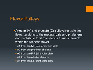
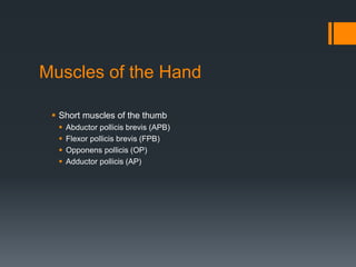
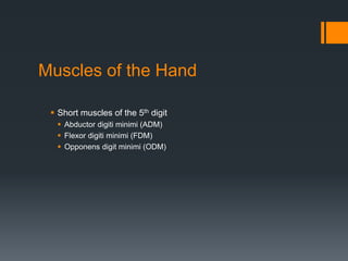
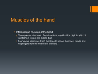
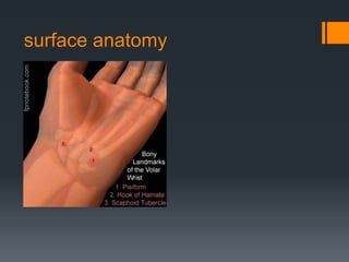
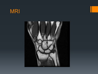
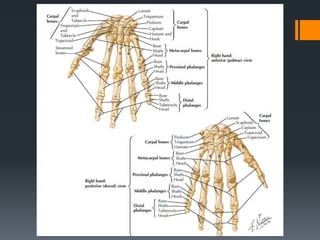
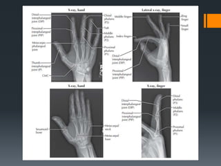
SlideShare a Scribd company logo
1 of 96
Clinical Examination
of the Hand and Wrist
• Campbell’s operative orthopaedics 11th edition
• Text book of orthopaedics & fractures 5th edition Dr B. Aalami Harandi
• Gray’s anatomy 2nd edition
• Clinical anatomy Richard S. Snell
Anatomy of the wrist
Distal radioulnar joint
Radiocarpal joint
Ulnocarpal joint
8 carpal bones (proximal and distal row and attached
ligaments)
Proximal:
1. Scaphoid
2. Lunate
3. Triquetrum
4. Pisiform (smallest)
Distal:
1. Trapezium
2. Trapezoid
3. Capitate (largest)
4. hamate
Variations
Fourth carpometacarpal articulation
Scaphotrapeziotrapezoid articulation
Capitolunate articulation
Hamatolunate articulation
Articulations
 Radiocarpal joints
 Triquetrum and triangular fibrocartilage
 Midcarpal articulations
 Distal row articulations with the matacarpals
1. Mobility in the thumb
2. Stability in the index and long finger metacarpals
3. Increased mobility in the ring and little finger
Triangular fibrocartilage complex
Ulnar collateral ligament
Dorsal and volar radioulnar ligament
Articular disc(Compressed with Pronation and
Extension Compressed with Ulnar deviation)
Meniscal homologue
Extensor carpi ulnaris sheat
Ulnolunate and ulnotriquetral ligament
Carpal Ligaments
The major ligaments of the wrist include the
palmar intrinsic ligaments, the volar extrinsic
and the dorsal extrinsic and intrinsic
ligaments
The extrinsic palmar ligaments provide the
majority of the wrist stability
The intrinsic ligaments serve as rotational
restraints, binding the proximal row into a unit of
rotational stability
Radiocarpal Joint
 Formed by the large articular concave surface of the distal
end of the radius, the scaphoid and lunate of the proximal
carpal row, and the TFCC
extensor retinaculum
The extensor retinaculum compartments,
from lateral to medial, contain the tendons of:
Abductor pollicis longus and extensor pollicis
brevis
Extensor carpi radialis longus and brevis
Extensor pollicis longus
Extensor digitorum and indicis
Extensor digiti minimi
Extensor carpi ulnaris
The Flexor Retinaculum
Transforms the carpal arch into a tunnel, through
which pass the median nerve and some of the
tendons of the hand
 Proximally, the retinaculum attaches to the tubercle of the
scaphoid and the pisiform
 Distally it attaches to the hook of the hamate, and the tubercle
of the trapezium
In the condition known as ‘carpal tunnel syndrome’
the median nerve is compressed in this relatively
unyielding space
Carpal Tunnel
Serves as a conduit for the median nerve and nine
flexor tendons
 The palmar radiocarpal ligament and the palmar ligament
complex form the floor of the canal
 The roof of the tunnel is formed by the flexor retinaculum
(transverse carpal ligament)
 The ulnar and radial borders are formed by carpal bones
(trapezium and hook of hamate respectively)
 Within the tunnel, the median nerve divides into a motor
branch and distal sensory branches
Tunnel of Guyon
A depression superficial to the flexor retinaculum,
located between the hook of the hamate and the
pisiform bones
 The palmar (volar) carpal ligament, palmaris brevis muscle,
and the palmar aponeurosis form its roof
 Its floor is formed by the flexor retinaculum (transverse
carpal ligament), pisohamate ligament, and pisometacarpal
ligament
The tunnel serves as a passage way for the ulnar
nerve and artery into the hand
Phalanges
Fourteen in number
Each consist of a base, shaft, and head
Two shallow depressions, which correspond to
the pulley-shaped heads of the adjacent
phalanges, mark the concave proximal bases
Two distinct convex condyles produce the pulley-
shaped configuration of the phalangeal heads
Metacarpophalangeal (MCP) Joints
of the 2nd-5th Fingers
The 2nd-5th metacarpals articulate with the
respective proximal phalanges in biaxial joints
The MCP joints allow flexion-extension and
medial-lateral deviation associated with a slight
degree of axial rotation
Carpometacarpal Joints
Articulation between the distal borders of the
distal carpal row bones and the bases of the
metacarpals
Stability of the CMC joints is provided by the
palmar and dorsal carpometacarpal and
intermetacarpal ligaments
First Carpometacarpal Joint
 Functionally the sellar (saddle-shaped) carpometacarpal
(CMC) joint is the most important joint of the thumb
 Consists of the articulation between the base of the first
metacarpal and the distal aspect of the trapezium
 Motions that can occur at this joint include flexion/extension,
adduction/abduction and opposition (which includes varying
amounts of flexion, internal rotation, and palmar adduction)
Metacarpophalangeal Joint of the
Thumb
 A hinge joint
 Consists of a convex surface on the head of the metacarpal, and
a concave surface on the base of the phalanx
Interphalangeal (IP) Joints
 Adjacent phalanges articulate in hinge joints that allow motion in
only one plane
 The congruency of the IP joint surfaces contributes greatly to
finger joint stability
 The proximal IP joint is a hinged joint capable of flexion and extension
 The distal IP joint has similar structures but less stability and allows
some hyperextension.
Palmar Aponeurosis
A dense fibrous structure continuous with the
palmaris longus tendon and fascia covering the
thenar and hypothenar muscles
Dupuytren’s contracture is a fibrotic condition of
the palmar aponeurosis that results in nodule
formation or scarring of the aponeurosis, and
which may ultimately cause finger flexion
contractures
Extensor Hood
A complex tendon, which covers the dorsal
aspect of the digits is formed from a combination
of the tendons of insertion from extensor
digitorum, extensor indicis, and extensor digiti
minimi
Creates a ‘cable’ system that provides a
mechanism for extending the MCP and IP joints,
and allows the lumbrical, and possibly
interosseous muscles, to assist in the flexion of
the MCP joints
Synovial Sheaths
 Long narrow balloons filled with synovial fluid, which wrap around
a tendon so that one part of the balloon wall (visceral layer) is
directly on the tendon, while the other part of the balloon wall
(parietal layer) is separate
Flexor Pulleys
Annular (A) and cruciate (C) pulleys restrain the
flexor tendons to the metacarpals and phalanges
and contribute to fibro-osseous tunnels through
which the tendons travel
 A1 from the MP joint and volar plate
 A2 from the proximal phalanx
 A3 from the PIP joint volar plate
 A4 from the middle phalanx
 A5 from the DIP joint volar plate
Muscles of the Hand
 Short muscles of the thumb
 Abductor pollicis brevis (APB)
 Flexor pollicis brevis (FPB)
 Opponens pollicis (OP)
 Adductor pollicis (AP)
Muscles of the Hand
 Short muscles of the 5th digit
 Abductor digiti minimi (ADM)
 Flexor digiti minimi (FDM)
 Opponens digit minimi (ODM)
Muscles of the hand
 Interosseous muscles of the hand
 Three palmar interossei. Each functions to adduct the digit, to which it
is attached, toward the middle digit
 Four dorsal interossei. Each functions to abduct the index, middle and
ring fingers from the mid-line of the hand
Muscles of the hand
 Lumbricales
 Function to perform the motion of IP joint extension with the MCP joint
held in extension
 Can assist in MCP flexion
Anatomic Snuff Box
A depression on the dorsal surface of the hand
at the base of the thumb, just distal to the radius
Formed by the tendons of the APL and EPB,
while the ulnar border is formed by the tendon of
the EPL
Along the floor of the snuffbox is the deep
branch of the radial artery and the tendinous
insertion of the ECRL. Underneath these
structures, the scaphoid and trapezium bones
are found
Neurology
 The three peripheral nerves that supply the skin and muscles of
the wrist and hand include the median, ulnar, and radial nerve
Vasculature of the wrist and hand
 The brachial artery bifurcates at the elbow into radial and ulnar
branches, which are the main arterial branches to the hand
 Vascular arches of the hand
 Dorsal arches
 Palmar arches
Biomechanics
 The wrist contains several segments whose combined
movements create a total range of motion that is greater than
the sum of its individual parts
Pronation
 Approximately 90° of forearm pronation is available
 During pronation, the concave ulnar notch of the radius glides around
the peripheral surface of the relatively fixed convex ulnar head
 Pronation is limited by the bony impaction between the radius and the
ulna
Supination
 Approximately 85-90° of forearm supination is available
 Supination is limited by the interosseous membrane, and the bony
impaction between the ulnar notch of the radius, and the ulnar styloid
process
Wrist flexion and extension
 The movements of flexion and extension of the wrist are shared
among the radiocarpal articulation, and the intercarpal
articulation, in varying proportions
Wrist flexion and extension
During wrist flexion, most of the motion occurs in
the midcarpal joint (60% or 40° versus 40% or
30° at the radiocarpal joint), and is associated
with slight ulnar deviation and supination of the
forearm
During wrist extension, most of the motion
occurs at the radiocarpal joint (66.5% or 40°
versus 33.5% or 20° at the midcarpal joint), and
is associated with slight radial deviation and
pronation of the forearm
Radial Deviation
 Radial deviation occurs primarily between the proximal and distal
rows of the carpal bones
 The motion of radial deviation is limited by impact of the scaphoid
onto the radial styloid, and ulnar collateral ligament
Ulnar deviation
 Ulnar deviation occurs primarily at the radiocarpal joint
 Ulnar deviation is limited by the radial collateral ligament
The hand
The hand accounts for about 90% of upper
limb function
The thumb is involved in 40-50% of hand function
The index finger is involved in about 20% of hand
function
The middle finger, which accounts for about 20%
of all hand function, is the strongest finger, and is
important for both precision and power functions
Thumb motions
Within the first CMC joint, the longitudinal
diameter of the articular surface of the trapezium
is generally concave from a palmar to dorsal
direction
The transverse diameter is generally convex
along a medial to lateral direction
The proximal articular surface of the first
metacarpal is reciprocally shaped to that of the
trapezium
Thumb flexion and extension
 Thumb flexion and extension occur around an anterior-posterior
axis in the frontal plane that is perpendicular to the sagittal plane
of finger flexion and extension
 In this plane, the metacarpal surface is concave, and the
trapezium surface is convex
Thumb abduction and adduction
 Thumb abduction and adduction occur around a medial-lateral
axis in the sagittal plane, that is perpendicular to the frontal plane
of finger abduction and adduction
 During thumb abduction and adduction, the convex metacarpal
surface moves on the concave trapezium
A number of grips have been
recognized:
Fist grip
Cylindrical grip
Ball grip
Hook grip
Ring grip
Pincer grip
Pliers grip
history
What is the cause of pain?
Mechanism of injury?
Previous history?
Location, duration and intensity of pain?
Creptitus, numbness, distortion in temperature?
Sounds or sensations?
Technique changes?
Weakness or fatigue?
What provides relief?
Observation
The clinician inspects for lacerations, surgical
scars, masses, localized swelling, or erythema
Scars should be examined for degree of
adherence, degree of maturation, hypertrophy
(excess collagen within boundary of wound), and
keloid (excess collagen that no longer conforms
to wound boundaries)
The location and type of edema should be noted
Examination
 AROM, then PROM with over pressure
 The gross motions of wrist, hand, finger and thumb flexion,
extension, and radial and ulnar deviation are tested, first actively
and then passively
 Any loss of motion compared with the contralateral,
asymptomatic wrist and hand should be noted
Palpation
 Palpation of the muscles, tendon, insertions, ligaments, capsules,
bones of the wrist and hand should occur as indicated, and be
compared with the uninvolved side
Pain provocation tests
 These tests are used to determine the cause of a painful or
dysfunctional motion by systematically testing each of the
articulations to see whether the maneuvers reproduce the
patient’s symptoms
Strength testing
Isometric tests are carried out in the extreme
range, and if positive, in the neutral range
These isometric tests must include the interossei
and lumbricales
The straight plane motions of wrist flexion,
extension, ulnar and radial deviation are tested
initially
Pain with any of these tests requires a more
thorough examination of the individual muscles
Examination
 Functional Assessment
 The functional range of motion for the hand is the range in which
the hand can perform most of its grip and other functional
activities
 A number of assessment tools are available
Examination
 Passive Physiological Mobility Testing
In each of the tests, the clinician
notes the quantity of motion as well
as the joint reaction (end feel).
The tests are always repeated on,
and compared to, the same joint in
the opposite extremity
Passive Accessory Mobility Tests
 In each of the tests, the clinician notes the quantity of accessory
joint motion as well as the joint reaction
 The tests are always repeated on, and compared to, the same
joint in the opposite extremity
Ligament Stability
 A number of tests are available to evaluate the ligamentous
stability of the forearm, wrist, hand and finger joints
Neurovascular Status
 Allen Test
 Tinel’s test for Carpal Tunnel Syndrome
Examination
 Sensibility Testing
 The assessment of sensibility of the hand is an important
component of every hand examination because sensation is
essential for precision movements and object manipulation
 Two types of sensibility are assessed
 Protective
 Functional
Examination
Special tests
 Carpal Shake test
 Sit to Stand test
 Ulnar Impaction test
 Finkelstein’s test
 Flexor digitorum superficialis (FDS) test
 Flexor digitorum profundus test
 Extensor Hood rupture test
 Froment’s sign
 Murphy’s sign
Examination
Diagnostic testing
Diagnostic testing of the forearm, wrist and hand
is limited to plain radiographs for most patients
Bony tenderness with a history of trauma or a
suspicion of bone or joint disruption indicates a
need for radiographs
Standard projections for the wrist are the
posteroanterior, lateral, and oblique
For the patient with a suspicion of a scaphoid
injury, a scaphoid view should be added
Radiographic Anatomy
Wrist AP
Adult Wrist - Lateral View
CT scan:
CT
Radius
ulna
scaphoid
Lunate
triquetrum
hamate
Capitate
Trapezoid Trapezium
surface anatomy
MRI
ROM
Distal Radioulnar joint
 Supination and Pronation – 80-90o
 Ulna moves posteriorly and laterally with pronation
Radiocarpal joint (and Ulnocarpal joint)
 Flexion (80-90o) and Extension (75-85o)
 Radial (20o) and Ulnar (35o) Deviation
Intercarpal joints
 Gliding
palpation
Bony and Soft Tissue Palpation
 Are they where they should be?
 Do they feel like they should feel?
Circulatory and Neurological Evaluation
 Hands should be felt for temperature
 Cold hands indicate decreased circulation
 Take pulse – radial artery
 Pinching fingernails can also help detect circulatory problems
(capillary refill)
 Hand’s neurological functioning should also be tested
(sensation and motor functioning)
Wrist and hand examination

More Related Content

What's hot (20)

Flat foot
Flat footFlat foot
Flat foot
 
Shoulder examination
Shoulder examination Shoulder examination
Shoulder examination
 
Trigger finger final
Trigger finger finalTrigger finger final
Trigger finger final
 
Ape thumb deformity to publish
Ape thumb deformity to publishApe thumb deformity to publish
Ape thumb deformity to publish
 
Patella fracture
Patella fracturePatella fracture
Patella fracture
 
Dupuytrens Contracture
Dupuytrens ContractureDupuytrens Contracture
Dupuytrens Contracture
 
Elbow examination
Elbow examinationElbow examination
Elbow examination
 
Radial nerve palsy
Radial nerve palsyRadial nerve palsy
Radial nerve palsy
 
supracondylar fracture humerus in children
supracondylar fracture humerus in childrensupracondylar fracture humerus in children
supracondylar fracture humerus in children
 
Ankle Sprains
Ankle SprainsAnkle Sprains
Ankle Sprains
 
De quervain’s
De quervain’sDe quervain’s
De quervain’s
 
Congenital vertical talus BY DR.NAVEEN RATHOR
Congenital vertical talus BY DR.NAVEEN RATHORCongenital vertical talus BY DR.NAVEEN RATHOR
Congenital vertical talus BY DR.NAVEEN RATHOR
 
Knee Injuries In Detail
Knee Injuries In Detail Knee Injuries In Detail
Knee Injuries In Detail
 
Clinical Examination of Hip
Clinical Examination of HipClinical Examination of Hip
Clinical Examination of Hip
 
Swan neck-deformity
Swan neck-deformitySwan neck-deformity
Swan neck-deformity
 
Wrist drop
Wrist dropWrist drop
Wrist drop
 
Hand evaluation
Hand evaluationHand evaluation
Hand evaluation
 
Meniscus injury
Meniscus injuryMeniscus injury
Meniscus injury
 
Trigger finger,causes,symptom,management
Trigger finger,causes,symptom,managementTrigger finger,causes,symptom,management
Trigger finger,causes,symptom,management
 
post polio residual paralysis
post polio residual paralysispost polio residual paralysis
post polio residual paralysis
 

Similar to Wrist and hand examination

A TO Z of Wrist complex by usman
A TO Z  of Wrist  complex by usmanA TO Z  of Wrist  complex by usman
A TO Z of Wrist complex by usmanUsman Farooq
 
UPPER LIMBS - WRIST AND HAND MAY 2022.pdf
UPPER LIMBS - WRIST AND HAND MAY 2022.pdfUPPER LIMBS - WRIST AND HAND MAY 2022.pdf
UPPER LIMBS - WRIST AND HAND MAY 2022.pdfManishaRao30
 
Structures of the Hand PPT
Structures of the Hand PPTStructures of the Hand PPT
Structures of the Hand PPTStacey Turner
 
Kin 191 B – Wrist, Hand And Finger Anatomy
Kin 191 B – Wrist, Hand And Finger AnatomyKin 191 B – Wrist, Hand And Finger Anatomy
Kin 191 B – Wrist, Hand And Finger AnatomyJLS10
 
anatomyofforearm-170105182843.pdf
anatomyofforearm-170105182843.pdfanatomyofforearm-170105182843.pdf
anatomyofforearm-170105182843.pdfsonalidas935894
 
Anatomy of forearm
Anatomy of forearmAnatomy of forearm
Anatomy of forearmsongao
 
Forearm anatomy complete
Forearm anatomy completeForearm anatomy complete
Forearm anatomy completeVedikaAgrawal13
 
Radius, Ulna, Elbow and Radioulnar Joint
Radius, Ulna, Elbow and Radioulnar JointRadius, Ulna, Elbow and Radioulnar Joint
Radius, Ulna, Elbow and Radioulnar JointSado Anatomist
 
Wrist Joint.....2021.pdf
Wrist Joint.....2021.pdfWrist Joint.....2021.pdf
Wrist Joint.....2021.pdfssusercabe92
 
805_Front-of-Forearm.pptx
805_Front-of-Forearm.pptx805_Front-of-Forearm.pptx
805_Front-of-Forearm.pptxGUNASEKARANM20
 
عملي تشريح سنة اولي ترم اول
عملي تشريح سنة اولي ترم اولعملي تشريح سنة اولي ترم اول
عملي تشريح سنة اولي ترم اولebrahim mahmoud
 
تشريح اولي عملي
تشريح اولي عمليتشريح اولي عملي
تشريح اولي عمليebrahim mahmoud
 
Anatomy of Upper extremity
Anatomy of Upper extremityAnatomy of Upper extremity
Anatomy of Upper extremitySunil Pahari
 
Elbow joint with applied anatomy
Elbow joint with applied anatomyElbow joint with applied anatomy
Elbow joint with applied anatomyHETA PATEL
 
Bones of hand and anatomical spaceas
Bones of hand and anatomical spaceasBones of hand and anatomical spaceas
Bones of hand and anatomical spaceasEimaan Ktk
 
Detailed Hand surgical anatomy by mohamed abdelhady
Detailed Hand surgical anatomy by mohamed abdelhadyDetailed Hand surgical anatomy by mohamed abdelhady
Detailed Hand surgical anatomy by mohamed abdelhadyMohamed Abdelhady
 

Similar to Wrist and hand examination (20)

A TO Z of Wrist complex by usman
A TO Z  of Wrist  complex by usmanA TO Z  of Wrist  complex by usman
A TO Z of Wrist complex by usman
 
Wrist joint complex
Wrist joint complexWrist joint complex
Wrist joint complex
 
UPPER LIMBS - WRIST AND HAND MAY 2022.pdf
UPPER LIMBS - WRIST AND HAND MAY 2022.pdfUPPER LIMBS - WRIST AND HAND MAY 2022.pdf
UPPER LIMBS - WRIST AND HAND MAY 2022.pdf
 
Structures of the Hand PPT
Structures of the Hand PPTStructures of the Hand PPT
Structures of the Hand PPT
 
Kin 191 B – Wrist, Hand And Finger Anatomy
Kin 191 B – Wrist, Hand And Finger AnatomyKin 191 B – Wrist, Hand And Finger Anatomy
Kin 191 B – Wrist, Hand And Finger Anatomy
 
anatomyofforearm-170105182843.pdf
anatomyofforearm-170105182843.pdfanatomyofforearm-170105182843.pdf
anatomyofforearm-170105182843.pdf
 
Anatomy of forearm
Anatomy of forearmAnatomy of forearm
Anatomy of forearm
 
Forearm anatomy complete
Forearm anatomy completeForearm anatomy complete
Forearm anatomy complete
 
Anatomy bones of upper limbs
Anatomy bones of upper limbsAnatomy bones of upper limbs
Anatomy bones of upper limbs
 
Radius, Ulna, Elbow and Radioulnar Joint
Radius, Ulna, Elbow and Radioulnar JointRadius, Ulna, Elbow and Radioulnar Joint
Radius, Ulna, Elbow and Radioulnar Joint
 
Wrist Joint.....2021.pdf
Wrist Joint.....2021.pdfWrist Joint.....2021.pdf
Wrist Joint.....2021.pdf
 
Anatomy of Hand
Anatomy of HandAnatomy of Hand
Anatomy of Hand
 
805_Front-of-Forearm.pptx
805_Front-of-Forearm.pptx805_Front-of-Forearm.pptx
805_Front-of-Forearm.pptx
 
عملي تشريح سنة اولي ترم اول
عملي تشريح سنة اولي ترم اولعملي تشريح سنة اولي ترم اول
عملي تشريح سنة اولي ترم اول
 
تشريح اولي عملي
تشريح اولي عمليتشريح اولي عملي
تشريح اولي عملي
 
Anatomy of Upper extremity
Anatomy of Upper extremityAnatomy of Upper extremity
Anatomy of Upper extremity
 
Elbow joint with applied anatomy
Elbow joint with applied anatomyElbow joint with applied anatomy
Elbow joint with applied anatomy
 
peripheral nerve injuries
peripheral nerve injuriesperipheral nerve injuries
peripheral nerve injuries
 
Bones of hand and anatomical spaceas
Bones of hand and anatomical spaceasBones of hand and anatomical spaceas
Bones of hand and anatomical spaceas
 
Detailed Hand surgical anatomy by mohamed abdelhady
Detailed Hand surgical anatomy by mohamed abdelhadyDetailed Hand surgical anatomy by mohamed abdelhady
Detailed Hand surgical anatomy by mohamed abdelhady
 

Recently uploaded

Kisan Call Centre - To harness potential of ICT in Agriculture by answer farm...
Kisan Call Centre - To harness potential of ICT in Agriculture by answer farm...Kisan Call Centre - To harness potential of ICT in Agriculture by answer farm...
Kisan Call Centre - To harness potential of ICT in Agriculture by answer farm...Krashi Coaching
 
The Most Excellent Way | 1 Corinthians 13
The Most Excellent Way | 1 Corinthians 13The Most Excellent Way | 1 Corinthians 13
The Most Excellent Way | 1 Corinthians 13Steve Thomason
 
CARE OF CHILD IN INCUBATOR..........pptx
CARE OF CHILD IN INCUBATOR..........pptxCARE OF CHILD IN INCUBATOR..........pptx
CARE OF CHILD IN INCUBATOR..........pptxGaneshChakor2
 
Alper Gobel In Media Res Media Component
Alper Gobel In Media Res Media ComponentAlper Gobel In Media Res Media Component
Alper Gobel In Media Res Media ComponentInMediaRes1
 
A Critique of the Proposed National Education Policy Reform
A Critique of the Proposed National Education Policy ReformA Critique of the Proposed National Education Policy Reform
A Critique of the Proposed National Education Policy ReformChameera Dedduwage
 
Accessible design: Minimum effort, maximum impact
Accessible design: Minimum effort, maximum impactAccessible design: Minimum effort, maximum impact
Accessible design: Minimum effort, maximum impactdawncurless
 
Organic Name Reactions for the students and aspirants of Chemistry12th.pptx
Organic Name Reactions  for the students and aspirants of Chemistry12th.pptxOrganic Name Reactions  for the students and aspirants of Chemistry12th.pptx
Organic Name Reactions for the students and aspirants of Chemistry12th.pptxVS Mahajan Coaching Centre
 
BASLIQ CURRENT LOOKBOOK LOOKBOOK(1) (1).pdf
BASLIQ CURRENT LOOKBOOK  LOOKBOOK(1) (1).pdfBASLIQ CURRENT LOOKBOOK  LOOKBOOK(1) (1).pdf
BASLIQ CURRENT LOOKBOOK LOOKBOOK(1) (1).pdfSoniaTolstoy
 
KSHARA STURA .pptx---KSHARA KARMA THERAPY (CAUSTIC THERAPY)————IMP.OF KSHARA ...
KSHARA STURA .pptx---KSHARA KARMA THERAPY (CAUSTIC THERAPY)————IMP.OF KSHARA ...KSHARA STURA .pptx---KSHARA KARMA THERAPY (CAUSTIC THERAPY)————IMP.OF KSHARA ...
KSHARA STURA .pptx---KSHARA KARMA THERAPY (CAUSTIC THERAPY)————IMP.OF KSHARA ...M56BOOKSTORE PRODUCT/SERVICE
 
mini mental status format.docx
mini    mental       status     format.docxmini    mental       status     format.docx
mini mental status format.docxPoojaSen20
 
SOCIAL AND HISTORICAL CONTEXT - LFTVD.pptx
SOCIAL AND HISTORICAL CONTEXT - LFTVD.pptxSOCIAL AND HISTORICAL CONTEXT - LFTVD.pptx
SOCIAL AND HISTORICAL CONTEXT - LFTVD.pptxiammrhaywood
 
Paris 2024 Olympic Geographies - an activity
Paris 2024 Olympic Geographies - an activityParis 2024 Olympic Geographies - an activity
Paris 2024 Olympic Geographies - an activityGeoBlogs
 
18-04-UA_REPORT_MEDIALITERAСY_INDEX-DM_23-1-final-eng.pdf
18-04-UA_REPORT_MEDIALITERAСY_INDEX-DM_23-1-final-eng.pdf18-04-UA_REPORT_MEDIALITERAСY_INDEX-DM_23-1-final-eng.pdf
18-04-UA_REPORT_MEDIALITERAСY_INDEX-DM_23-1-final-eng.pdfssuser54595a
 
MENTAL STATUS EXAMINATION format.docx
MENTAL     STATUS EXAMINATION format.docxMENTAL     STATUS EXAMINATION format.docx
MENTAL STATUS EXAMINATION format.docxPoojaSen20
 
POINT- BIOCHEMISTRY SEM 2 ENZYMES UNIT 5.pptx
POINT- BIOCHEMISTRY SEM 2 ENZYMES UNIT 5.pptxPOINT- BIOCHEMISTRY SEM 2 ENZYMES UNIT 5.pptx
POINT- BIOCHEMISTRY SEM 2 ENZYMES UNIT 5.pptxSayali Powar
 
Interactive Powerpoint_How to Master effective communication
Interactive Powerpoint_How to Master effective communicationInteractive Powerpoint_How to Master effective communication
Interactive Powerpoint_How to Master effective communicationnomboosow
 
Employee wellbeing at the workplace.pptx
Employee wellbeing at the workplace.pptxEmployee wellbeing at the workplace.pptx
Employee wellbeing at the workplace.pptxNirmalaLoungPoorunde1
 
Enzyme, Pharmaceutical Aids, Miscellaneous Last Part of Chapter no 5th.pdf
Enzyme, Pharmaceutical Aids, Miscellaneous Last Part of Chapter no 5th.pdfEnzyme, Pharmaceutical Aids, Miscellaneous Last Part of Chapter no 5th.pdf
Enzyme, Pharmaceutical Aids, Miscellaneous Last Part of Chapter no 5th.pdfSumit Tiwari
 

Recently uploaded (20)

Kisan Call Centre - To harness potential of ICT in Agriculture by answer farm...
Kisan Call Centre - To harness potential of ICT in Agriculture by answer farm...Kisan Call Centre - To harness potential of ICT in Agriculture by answer farm...
Kisan Call Centre - To harness potential of ICT in Agriculture by answer farm...
 
The Most Excellent Way | 1 Corinthians 13
The Most Excellent Way | 1 Corinthians 13The Most Excellent Way | 1 Corinthians 13
The Most Excellent Way | 1 Corinthians 13
 
CARE OF CHILD IN INCUBATOR..........pptx
CARE OF CHILD IN INCUBATOR..........pptxCARE OF CHILD IN INCUBATOR..........pptx
CARE OF CHILD IN INCUBATOR..........pptx
 
Alper Gobel In Media Res Media Component
Alper Gobel In Media Res Media ComponentAlper Gobel In Media Res Media Component
Alper Gobel In Media Res Media Component
 
A Critique of the Proposed National Education Policy Reform
A Critique of the Proposed National Education Policy ReformA Critique of the Proposed National Education Policy Reform
A Critique of the Proposed National Education Policy Reform
 
Accessible design: Minimum effort, maximum impact
Accessible design: Minimum effort, maximum impactAccessible design: Minimum effort, maximum impact
Accessible design: Minimum effort, maximum impact
 
Organic Name Reactions for the students and aspirants of Chemistry12th.pptx
Organic Name Reactions  for the students and aspirants of Chemistry12th.pptxOrganic Name Reactions  for the students and aspirants of Chemistry12th.pptx
Organic Name Reactions for the students and aspirants of Chemistry12th.pptx
 
BASLIQ CURRENT LOOKBOOK LOOKBOOK(1) (1).pdf
BASLIQ CURRENT LOOKBOOK  LOOKBOOK(1) (1).pdfBASLIQ CURRENT LOOKBOOK  LOOKBOOK(1) (1).pdf
BASLIQ CURRENT LOOKBOOK LOOKBOOK(1) (1).pdf
 
KSHARA STURA .pptx---KSHARA KARMA THERAPY (CAUSTIC THERAPY)————IMP.OF KSHARA ...
KSHARA STURA .pptx---KSHARA KARMA THERAPY (CAUSTIC THERAPY)————IMP.OF KSHARA ...KSHARA STURA .pptx---KSHARA KARMA THERAPY (CAUSTIC THERAPY)————IMP.OF KSHARA ...
KSHARA STURA .pptx---KSHARA KARMA THERAPY (CAUSTIC THERAPY)————IMP.OF KSHARA ...
 
TataKelola dan KamSiber Kecerdasan Buatan v022.pdf
TataKelola dan KamSiber Kecerdasan Buatan v022.pdfTataKelola dan KamSiber Kecerdasan Buatan v022.pdf
TataKelola dan KamSiber Kecerdasan Buatan v022.pdf
 
mini mental status format.docx
mini    mental       status     format.docxmini    mental       status     format.docx
mini mental status format.docx
 
SOCIAL AND HISTORICAL CONTEXT - LFTVD.pptx
SOCIAL AND HISTORICAL CONTEXT - LFTVD.pptxSOCIAL AND HISTORICAL CONTEXT - LFTVD.pptx
SOCIAL AND HISTORICAL CONTEXT - LFTVD.pptx
 
Paris 2024 Olympic Geographies - an activity
Paris 2024 Olympic Geographies - an activityParis 2024 Olympic Geographies - an activity
Paris 2024 Olympic Geographies - an activity
 
18-04-UA_REPORT_MEDIALITERAСY_INDEX-DM_23-1-final-eng.pdf
18-04-UA_REPORT_MEDIALITERAСY_INDEX-DM_23-1-final-eng.pdf18-04-UA_REPORT_MEDIALITERAСY_INDEX-DM_23-1-final-eng.pdf
18-04-UA_REPORT_MEDIALITERAСY_INDEX-DM_23-1-final-eng.pdf
 
Código Creativo y Arte de Software | Unidad 1
Código Creativo y Arte de Software | Unidad 1Código Creativo y Arte de Software | Unidad 1
Código Creativo y Arte de Software | Unidad 1
 
MENTAL STATUS EXAMINATION format.docx
MENTAL     STATUS EXAMINATION format.docxMENTAL     STATUS EXAMINATION format.docx
MENTAL STATUS EXAMINATION format.docx
 
POINT- BIOCHEMISTRY SEM 2 ENZYMES UNIT 5.pptx
POINT- BIOCHEMISTRY SEM 2 ENZYMES UNIT 5.pptxPOINT- BIOCHEMISTRY SEM 2 ENZYMES UNIT 5.pptx
POINT- BIOCHEMISTRY SEM 2 ENZYMES UNIT 5.pptx
 
Interactive Powerpoint_How to Master effective communication
Interactive Powerpoint_How to Master effective communicationInteractive Powerpoint_How to Master effective communication
Interactive Powerpoint_How to Master effective communication
 
Employee wellbeing at the workplace.pptx
Employee wellbeing at the workplace.pptxEmployee wellbeing at the workplace.pptx
Employee wellbeing at the workplace.pptx
 
Enzyme, Pharmaceutical Aids, Miscellaneous Last Part of Chapter no 5th.pdf
Enzyme, Pharmaceutical Aids, Miscellaneous Last Part of Chapter no 5th.pdfEnzyme, Pharmaceutical Aids, Miscellaneous Last Part of Chapter no 5th.pdf
Enzyme, Pharmaceutical Aids, Miscellaneous Last Part of Chapter no 5th.pdf
 

Wrist and hand examination

  • 1. Clinical Examination of the Hand and Wrist • Campbell’s operative orthopaedics 11th edition • Text book of orthopaedics & fractures 5th edition Dr B. Aalami Harandi • Gray’s anatomy 2nd edition • Clinical anatomy Richard S. Snell
  • 2. Anatomy of the wrist Distal radioulnar joint Radiocarpal joint Ulnocarpal joint 8 carpal bones (proximal and distal row and attached ligaments)
  • 3. Proximal: 1. Scaphoid 2. Lunate 3. Triquetrum 4. Pisiform (smallest) Distal: 1. Trapezium 2. Trapezoid 3. Capitate (largest) 4. hamate
  • 4.
  • 5. Variations Fourth carpometacarpal articulation Scaphotrapeziotrapezoid articulation Capitolunate articulation Hamatolunate articulation
  • 6. Articulations  Radiocarpal joints  Triquetrum and triangular fibrocartilage  Midcarpal articulations  Distal row articulations with the matacarpals 1. Mobility in the thumb 2. Stability in the index and long finger metacarpals 3. Increased mobility in the ring and little finger
  • 7. Triangular fibrocartilage complex Ulnar collateral ligament Dorsal and volar radioulnar ligament Articular disc(Compressed with Pronation and Extension Compressed with Ulnar deviation) Meniscal homologue Extensor carpi ulnaris sheat Ulnolunate and ulnotriquetral ligament
  • 8. Carpal Ligaments The major ligaments of the wrist include the palmar intrinsic ligaments, the volar extrinsic and the dorsal extrinsic and intrinsic ligaments The extrinsic palmar ligaments provide the majority of the wrist stability The intrinsic ligaments serve as rotational restraints, binding the proximal row into a unit of rotational stability
  • 9. Radiocarpal Joint  Formed by the large articular concave surface of the distal end of the radius, the scaphoid and lunate of the proximal carpal row, and the TFCC
  • 10. extensor retinaculum The extensor retinaculum compartments, from lateral to medial, contain the tendons of: Abductor pollicis longus and extensor pollicis brevis Extensor carpi radialis longus and brevis Extensor pollicis longus Extensor digitorum and indicis Extensor digiti minimi Extensor carpi ulnaris
  • 11. The Flexor Retinaculum Transforms the carpal arch into a tunnel, through which pass the median nerve and some of the tendons of the hand  Proximally, the retinaculum attaches to the tubercle of the scaphoid and the pisiform  Distally it attaches to the hook of the hamate, and the tubercle of the trapezium In the condition known as ‘carpal tunnel syndrome’ the median nerve is compressed in this relatively unyielding space
  • 12. Carpal Tunnel Serves as a conduit for the median nerve and nine flexor tendons  The palmar radiocarpal ligament and the palmar ligament complex form the floor of the canal  The roof of the tunnel is formed by the flexor retinaculum (transverse carpal ligament)  The ulnar and radial borders are formed by carpal bones (trapezium and hook of hamate respectively)  Within the tunnel, the median nerve divides into a motor branch and distal sensory branches
  • 13. Tunnel of Guyon A depression superficial to the flexor retinaculum, located between the hook of the hamate and the pisiform bones  The palmar (volar) carpal ligament, palmaris brevis muscle, and the palmar aponeurosis form its roof  Its floor is formed by the flexor retinaculum (transverse carpal ligament), pisohamate ligament, and pisometacarpal ligament The tunnel serves as a passage way for the ulnar nerve and artery into the hand
  • 14. Phalanges Fourteen in number Each consist of a base, shaft, and head Two shallow depressions, which correspond to the pulley-shaped heads of the adjacent phalanges, mark the concave proximal bases Two distinct convex condyles produce the pulley- shaped configuration of the phalangeal heads
  • 15. Metacarpophalangeal (MCP) Joints of the 2nd-5th Fingers The 2nd-5th metacarpals articulate with the respective proximal phalanges in biaxial joints The MCP joints allow flexion-extension and medial-lateral deviation associated with a slight degree of axial rotation
  • 16. Carpometacarpal Joints Articulation between the distal borders of the distal carpal row bones and the bases of the metacarpals Stability of the CMC joints is provided by the palmar and dorsal carpometacarpal and intermetacarpal ligaments
  • 17. First Carpometacarpal Joint  Functionally the sellar (saddle-shaped) carpometacarpal (CMC) joint is the most important joint of the thumb  Consists of the articulation between the base of the first metacarpal and the distal aspect of the trapezium  Motions that can occur at this joint include flexion/extension, adduction/abduction and opposition (which includes varying amounts of flexion, internal rotation, and palmar adduction)
  • 18. Metacarpophalangeal Joint of the Thumb  A hinge joint  Consists of a convex surface on the head of the metacarpal, and a concave surface on the base of the phalanx
  • 19. Interphalangeal (IP) Joints  Adjacent phalanges articulate in hinge joints that allow motion in only one plane  The congruency of the IP joint surfaces contributes greatly to finger joint stability  The proximal IP joint is a hinged joint capable of flexion and extension  The distal IP joint has similar structures but less stability and allows some hyperextension.
  • 20. Palmar Aponeurosis A dense fibrous structure continuous with the palmaris longus tendon and fascia covering the thenar and hypothenar muscles Dupuytren’s contracture is a fibrotic condition of the palmar aponeurosis that results in nodule formation or scarring of the aponeurosis, and which may ultimately cause finger flexion contractures
  • 21. Extensor Hood A complex tendon, which covers the dorsal aspect of the digits is formed from a combination of the tendons of insertion from extensor digitorum, extensor indicis, and extensor digiti minimi Creates a ‘cable’ system that provides a mechanism for extending the MCP and IP joints, and allows the lumbrical, and possibly interosseous muscles, to assist in the flexion of the MCP joints
  • 22. Synovial Sheaths  Long narrow balloons filled with synovial fluid, which wrap around a tendon so that one part of the balloon wall (visceral layer) is directly on the tendon, while the other part of the balloon wall (parietal layer) is separate
  • 23. Flexor Pulleys Annular (A) and cruciate (C) pulleys restrain the flexor tendons to the metacarpals and phalanges and contribute to fibro-osseous tunnels through which the tendons travel  A1 from the MP joint and volar plate  A2 from the proximal phalanx  A3 from the PIP joint volar plate  A4 from the middle phalanx  A5 from the DIP joint volar plate
  • 24. Muscles of the Hand  Short muscles of the thumb  Abductor pollicis brevis (APB)  Flexor pollicis brevis (FPB)  Opponens pollicis (OP)  Adductor pollicis (AP)
  • 25. Muscles of the Hand  Short muscles of the 5th digit  Abductor digiti minimi (ADM)  Flexor digiti minimi (FDM)  Opponens digit minimi (ODM)
  • 26. Muscles of the hand  Interosseous muscles of the hand  Three palmar interossei. Each functions to adduct the digit, to which it is attached, toward the middle digit  Four dorsal interossei. Each functions to abduct the index, middle and ring fingers from the mid-line of the hand
  • 27. Muscles of the hand  Lumbricales  Function to perform the motion of IP joint extension with the MCP joint held in extension  Can assist in MCP flexion
  • 28. Anatomic Snuff Box A depression on the dorsal surface of the hand at the base of the thumb, just distal to the radius Formed by the tendons of the APL and EPB, while the ulnar border is formed by the tendon of the EPL Along the floor of the snuffbox is the deep branch of the radial artery and the tendinous insertion of the ECRL. Underneath these structures, the scaphoid and trapezium bones are found
  • 29. Neurology  The three peripheral nerves that supply the skin and muscles of the wrist and hand include the median, ulnar, and radial nerve
  • 30. Vasculature of the wrist and hand  The brachial artery bifurcates at the elbow into radial and ulnar branches, which are the main arterial branches to the hand  Vascular arches of the hand  Dorsal arches  Palmar arches
  • 31. Biomechanics  The wrist contains several segments whose combined movements create a total range of motion that is greater than the sum of its individual parts
  • 32. Pronation  Approximately 90° of forearm pronation is available  During pronation, the concave ulnar notch of the radius glides around the peripheral surface of the relatively fixed convex ulnar head  Pronation is limited by the bony impaction between the radius and the ulna
  • 33. Supination  Approximately 85-90° of forearm supination is available  Supination is limited by the interosseous membrane, and the bony impaction between the ulnar notch of the radius, and the ulnar styloid process
  • 34. Wrist flexion and extension  The movements of flexion and extension of the wrist are shared among the radiocarpal articulation, and the intercarpal articulation, in varying proportions
  • 35. Wrist flexion and extension During wrist flexion, most of the motion occurs in the midcarpal joint (60% or 40° versus 40% or 30° at the radiocarpal joint), and is associated with slight ulnar deviation and supination of the forearm During wrist extension, most of the motion occurs at the radiocarpal joint (66.5% or 40° versus 33.5% or 20° at the midcarpal joint), and is associated with slight radial deviation and pronation of the forearm
  • 36. Radial Deviation  Radial deviation occurs primarily between the proximal and distal rows of the carpal bones  The motion of radial deviation is limited by impact of the scaphoid onto the radial styloid, and ulnar collateral ligament
  • 37. Ulnar deviation  Ulnar deviation occurs primarily at the radiocarpal joint  Ulnar deviation is limited by the radial collateral ligament
  • 38. The hand The hand accounts for about 90% of upper limb function The thumb is involved in 40-50% of hand function The index finger is involved in about 20% of hand function The middle finger, which accounts for about 20% of all hand function, is the strongest finger, and is important for both precision and power functions
  • 39. Thumb motions Within the first CMC joint, the longitudinal diameter of the articular surface of the trapezium is generally concave from a palmar to dorsal direction The transverse diameter is generally convex along a medial to lateral direction The proximal articular surface of the first metacarpal is reciprocally shaped to that of the trapezium
  • 40. Thumb flexion and extension  Thumb flexion and extension occur around an anterior-posterior axis in the frontal plane that is perpendicular to the sagittal plane of finger flexion and extension  In this plane, the metacarpal surface is concave, and the trapezium surface is convex
  • 41. Thumb abduction and adduction  Thumb abduction and adduction occur around a medial-lateral axis in the sagittal plane, that is perpendicular to the frontal plane of finger abduction and adduction  During thumb abduction and adduction, the convex metacarpal surface moves on the concave trapezium
  • 42. A number of grips have been recognized: Fist grip Cylindrical grip Ball grip Hook grip Ring grip Pincer grip Pliers grip
  • 43. history What is the cause of pain? Mechanism of injury? Previous history? Location, duration and intensity of pain? Creptitus, numbness, distortion in temperature? Sounds or sensations? Technique changes? Weakness or fatigue? What provides relief?
  • 44. Observation The clinician inspects for lacerations, surgical scars, masses, localized swelling, or erythema Scars should be examined for degree of adherence, degree of maturation, hypertrophy (excess collagen within boundary of wound), and keloid (excess collagen that no longer conforms to wound boundaries) The location and type of edema should be noted
  • 45. Examination  AROM, then PROM with over pressure  The gross motions of wrist, hand, finger and thumb flexion, extension, and radial and ulnar deviation are tested, first actively and then passively  Any loss of motion compared with the contralateral, asymptomatic wrist and hand should be noted
  • 46. Palpation  Palpation of the muscles, tendon, insertions, ligaments, capsules, bones of the wrist and hand should occur as indicated, and be compared with the uninvolved side
  • 47. Pain provocation tests  These tests are used to determine the cause of a painful or dysfunctional motion by systematically testing each of the articulations to see whether the maneuvers reproduce the patient’s symptoms
  • 48. Strength testing Isometric tests are carried out in the extreme range, and if positive, in the neutral range These isometric tests must include the interossei and lumbricales The straight plane motions of wrist flexion, extension, ulnar and radial deviation are tested initially Pain with any of these tests requires a more thorough examination of the individual muscles
  • 49. Examination  Functional Assessment  The functional range of motion for the hand is the range in which the hand can perform most of its grip and other functional activities  A number of assessment tools are available
  • 50. Examination  Passive Physiological Mobility Testing In each of the tests, the clinician notes the quantity of motion as well as the joint reaction (end feel). The tests are always repeated on, and compared to, the same joint in the opposite extremity
  • 51. Passive Accessory Mobility Tests  In each of the tests, the clinician notes the quantity of accessory joint motion as well as the joint reaction  The tests are always repeated on, and compared to, the same joint in the opposite extremity
  • 52. Ligament Stability  A number of tests are available to evaluate the ligamentous stability of the forearm, wrist, hand and finger joints
  • 53. Neurovascular Status  Allen Test  Tinel’s test for Carpal Tunnel Syndrome
  • 54. Examination  Sensibility Testing  The assessment of sensibility of the hand is an important component of every hand examination because sensation is essential for precision movements and object manipulation  Two types of sensibility are assessed  Protective  Functional
  • 55. Examination Special tests  Carpal Shake test  Sit to Stand test  Ulnar Impaction test  Finkelstein’s test  Flexor digitorum superficialis (FDS) test  Flexor digitorum profundus test  Extensor Hood rupture test  Froment’s sign  Murphy’s sign
  • 56. Examination Diagnostic testing Diagnostic testing of the forearm, wrist and hand is limited to plain radiographs for most patients Bony tenderness with a history of trauma or a suspicion of bone or joint disruption indicates a need for radiographs Standard projections for the wrist are the posteroanterior, lateral, and oblique For the patient with a suspicion of a scaphoid injury, a scaphoid view should be added
  • 57.
  • 58.
  • 59.
  • 60.
  • 61.
  • 63. Adult Wrist - Lateral View
  • 64.
  • 68. MRI
  • 69.
  • 70.
  • 71.
  • 72.
  • 73.
  • 74. ROM Distal Radioulnar joint  Supination and Pronation – 80-90o  Ulna moves posteriorly and laterally with pronation Radiocarpal joint (and Ulnocarpal joint)  Flexion (80-90o) and Extension (75-85o)  Radial (20o) and Ulnar (35o) Deviation Intercarpal joints  Gliding
  • 75.
  • 76. palpation Bony and Soft Tissue Palpation  Are they where they should be?  Do they feel like they should feel? Circulatory and Neurological Evaluation  Hands should be felt for temperature  Cold hands indicate decreased circulation  Take pulse – radial artery  Pinching fingernails can also help detect circulatory problems (capillary refill)  Hand’s neurological functioning should also be tested (sensation and motor functioning)