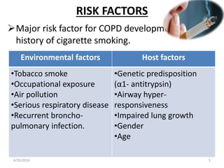
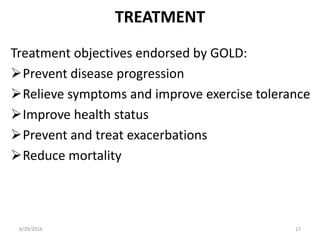
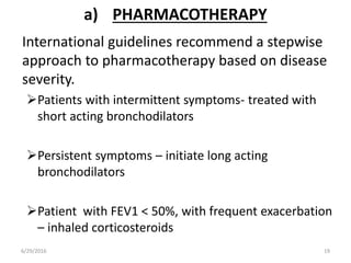
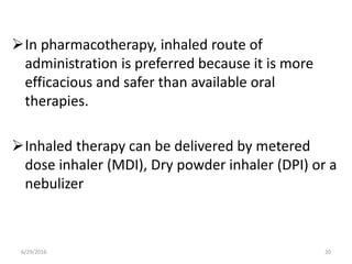
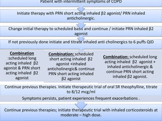
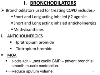
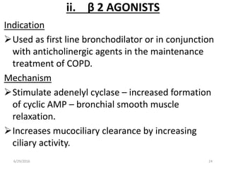
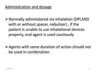
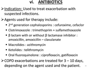
CHRONIC OBSTRUCTIVE PULMONARY DISEASE (COPD)
SHAHANAS CK
FIRST YEAR M.PHARM
PHARMACY PRACTICE
6/29/2016 1
DEFINITION
Global initiative for chronic obstructive lung
diseases defines COPD as:
“Disease characterized by airflow limitation
that is not fully reversible, usually progressive
and associated with abnormal inflammatory
response of the lungs to noxious particles or
gases.”
Common conditions associated with COPD are:
• Chronic bronchitis
• Emphysema
6/29/2016 2
CHRONIC BRONCHITIS:
Chronic bronchitis is associated with chronic or
recurrent excessive mucus secretion in to the
bronchial tree with cough that is present on most
days for at least 2 consecutive years in a patient, in
whom other causes of chronic cough have been
excluded.
EMPHYSEMA:
Abnormal permanent enlargement of airspaces
distal to the terminal bronchioles accompanied by
destruction of their walls without obvious fibrosis.
6/29/2016 3
EPIDEMIOLOGY
It is the 6th leading cause of death in the world.
Mortality rate is higher in males than females, and
in whites than blacks.
COPD is the 2nd leading cause of disability in the
united states.
In India prevalence rate indicates that about 5%
male and 2.7% female over the age of 30 suffer
from COPD.
6/29/2016 4
RISK FACTORS
Major risk factor for COPD development is a
history of cigarette smoking.
Environmental factors Host factors
•Tobacco smoke
•Occupational exposure
•Air pollution
•Serious respiratory disease
•Recurrent broncho-
pulmonary infection.
•Genetic predisposition
(α1- antitrypsin)
•Airway hyper-
responsiveness
•Impaired lung growth
•Gender
•Age
6/29/2016 5
PATHOGENESIS
Exposure to noxious chemical & gases – ↑sed
bronchial reactivity – inflammation
Chronic inflammation - remodeling & narrowing of
airway lumen- fixed / irriversible airflow
obstruction
6/29/2016 6
Four areas of lungs are affected:
6/29/2016 7
Central airways
• Inflammatory
exudates - ↑sed
number & size of
goblet cells &
mucus glands –
mucus
hypersecretion
• Airway cilia
destroyed - ↓sed
clearance of
mucus.
Peripheral
airways
• Thickenin
g of
smooth
muscles
&
connectiv
e tissues.
Lung
parenchyma
• Air spaces
enlarged,
attachmen
ts lost –
alveolar
collapse.
Pulmonary
vasculature
• Thickenin
g of
pulmonar
y vessels

6/29/2016 8
• Neutrophils
• CD8+ lymphocytes
• Macrophages
Prominent
cells of
inflammation:
• Tumor necrosis factor- α
• Interleukin – 8
• Leukotriene –B4
Inflammatory
mediators
involved:
Balance in antiprotease (protective) and
protease (aggressive) activity in the lungs is
disturbed in COPD – injury and damage to
normal lung tissue.
Noxious chemicals and gases increases
oxidative stress – stimulate inflammatory cells
to release additional proteases.
6/29/2016 9
PATHOPHYSIOLOGY
Pathologic changes in COPD are exhibited as:
Air flow limitation
Reduced ratio of FEV1 to FVC
Increased functional residual capacity due to air
trapping and thoracic hyper inflation.
Mucus plugging, chronic airflow limitation and
thoracic hyper inflation disturb normal ventilatory
dynamics.
6/29/2016 10
6/29/2016 11
• Abnormality of gas
exchange: Changes in
pulmonary vasculature
• Pulmonary hypertension:
Elevated mean
pulmonary artery
pressure and pulmonary
vascular resistance
• Hypertrophy of right
ventricle: Development
of cor pulmonale.
Consequences
of advanced
disease
 Mucus plugging – colonization of
bacteria – contribute to chronic
inflammation.
 Factors contributes to airflow
obstruction.
 Increased mucus
 Airway wall fibrosis
 Alveolar collapse
 Normal elastic recoil of lung
during exhalation is lost in COPD-
patient use abdominal and chest
muscle to force air out- further
collapse of airways & air trapping
– thoracic hyper inflation6/29/2016 12
EXACERBATION
A change in patient’s baseline symptoms
(dyspnoea, cough, sputum production) beyond
day to day variability sufficient to warrant a
change in management.
Eosinophils are prominent during
exacerbation.
Typically have an infectious etiology, either
bacterial or viral.
‘haemophylus influenzae’, ‘streptococcus
pneumoniae’, ‘moraxella catarrhalis’ etc are
responsible for most of bacterial respiratory
infections.6/29/2016 13
CLINICAL PRESENTATION
SYMPTOMS
 Cough
 Sputum production
 Dyspnea
PHYSICAL EXAMINATION
 Cyanosis of mucosal membrane
 Barrel chest
 Increased resting respiratory rate
 Shallow breathing
 Pursed lips during expiration
 Use of accessory respiratory
muscles
6/29/2016 14
DIAGNOSTIC TEST RESULTS:-
6/29/2016 15
• Post bronchodilator FEV1/FVC < 70% -
COPD.
Spirometry
• Identifies thoracic hyper inflation, barrel
chest, and increased bronchovascular
markings.
Chest X-ray
• To exclude asthma if there is a doubt about
diagnosis.
Serial domiciliary peak
flow measurements
• Particularly with early onset disease or a
minimal smoking/family history
α1-Antitrypsin
• To identify organism if persistantly present
and purulent .
Sputum culture
• To reveal right ventricular hypertrophy and
changes consistent with cor pulmonale
ECG
Severity classification of COPD
6/29/2016 16
Severity stage Classification Predicted FEV1
1 Mild ≥80%
2 Moderate ≥50% & <80%
3 Severe ≥30% & < 50%
4 Very severe <30% or < 50%
with respiratory
failure
TREATMENT
Treatment objectives endorsed by GOLD:
Prevent disease progression
Relieve symptoms and improve exercise tolerance
Improve health status
Prevent and treat exacerbations
Reduce mortality
6/29/2016 17
THERAPEUTIC PLAN
1. Approaches to treatment
a) Pharmacotherapy
b) Non pharmacologic approach
c) Surgical management
d) Managing exacerbations
2. Improving outcomes
6/29/2016 18
a) PHARMACOTHERAPY
International guidelines recommend a stepwise
approach to pharmacotherapy based on disease
severity.
Patients with intermittent symptoms- treated with
short acting bronchodilators
Persistent symptoms – initiate long acting
bronchodilators
Patient with FEV1 < 50%, with frequent exacerbation
– inhaled corticosteroids
6/29/2016 19
In pharmacotherapy, inhaled route of
administration is preferred because it is more
efficacious and safer than available oral
therapies.
Inhaled therapy can be delivered by metered
dose inhaler (MDI), Dry powder inhaler (DPI) or a
nebulizer
6/29/2016 20
Continue previous therapies; initiate therapeutic trial with inhaled corticosteroids at
moderate – high dose.
Continue previous therapies. Initiate therapeutic trial of oral SR theophylline, titrate
to 8/12 mcg/ml
Symptoms persists, patient experiences frequent exacerbations .
If not previously done initiate and titrate inhaled anti cholinergics to 6 puffs QID
Combination
:scheduled long
acting inhaled β2
agonist & PRN short
acting inhaled β2
agonist
Combination: scheduled
short acting inhaled β2
agonist +inhaled
anticholinergic& continue
PRN short acting inhaled
β2 agonist
Combination: scheduled long
acting inhaled β2 agonist +
inhaled anticholinergic &
continue PRN short acting
inhaled β2 agonist.
Change initial therapy to scheduled basis and continue / initiate PRN inhaled β2
agonist
Initiate therapy with PRN short acting inhaled β2 agonist/ PRN inhaled
anticholinergic.
Patient with intermittent symptoms of COPD
I. BRONCHODILATORS
 Bronchodilators used for treating COPD includes:-
Short and Long acting inhaled β2 agonist
Short and Long acting inhaled anticholinergics
Methylxanthines
i. ANTICHOLINERGICS
 Ipratropium bromide
 Tiotropium bromide
 MOA
 blocks Ach – ↓ses cyclic GMP – privent bronchial
smooth muscle contraction.
 Reduce sputum volume.6/29/2016 22
6/29/2016 23
Ipratropium
Initial MDI dosing – 40μg (2 inhalations)
QID
Dosing can be ↑sed to – 4 inhalations
QID
Administered through MDI with or
without spacer
Ipratropium bromide solution: dosing
500μg /2.5mL or more via nebulizer QID.
It has a slower onset of action, and effect
last for about 4-6 hours.
Patient complaints
Dry mouth
Nausea
Occasional metallic taste
Tiotropium
• It should be administered once
daily only via a handihaler
device , which delivers 10 μg of
tiotropium.
• Patient should not be placed
on both ipratropium and
tiotropium due to increased
risk of anticholinergic side
effects.
• It has a slower onset of action
within 30 minutes, with a peak
effect in 3 hours and persists
for about 24 hours
ii. β 2 AGONISTS
Indication
Used as first line bronchodilator or in conjunction
with anticholinergic agents in the maintenance
treatment of COPD.
Mechanism
Stimulate adenelyl cyclase – increased formation
of cyclic AMP – bronchial smooth muscle
relaxation.
Increases mucociliary clearance by increasing
ciliary activity.
6/29/2016 24
Administration and dosage
Normally administered via inhalation (DPI,MDI
with or without spacer, nebulizer) , if the
patient is unable to use inhalational devices
properly, oral agent is used cautiously
Agents with same duration of action should not
be used in combination.
6/29/2016 25
Short acting agents
Salbutamol
• Available dosage
forms:100, 200μg MDI&
DPI, 5mg/mL solution for
inhalation, 5mg pill &
0.24%syrup
• Duration of action: 4 – 6
hours
Terbutalin
• Avalable dosage forms:
400, 500 μg MDI & DPI &
2.5, 5 mg pills
• Duration of action : 4 – 6
hours
6/29/2016 26
Long acting agents
• Available dosage forms :
4.5, 12 μg MDI & DPI
• Duration of action: 12
hours
Salmeterol
• Available dosage forms :
25, 50 μg MDI & DPI
• Duration of action: 12 hoursFormoterol
6/29/2016 27
iii.METHYLXANTHINES
THEOPHYLLINE
 Theophylline compounds added to the drug regimen after
an unsuccessful trial of ipratropium bromide and β
adrenergics.
6/29/2016 28
• Inhibition of phosphodiesterase –
increase cyclic AMP level
• Inhibition of calcium influx in to smooth
muscle.
• Prostaglandin antagonism
• Stimulation of endogenous
catecholamines
• Adenosine receptor antagonism
• Inhibition of release of mediators from
mast cells and leukocytes
Mechanism
of action
It produce
bronchodilatio
n through:-
It also Increases
– mucociliary clearance
– stimulate respiratory drive
– enhance diaphragmatic contractility
– Improves ventricular ejection fraction
– stimulate renal diuresis.
ADMINISTRATION AND DOSAGE
It is available as 100 – 600mg pills, having variable
duration of action up to 24 hours.
a trial of 1-2 months with serum concentration
maintained at 5-12μg/mL and maximized.
Sustained release oral preparations are
appropriate for long term management of COPD.
6/29/2016 29
iv. CORTICOSTEROIDS
INDICATION:
Systemic corticosteroids: oral – for acute COPD
exacerbations
Inhaled corticosteroids: given alone and in
combination with long acting β2 agonist.
Mechanism of action
Reduce capillary permeability – reduce mucus.
Inhibit release of proteolytic enzymes from
leukocytes
Inhibit prostaglandins
6/29/2016 30
Agents used:
6/29/2016 31
systemic
• Prednisone: available as 5 – 60mg pills
• Methyl prednisolone: available as 4, 8 ,16 mg
pills
Inhaled
• Beclomethasone:50-400μg (MDI & DPI), 0.2-0.4 mg/ml
(solution for inhalation)
• Budesonide: 100, 200, 400μg (DPI), 0.20, 0.25, 0.5 mg/ml
soln for inhalation
• Fluticasone: 50-500μg (MDI & DPI)
Combinati
on with
long
acting β2
agonist
• Formoterol/beclometasone: 6/100 μg(MDI)
• Formoterol/budesonide: 4.5/160 μg (MDI), 9/320μg (DPI)
• Formoterol/mometasone:10/200, 10/400 μg (MDI)
• Salmeterol/Fluticasone: 50/100, 250, 500 μg(DPI)
Long term adverse effect:
Osteoporosis
Muscular atrophy
Thinning of skin
Development of cataracts
Adrenal suppression.
6/29/2016 32
v. α1 ANTITRYPSIN REPLACEMENT THERAPY
Used as an augmentation therapy in patient
with inherited AAT deficiency.
To maintain the serum concentrations above
the protective threshold.
Consists of weekly infusion of pooled human
AAT to maintain AAT plasma level greater than
10 micromolars.
6/29/2016 33
vi. ANTIBIOTICS
Indication: Used to treat exacerbation with
suspected infections.
Agents used for therapy include:
 2nd generation cephalosporins : cefuroxime, cefaclor
 Clotrimoxazole : trimethoprim + sulfamethoxazole
 β lactam with or without β lactamase inhibitor :
amoxicillin, amoxicillin – clavulanate
 Macrolides : azithromycin
 Ketolides : telithromycin
 Oral fluoroquinolone : ciprofloxacin, gatifloxacin
COPD exacerbations are treated for 3 – 10 days,
depending on the agent used and the patient.6/29/2016 34
vii.PHOSPHODIESTERASE 4 INHIBITORS
The principal action of phosphodiesterase-4
inhibitors is to reduce inflammation by inhibiting
the breakdown of intracellular cyclic AMP.
Roflumilast
 It is a once daily oral medication with no direct
bronchodilator activity, although it has been
shown to improve FEV1in patients treated with
salmeterol or tiotropium.
Dose: Roflumilast - 500 mcg (Pill)
6/29/2016 35
Adverse effects.
Phosphodiesterase-4 inhibitors have more
adverse effects than inhaled medications for
COPD
The most frequent adverse effects are
 nausea,
 reduced appetite,
 abdominal pain,
 diarrhea,
 sleep disturbances
 headache
6/29/2016 36
6/29/2016 37
MUCOLYTICS
• Improve sputum
clearance and
disrupt mucus plugs
• Eg :- Iodinated
glycerol
EXPECTORANTS
• Guaifenesin may
be used
ANTIOXIDANTS
• N- acetyl cysteine
may reduce
exacerbation
frequency
b) NON PHARMACOLOGICAL THERAPY
1. TOBACCO CESSATION STRATEGIES.
 The 5 ‘A’ system is commonly used to evaluate
and assist smokers.
1. ASK : Use systematic approach to identify
tobacco users
2. ADVICE : Urge all tobacco users to quit
3. ASSESS : Determine willingness to make a
cessation attempt
4. ASSIST : Provide support for quit smoking
5. ARRANGE : Schedule follow up and monitor.
6/29/2016 38
Drugs for smoking cessation.
6/29/2016 39
AGENT USUAL DOSE DURATION COMMON
COMPLAINTS
Bupropion SR 150 mg OD- oral 12 weeks – 6
months
Insomnia, dry
mouth
Nicotine gum 2-4 mg gum
PRN
12 weeks Sore mouth,
dyspepsia
Nicotine inhaler 6-16 catridges
/day
Up to 6 months Sore mouth,
sore throat
Nicotine nasal
spray
8-40 doses / day 3 – 6 months Nasal irritation
Nicotine
patches
7 – 21 mg every
24 hours
Up to 8 weeks Skin reaction,
insomnia
2. PULMONARY REHABILITATION
 Pulmonary rehabilitation program includes
 Patient education
 Exercise training
 Psychological support
 Nutritional therapy
Along with smoking cessation & optimal medical
treatment
6/29/2016 40
For the lungs to get more air
PURSED-LIP BREATHING
(like breathing out slowly into a
straw)
6/29/2016 41
INHALE EXHALE
6/29/2016 42
Sit comfortably
&
relax your shoulders
Put one hand on your
abdomen. Now inhale
slowly through your
nose. (Push your
abdomen out while you
breathe in)
Then push in your
abdominal muscles
and breathe out using
the pursed-lip
technique
For the lungs to get more air
DIAPHRAGMATIC BREATHING
3. IMMUNIZATIONS
 Annual influenza vaccination is recommended by
american thoracic society
 GOLD guidelines recommend pneumococcal
vaccine for all COPD patient with age 65 years
and older.
4. OXYGEN THERAPY
 Commonly used in two situations
 Acute exacerbation of COPD with drop in Pa O2 <
55mmHg.
 Patients who are chronically hypoxemic.
 Goal: correct arterial hypoxemia and prevent
secondary organ damage.6/29/2016 43
5. CHEST PHYSIOTHERAPY.
 Loosens secretions, helps re expand the lungs,
increases efficacy of respiratory muscle use.
 Techniques used:-
 Deep breathing
 Coughing
 Postural drainage
 Chest percussion and vibration
6/29/2016 44
c) SURGICAL MANAGEMENT
BULLECTOMY
 Bullae are enlarged airspaces that do not
contribute to ventilation but occupy space in
the thorax, these areas may be surgically
excised – reduce obstruction – improve
functional lung volume
6/29/2016 45
LUNG VOLUME REDUCTION SURGERY.
It involves the removal of a portion of the diseased
lung parenchyma. this allows the functional tissue
to expand.
LUNG TRANSPLANTATION
Lung transplantation is an option in severe
emphysema and other options have failed.
Usually one lung is transplanted because the
survival rate has proved to be higher for people
with single-lung transplants than for people with
double-lung transplants6/29/2016 46
d) MANAGING EXACERBATIONS
General management of exacerbation include:
 Supplemental oxygen therapy
 Intensification of bronchodilator regimen
 Systemic corticosteroid therapy
 Antimicrobial therapy
 Ventilator support
6/29/2016 47
6/29/2016 48
LEVEL 1
• Treated with
intensification of
bronchodilator therapy
– dosage of short acting
bronchodilator is
increased
• Administered either by
MDI or nebulizer
• Treatment also include
the use of systemic
corticosteroids
(prednisone )
LEVEL 2
• Treated on
an inpatient
basis,
treatment is
similar to
that of level
1
exacerbation
s
LEVEL 3
• Require
ventilator
support.
• Aggressive
bronchodilator
therapy is used
• Corticosteroid
therapy and
broad spectrum
antimicrobial
therapy may
used.
2. IMPROVING OUTCOMES
i. PATIENT EDUCATION
 Counseling can help the patient to develop self
management skills, improve coping abilities and
improve overall health status.
 For inhalation therapies, the patient must
understand how to use the various delivery
devices correctly.
6/29/2016 49
REFERENCES
1) Appliedtherapeutics, theclinical useofdrugs:8th edition
byMaryAnneKodaKimbleetal;page:24.1–24.25.
2) Textbookoftherapeutics, druganddisease
management, 8th edition, byEricTHerfindaletal;
page:919–937
3) Pharmacotherapy apathophysiologicapproachby
Joseph.T.Dipiroetal;7th edition, pageNo:495–514.
4) www.medscape.com
5) GlobalInitiative forChronic Obstructive LungDisease:
Globalstrategyforthediagnosis, management, and
preventionofchronicobstructive pulmonary disease
updated2015.6/29/2016 50
6/29/2016 51

Chronic obstructive pulmonary disease (copd)

  • 1.
    CHRONIC OBSTRUCTIVE PULMONARYDISEASE (COPD) SHAHANAS CK FIRST YEAR M.PHARM PHARMACY PRACTICE 6/29/2016 1
  • 2.
    DEFINITION Global initiative forchronic obstructive lung diseases defines COPD as: “Disease characterized by airflow limitation that is not fully reversible, usually progressive and associated with abnormal inflammatory response of the lungs to noxious particles or gases.” Common conditions associated with COPD are: • Chronic bronchitis • Emphysema 6/29/2016 2
  • 3.
    CHRONIC BRONCHITIS: Chronic bronchitisis associated with chronic or recurrent excessive mucus secretion in to the bronchial tree with cough that is present on most days for at least 2 consecutive years in a patient, in whom other causes of chronic cough have been excluded. EMPHYSEMA: Abnormal permanent enlargement of airspaces distal to the terminal bronchioles accompanied by destruction of their walls without obvious fibrosis. 6/29/2016 3
  • 4.
    EPIDEMIOLOGY It is the6th leading cause of death in the world. Mortality rate is higher in males than females, and in whites than blacks. COPD is the 2nd leading cause of disability in the united states. In India prevalence rate indicates that about 5% male and 2.7% female over the age of 30 suffer from COPD. 6/29/2016 4
  • 5.
    RISK FACTORS Major riskfactor for COPD development is a history of cigarette smoking. Environmental factors Host factors •Tobacco smoke •Occupational exposure •Air pollution •Serious respiratory disease •Recurrent broncho- pulmonary infection. •Genetic predisposition (α1- antitrypsin) •Airway hyper- responsiveness •Impaired lung growth •Gender •Age 6/29/2016 5
  • 6.
    PATHOGENESIS Exposure to noxiouschemical & gases – ↑sed bronchial reactivity – inflammation Chronic inflammation - remodeling & narrowing of airway lumen- fixed / irriversible airflow obstruction 6/29/2016 6
  • 7.
    Four areas oflungs are affected: 6/29/2016 7 Central airways • Inflammatory exudates - ↑sed number & size of goblet cells & mucus glands – mucus hypersecretion • Airway cilia destroyed - ↓sed clearance of mucus. Peripheral airways • Thickenin g of smooth muscles & connectiv e tissues. Lung parenchyma • Air spaces enlarged, attachmen ts lost – alveolar collapse. Pulmonary vasculature • Thickenin g of pulmonar y vessels
  • 8.
     6/29/2016 8 • Neutrophils •CD8+ lymphocytes • Macrophages Prominent cells of inflammation: • Tumor necrosis factor- α • Interleukin – 8 • Leukotriene –B4 Inflammatory mediators involved:
  • 9.
    Balance in antiprotease(protective) and protease (aggressive) activity in the lungs is disturbed in COPD – injury and damage to normal lung tissue. Noxious chemicals and gases increases oxidative stress – stimulate inflammatory cells to release additional proteases. 6/29/2016 9
  • 10.
    PATHOPHYSIOLOGY Pathologic changes inCOPD are exhibited as: Air flow limitation Reduced ratio of FEV1 to FVC Increased functional residual capacity due to air trapping and thoracic hyper inflation. Mucus plugging, chronic airflow limitation and thoracic hyper inflation disturb normal ventilatory dynamics. 6/29/2016 10
  • 11.
    6/29/2016 11 • Abnormalityof gas exchange: Changes in pulmonary vasculature • Pulmonary hypertension: Elevated mean pulmonary artery pressure and pulmonary vascular resistance • Hypertrophy of right ventricle: Development of cor pulmonale. Consequences of advanced disease
  • 12.
     Mucus plugging– colonization of bacteria – contribute to chronic inflammation.  Factors contributes to airflow obstruction.  Increased mucus  Airway wall fibrosis  Alveolar collapse  Normal elastic recoil of lung during exhalation is lost in COPD- patient use abdominal and chest muscle to force air out- further collapse of airways & air trapping – thoracic hyper inflation6/29/2016 12
  • 13.
    EXACERBATION A change inpatient’s baseline symptoms (dyspnoea, cough, sputum production) beyond day to day variability sufficient to warrant a change in management. Eosinophils are prominent during exacerbation. Typically have an infectious etiology, either bacterial or viral. ‘haemophylus influenzae’, ‘streptococcus pneumoniae’, ‘moraxella catarrhalis’ etc are responsible for most of bacterial respiratory infections.6/29/2016 13
  • 14.
    CLINICAL PRESENTATION SYMPTOMS  Cough Sputum production  Dyspnea PHYSICAL EXAMINATION  Cyanosis of mucosal membrane  Barrel chest  Increased resting respiratory rate  Shallow breathing  Pursed lips during expiration  Use of accessory respiratory muscles 6/29/2016 14
  • 15.
    DIAGNOSTIC TEST RESULTS:- 6/29/201615 • Post bronchodilator FEV1/FVC < 70% - COPD. Spirometry • Identifies thoracic hyper inflation, barrel chest, and increased bronchovascular markings. Chest X-ray • To exclude asthma if there is a doubt about diagnosis. Serial domiciliary peak flow measurements • Particularly with early onset disease or a minimal smoking/family history α1-Antitrypsin • To identify organism if persistantly present and purulent . Sputum culture • To reveal right ventricular hypertrophy and changes consistent with cor pulmonale ECG
  • 16.
    Severity classification ofCOPD 6/29/2016 16 Severity stage Classification Predicted FEV1 1 Mild ≥80% 2 Moderate ≥50% & <80% 3 Severe ≥30% & < 50% 4 Very severe <30% or < 50% with respiratory failure
  • 17.
    TREATMENT Treatment objectives endorsedby GOLD: Prevent disease progression Relieve symptoms and improve exercise tolerance Improve health status Prevent and treat exacerbations Reduce mortality 6/29/2016 17
  • 18.
    THERAPEUTIC PLAN 1. Approachesto treatment a) Pharmacotherapy b) Non pharmacologic approach c) Surgical management d) Managing exacerbations 2. Improving outcomes 6/29/2016 18
  • 19.
    a) PHARMACOTHERAPY International guidelinesrecommend a stepwise approach to pharmacotherapy based on disease severity. Patients with intermittent symptoms- treated with short acting bronchodilators Persistent symptoms – initiate long acting bronchodilators Patient with FEV1 < 50%, with frequent exacerbation – inhaled corticosteroids 6/29/2016 19
  • 20.
    In pharmacotherapy, inhaledroute of administration is preferred because it is more efficacious and safer than available oral therapies. Inhaled therapy can be delivered by metered dose inhaler (MDI), Dry powder inhaler (DPI) or a nebulizer 6/29/2016 20
  • 21.
    Continue previous therapies;initiate therapeutic trial with inhaled corticosteroids at moderate – high dose. Continue previous therapies. Initiate therapeutic trial of oral SR theophylline, titrate to 8/12 mcg/ml Symptoms persists, patient experiences frequent exacerbations . If not previously done initiate and titrate inhaled anti cholinergics to 6 puffs QID Combination :scheduled long acting inhaled β2 agonist & PRN short acting inhaled β2 agonist Combination: scheduled short acting inhaled β2 agonist +inhaled anticholinergic& continue PRN short acting inhaled β2 agonist Combination: scheduled long acting inhaled β2 agonist + inhaled anticholinergic & continue PRN short acting inhaled β2 agonist. Change initial therapy to scheduled basis and continue / initiate PRN inhaled β2 agonist Initiate therapy with PRN short acting inhaled β2 agonist/ PRN inhaled anticholinergic. Patient with intermittent symptoms of COPD
  • 22.
    I. BRONCHODILATORS  Bronchodilatorsused for treating COPD includes:- Short and Long acting inhaled β2 agonist Short and Long acting inhaled anticholinergics Methylxanthines i. ANTICHOLINERGICS  Ipratropium bromide  Tiotropium bromide  MOA  blocks Ach – ↓ses cyclic GMP – privent bronchial smooth muscle contraction.  Reduce sputum volume.6/29/2016 22
  • 23.
    6/29/2016 23 Ipratropium Initial MDIdosing – 40μg (2 inhalations) QID Dosing can be ↑sed to – 4 inhalations QID Administered through MDI with or without spacer Ipratropium bromide solution: dosing 500μg /2.5mL or more via nebulizer QID. It has a slower onset of action, and effect last for about 4-6 hours. Patient complaints Dry mouth Nausea Occasional metallic taste Tiotropium • It should be administered once daily only via a handihaler device , which delivers 10 μg of tiotropium. • Patient should not be placed on both ipratropium and tiotropium due to increased risk of anticholinergic side effects. • It has a slower onset of action within 30 minutes, with a peak effect in 3 hours and persists for about 24 hours
  • 24.
    ii. β 2AGONISTS Indication Used as first line bronchodilator or in conjunction with anticholinergic agents in the maintenance treatment of COPD. Mechanism Stimulate adenelyl cyclase – increased formation of cyclic AMP – bronchial smooth muscle relaxation. Increases mucociliary clearance by increasing ciliary activity. 6/29/2016 24
  • 25.
    Administration and dosage Normallyadministered via inhalation (DPI,MDI with or without spacer, nebulizer) , if the patient is unable to use inhalational devices properly, oral agent is used cautiously Agents with same duration of action should not be used in combination. 6/29/2016 25
  • 26.
    Short acting agents Salbutamol •Available dosage forms:100, 200μg MDI& DPI, 5mg/mL solution for inhalation, 5mg pill & 0.24%syrup • Duration of action: 4 – 6 hours Terbutalin • Avalable dosage forms: 400, 500 μg MDI & DPI & 2.5, 5 mg pills • Duration of action : 4 – 6 hours 6/29/2016 26
  • 27.
    Long acting agents •Available dosage forms : 4.5, 12 μg MDI & DPI • Duration of action: 12 hours Salmeterol • Available dosage forms : 25, 50 μg MDI & DPI • Duration of action: 12 hoursFormoterol 6/29/2016 27
  • 28.
    iii.METHYLXANTHINES THEOPHYLLINE  Theophylline compoundsadded to the drug regimen after an unsuccessful trial of ipratropium bromide and β adrenergics. 6/29/2016 28 • Inhibition of phosphodiesterase – increase cyclic AMP level • Inhibition of calcium influx in to smooth muscle. • Prostaglandin antagonism • Stimulation of endogenous catecholamines • Adenosine receptor antagonism • Inhibition of release of mediators from mast cells and leukocytes Mechanism of action It produce bronchodilatio n through:-
  • 29.
    It also Increases –mucociliary clearance – stimulate respiratory drive – enhance diaphragmatic contractility – Improves ventricular ejection fraction – stimulate renal diuresis. ADMINISTRATION AND DOSAGE It is available as 100 – 600mg pills, having variable duration of action up to 24 hours. a trial of 1-2 months with serum concentration maintained at 5-12μg/mL and maximized. Sustained release oral preparations are appropriate for long term management of COPD. 6/29/2016 29
  • 30.
    iv. CORTICOSTEROIDS INDICATION: Systemic corticosteroids:oral – for acute COPD exacerbations Inhaled corticosteroids: given alone and in combination with long acting β2 agonist. Mechanism of action Reduce capillary permeability – reduce mucus. Inhibit release of proteolytic enzymes from leukocytes Inhibit prostaglandins 6/29/2016 30
  • 31.
    Agents used: 6/29/2016 31 systemic •Prednisone: available as 5 – 60mg pills • Methyl prednisolone: available as 4, 8 ,16 mg pills Inhaled • Beclomethasone:50-400μg (MDI & DPI), 0.2-0.4 mg/ml (solution for inhalation) • Budesonide: 100, 200, 400μg (DPI), 0.20, 0.25, 0.5 mg/ml soln for inhalation • Fluticasone: 50-500μg (MDI & DPI) Combinati on with long acting β2 agonist • Formoterol/beclometasone: 6/100 μg(MDI) • Formoterol/budesonide: 4.5/160 μg (MDI), 9/320μg (DPI) • Formoterol/mometasone:10/200, 10/400 μg (MDI) • Salmeterol/Fluticasone: 50/100, 250, 500 μg(DPI)
  • 32.
    Long term adverseeffect: Osteoporosis Muscular atrophy Thinning of skin Development of cataracts Adrenal suppression. 6/29/2016 32
  • 33.
    v. α1 ANTITRYPSINREPLACEMENT THERAPY Used as an augmentation therapy in patient with inherited AAT deficiency. To maintain the serum concentrations above the protective threshold. Consists of weekly infusion of pooled human AAT to maintain AAT plasma level greater than 10 micromolars. 6/29/2016 33
  • 34.
    vi. ANTIBIOTICS Indication: Usedto treat exacerbation with suspected infections. Agents used for therapy include:  2nd generation cephalosporins : cefuroxime, cefaclor  Clotrimoxazole : trimethoprim + sulfamethoxazole  β lactam with or without β lactamase inhibitor : amoxicillin, amoxicillin – clavulanate  Macrolides : azithromycin  Ketolides : telithromycin  Oral fluoroquinolone : ciprofloxacin, gatifloxacin COPD exacerbations are treated for 3 – 10 days, depending on the agent used and the patient.6/29/2016 34
  • 35.
    vii.PHOSPHODIESTERASE 4 INHIBITORS Theprincipal action of phosphodiesterase-4 inhibitors is to reduce inflammation by inhibiting the breakdown of intracellular cyclic AMP. Roflumilast  It is a once daily oral medication with no direct bronchodilator activity, although it has been shown to improve FEV1in patients treated with salmeterol or tiotropium. Dose: Roflumilast - 500 mcg (Pill) 6/29/2016 35
  • 36.
    Adverse effects. Phosphodiesterase-4 inhibitorshave more adverse effects than inhaled medications for COPD The most frequent adverse effects are  nausea,  reduced appetite,  abdominal pain,  diarrhea,  sleep disturbances  headache 6/29/2016 36
  • 37.
    6/29/2016 37 MUCOLYTICS • Improvesputum clearance and disrupt mucus plugs • Eg :- Iodinated glycerol EXPECTORANTS • Guaifenesin may be used ANTIOXIDANTS • N- acetyl cysteine may reduce exacerbation frequency
  • 38.
    b) NON PHARMACOLOGICALTHERAPY 1. TOBACCO CESSATION STRATEGIES.  The 5 ‘A’ system is commonly used to evaluate and assist smokers. 1. ASK : Use systematic approach to identify tobacco users 2. ADVICE : Urge all tobacco users to quit 3. ASSESS : Determine willingness to make a cessation attempt 4. ASSIST : Provide support for quit smoking 5. ARRANGE : Schedule follow up and monitor. 6/29/2016 38
  • 39.
    Drugs for smokingcessation. 6/29/2016 39 AGENT USUAL DOSE DURATION COMMON COMPLAINTS Bupropion SR 150 mg OD- oral 12 weeks – 6 months Insomnia, dry mouth Nicotine gum 2-4 mg gum PRN 12 weeks Sore mouth, dyspepsia Nicotine inhaler 6-16 catridges /day Up to 6 months Sore mouth, sore throat Nicotine nasal spray 8-40 doses / day 3 – 6 months Nasal irritation Nicotine patches 7 – 21 mg every 24 hours Up to 8 weeks Skin reaction, insomnia
  • 40.
    2. PULMONARY REHABILITATION Pulmonary rehabilitation program includes  Patient education  Exercise training  Psychological support  Nutritional therapy Along with smoking cessation & optimal medical treatment 6/29/2016 40
  • 41.
    For the lungsto get more air PURSED-LIP BREATHING (like breathing out slowly into a straw) 6/29/2016 41 INHALE EXHALE
  • 42.
    6/29/2016 42 Sit comfortably & relaxyour shoulders Put one hand on your abdomen. Now inhale slowly through your nose. (Push your abdomen out while you breathe in) Then push in your abdominal muscles and breathe out using the pursed-lip technique For the lungs to get more air DIAPHRAGMATIC BREATHING
  • 43.
    3. IMMUNIZATIONS  Annualinfluenza vaccination is recommended by american thoracic society  GOLD guidelines recommend pneumococcal vaccine for all COPD patient with age 65 years and older. 4. OXYGEN THERAPY  Commonly used in two situations  Acute exacerbation of COPD with drop in Pa O2 < 55mmHg.  Patients who are chronically hypoxemic.  Goal: correct arterial hypoxemia and prevent secondary organ damage.6/29/2016 43
  • 44.
    5. CHEST PHYSIOTHERAPY. Loosens secretions, helps re expand the lungs, increases efficacy of respiratory muscle use.  Techniques used:-  Deep breathing  Coughing  Postural drainage  Chest percussion and vibration 6/29/2016 44
  • 45.
    c) SURGICAL MANAGEMENT BULLECTOMY Bullae are enlarged airspaces that do not contribute to ventilation but occupy space in the thorax, these areas may be surgically excised – reduce obstruction – improve functional lung volume 6/29/2016 45
  • 46.
    LUNG VOLUME REDUCTIONSURGERY. It involves the removal of a portion of the diseased lung parenchyma. this allows the functional tissue to expand. LUNG TRANSPLANTATION Lung transplantation is an option in severe emphysema and other options have failed. Usually one lung is transplanted because the survival rate has proved to be higher for people with single-lung transplants than for people with double-lung transplants6/29/2016 46
  • 47.
    d) MANAGING EXACERBATIONS Generalmanagement of exacerbation include:  Supplemental oxygen therapy  Intensification of bronchodilator regimen  Systemic corticosteroid therapy  Antimicrobial therapy  Ventilator support 6/29/2016 47
  • 48.
    6/29/2016 48 LEVEL 1 •Treated with intensification of bronchodilator therapy – dosage of short acting bronchodilator is increased • Administered either by MDI or nebulizer • Treatment also include the use of systemic corticosteroids (prednisone ) LEVEL 2 • Treated on an inpatient basis, treatment is similar to that of level 1 exacerbation s LEVEL 3 • Require ventilator support. • Aggressive bronchodilator therapy is used • Corticosteroid therapy and broad spectrum antimicrobial therapy may used.
  • 49.
    2. IMPROVING OUTCOMES i.PATIENT EDUCATION  Counseling can help the patient to develop self management skills, improve coping abilities and improve overall health status.  For inhalation therapies, the patient must understand how to use the various delivery devices correctly. 6/29/2016 49
  • 50.
    REFERENCES 1) Appliedtherapeutics, theclinicaluseofdrugs:8th edition byMaryAnneKodaKimbleetal;page:24.1–24.25. 2) Textbookoftherapeutics, druganddisease management, 8th edition, byEricTHerfindaletal; page:919–937 3) Pharmacotherapy apathophysiologicapproachby Joseph.T.Dipiroetal;7th edition, pageNo:495–514. 4) www.medscape.com 5) GlobalInitiative forChronic Obstructive LungDisease: Globalstrategyforthediagnosis, management, and preventionofchronicobstructive pulmonary disease updated2015.6/29/2016 50
  • 51.