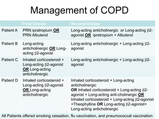
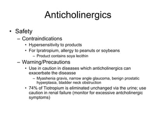
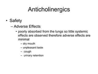
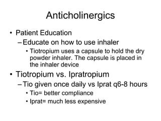
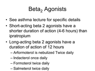
Chronic Obstructive
Pulmonary Disease (COPD)
Mikael Jones, Pharm.D., BCPS
NUR 652
COPD
• Disease state characterized by airflow limitation
• Airflow limitation is
– not fully reversible
– Usually progressive
– Associated with abnormal inflammatory response of the lungs to
noxious particles or gases
– Caused by a mixture of small airway disease (obstructive bronchiolitis)
and parenchymal destruction (emphysema)
• Relative contribution of each varies from patient to patient
• Chronic Inflammation is present and causes remodeling and narrowing of
small airways
– Destruction of the parenchyma
• Loss of alveolar attachments to the small airways
• Decreases lung elastic recoil
– The above changes lead to a diminishment in the ability of the airways
to remain open during expiration
• In the past COPD has been viewed as the overlapping of Chronic
Bronchitis and Emphysema
– However movement away from this view
Obstructive vs Restrictive
Pulmonary Disease
• Obstructive diseases impair exhalation
– Asthma, bronchitis, emphysema
– Obstructive diseases reduce FEV/FVC ratio
• vital capacity does not change (may even increase)
• Restrictive diseases impair inhalation
– Pneumonia, pulmonary fibroses (black lung)
– Due to fluid accumulation, loss of elasticity
– Restrictive diseases reduce volume (vital capacity) but do
not affect FEV/FVC ratio
COPD: Epidemiology
• Affects >14 million adults in U.S. (5% of
population)
– 12 million with chronic bronchitis (~90%)
– 1.65 million with emphysema (~12%)
– 4th Leading cause of mortality
• 80-90% of COPD is linked to cigarette smoking
– 15-20% of smokers develop COPD
– Other risk factors include occupational
exposures (dust, smoke, mining, textile
manufacturing), and air pollution
Chronic Bronchitis: Clinical
Characteristics
• Chronic bronchitis is characterized by
the excessive production of mucus with
productive cough
– Blockade of bronchioles by mucus,
inflammation and edema
• Presentation
– Early onset (20-30 yrs) with cigarette
cough, morning cough
– Diagnosis usually occurs at 50-60 yrs
– Smoking is the most significant risk factor
– Repeated respiratory infections are common
– Productive cough, copious, often purulent sputum, late
onset dyspnea
– Patient is typically obese
Chronic Bronchitis:
Pathophysiology
• Repeated irritation and inflammation leads to
bronchiolar smooth muscle hypertrophy and
reduction in airway diameters
– Increase in size and number of submucosal mucus glands
and goblet cells
• Goblet cells proliferate in bronchioli, which normally do not contain
mucus-secreting cells
• Mucus is thick and difficult to clear from airways
– Ciliary action is impaired by inflammation, resulting in poor
clearance of mucus, leading to repeated bacterial
infections
• Hypoventilation common
– Elevated blood CO2 and reduced O2, cyanosis
• Often termed “blue bloaters”
– Loss of respiratory drive unexplained
• Death results from right heart failure (cor pulmonale)
due to pulmonary hypertension, or respiratory failure
Emphysema: Clinical
Characteristics
• Emphysema results from acinar
distension and destruction of
alveolar membranes
– Loss of surface area for gas exchange
• Later onset than CB (30-40 yrs)
– Diagnosis usually occurs at 60-70 yrs
– Minimal history of cough
• Chief complaint is shortness of
breath
– Patients typically hyperventilate to
maintain normal blood gases
– “pink puffers”
• Environmental risk factors:
smoking, dust inhalation, pollution
• Patients are typically thin,
sometimes “barrel-chested”
Differentiation of Asthma, Chronic
Bronchitis, and Emphysema
Symptom Asthma Bronchitis Emphysema
Principal
complaint
Intermittent
bronchospasm
Chronic cough,
copious sputum
Dyspnea
Reversible with
β2-agonists
Yes No No
Allergic
component
Frequently No No
Inflammation Yes Yes Alveoli only
Sputum
production
During attacks
Copious,
continuous
Little
Cough After attack
Constant,
productive
No
Age of onset
Childhood
(allergic); adult
(nonallergic)
20-30 yr
(Diagnosis at 50
yr+)
30-40 yr
(Diagnosis at 60
yr+)
Body build Varied Obese
Thin, barrel-
chested
Hypoxic During attack Yes No
Four Components of COPD
Management
• Assess and monitor the disease
• Reduce Risk Factors
• Manage stable COPD
• Manage Exacerbations
Assess and Monitor Disease
• Diagnosis based on
– History of exposure to risk factors
– The presence of airflow limitation that is not
fully reversible, with or without the presence
of symptoms
• Therefore consider workup for patients who have
chronic cough and sputum production even in the
absence of dyspnea
• Spirometry is used to diagnosis COPD
Assess and Monitor Disease
Spirometry
0
5
1
4
2
3
Liter
1 6
5
4
3
2
FVC
FVC
FEV1
FEV1
Normal
COPD
3.900
5.200
2.350
4.150 80 %
60 %
Normal
COPD
FVC
FEV1 FVC
FEV1/
Seconds
Assess and Monitor Disease
• Severity of COPD
– Gold 1 Mild
• FEV1 80% predicted
– Gold II Moderate
• FEV1 50-79% predicted
– Gold III Severe
• FEV1 30-49% predicted
– Gold IV Very Severe
• FEV1 <30%
Assess and Monitor Disease
• Staging of COPD patients uses a
combination of Spirometetry and patient
report of symptom severity
• Number of exacerbations per year
• Modified Medical Research Council
Questionnaire for Assessing the
Severity of Breathlessness (mMRC)
– Used to capture symptom impact on
function
Assess and Monitor Disease
• Modified Medical Research Council Questionnaire for Assessing the
Severity of Breathlessness (mMRC)
Assess and Monitor Disease
• Use Spirometry and
mMRC to place a
patient into 1 of 4
categories
Assess and Monitor Disease
• Patient Group A- Low Risk, Less
symptoms
– Gold 1 or 2 and/or 0-1 exacerbations per year
and mMRc 0-1
• Patient Group B-Low Risk, More
symptoms
– Gold 1 or 2 and/or 0-1 exacerbations per year
and mMRc ≥2
Assess and Monitor Disease
• Patient Group C High Risk, Less
Symptoms
– Gold 3 or 4 and/or ≥2 exacerbations per year
and mMRc 0-1
• Patient Group D-Low Risk, More
symptoms
– Gold 3 or 4 and/or ≥2 exacerbations per year
and mMRc ≥2
Risk Factor Reduction
• Reduction exposure to
– Tobacco smoke
– Occupational dusts and chemicals
– Indoor and outdoor pollutants
• SMOKING CESSATION
– Reduces risk of developing COPD
– Stops its progression!
– Offer smoking cessation to every patent
Management of COPD
• General Concepts
– Smoking Cessation
– Exercise training
– Pharmacotherapy
• Does not modify long-term decline in lung function
• Medications decrease symptoms and/or complicatons
• Bronchodilators
– Can be given PRN or scheduled to prevent or reduce
symptoms
– Inhaled preferred
– In contrast to asthma, fully normal pulmonary
function cannot be attained
Management of COPD
First Choice Second Choice
Patient A PRN ipratropium OR
PRN Albuterol
Long-acting anticholinergic or Long-acting 2-
agonist OR ipratropium + Albuterol
Patient B Long-acting
anticholinergic OR Long-
acting 2-agonist
Long-acting anticholinergic + Long-acting 2-
agonist
Patient C Inhaled corticosteroid +
Long-acting 2-agonist
OR Long-acting
anticholinergic
Long-acting anticholinergic + Long-acting 2-
agonist
Patient D Inhaled corticosteroid +
Long-acting 2-agonist
OR Long-acting
anticholinergic
Inhaled corticosteroid + Long-acting
anticholinergic
OR Inhaled corticosteroid + Long-acting 2-
agonist + Long-acting anti-cholinergic OR
Inhaled corticosteroid + Long-acting 2-agonist
+Theophylline OR Long-acting 2-agonist+
Long-acting anticholinergic
All Patients offered smoking cessation, flu vaccination, and pneumococcal vaccination
Pharmacotherapy
• Principle Therapies
– Beta2 agonists
– Anticholinergics
– Theophylline
– Combination of above
• Choice between above agents depends on
patient individual response in terms of
safety and effectiveness
Pharmacotherapy
• Long acting bronchodilators are more effective
and convenient than short acting, but much
more expensive
• Combining drugs with different mechanisms and
durations of action may increase the degree of
bronchodilation for equivalent or lesser side
effects.
• Theophylline is effective in COPD, but due to its
potential toxicity, inhaled bronchodilators are
preferred when available.
Anticholinergics
• Available agents
– Ipratropium bromide
• Inhaler and nebulized solution
– Tiotropium
• Dry powder inhlaer
– Aclidinium
• Clinical Pharmacology
– anticholinergic agent that blocks the effects of
acetylcholine at muscarinic receptors in the airways,
reducing vagal-mediated bronchoconstriction
– Tiotropium has a duration of action of more than 24
hours
– Ipratropium lasts up to 8 hours from administration
– Aclidinium is does every 12 hours
Anticholinergics
• Safety
– Contraindications
• Hypersensitivity to products
• For Ipratropium, allergy to peanuts or soybeans
– Product contains soya lecithin
– Warning/Precautions
• Use in caution in diseases which anticholinergics can
exacerbate the diseasse
– Myashenia gravis, narrow angle glaucoma, benign prostatic
hyperplasia, bladder neck obstruction
• 74% of Tiotropium is eliminated unchanged via the urine; use
caution in renal failure (monitor for excessive antcholinergic
symptoms)
Anticholinergics
• Safety
– Adverse Effects
• poorly absorbed from the lungs so little systemic
effects are observed therefore adverse effects are
minimal
– dry mouth
– unpleasant taste
– cough
– urinary retention
Anticholinergics
• Patient Education
–Educate on how to use inhaler
• Tiotropium uses a capsule to hold the dry
powder inhaler. The capsule is placed in
the inhaler device
• Tiotropium vs. Ipratropium
–Tio given once daily vs Iprat q6-8 hours
• Tio= better compliance
• Iprat= much less expensive
Beta2 Agonists
• See asthma lecture for specific details
• Short-acting beta 2 agonists have a
shorter duration of action (4-6 hours) than
ipratropium
• Long-acting beta 2 agonists have a
duration of action of 12 hours
– Arformoterol is nebulized Twice daily
– Indacterol once daily
– Formoterol twice daily
– Salmeterol twice daily
Theophylline
• See Asthma handout
• Use is limited by toxicity
• Inhaled anticholinergics or beta agonists
are preferred over theophylline
Roflumilast
• Sectively inhibit phosphodiesterase-4
(PDE4) leading to an accumulation of
cyclic AMP (cAMP) within inflammatory
and structural cells
• Anti-inflammatory effects include
– suppression of cytokine release
– inhibition of lung infiltration by neutrophils and
other leukocytes.
Roflumilast
• Used as adjunctive therapy in severe
COPD associated with chronic bronchitis
• Dosed 500mg po daily
• Adverse effects
– Weight loss/ Diarrhea
– Insomnia
– Anxiety, depression
• Avoid in patients with history of depression
with suicidal behavior/ideation
Inhaled Corticosteroids
• See asthma lecture
• Reserved for patients in Patient
Categories C and D
• May reduce exacerbations
COPD Exacerbations
• Most common causes
– Infection of the
tracheobronchial tree
– Air pollution
– Unknown Factors
• Presentation
– Increased breathlessness
– Increased sputum
production
• Change in sputum quality
– Malaise, insomnia, fatigue
• Signs of Severe
exacerbation
– Use of accessory
respiratory muscles.
– Paradoxical chest wall
movements.
– Worsening or new onset
central cyanosis.
– Development of peripheral
edema.
– Hemodynamic instability.
– Signs of right heart failure.
– Reduced alertness.
COPD Exacerbations

Unit_6_COPD_1_.ppt

  • 1.
    Chronic Obstructive Pulmonary Disease(COPD) Mikael Jones, Pharm.D., BCPS NUR 652
  • 2.
    COPD • Disease statecharacterized by airflow limitation • Airflow limitation is – not fully reversible – Usually progressive – Associated with abnormal inflammatory response of the lungs to noxious particles or gases – Caused by a mixture of small airway disease (obstructive bronchiolitis) and parenchymal destruction (emphysema) • Relative contribution of each varies from patient to patient • Chronic Inflammation is present and causes remodeling and narrowing of small airways – Destruction of the parenchyma • Loss of alveolar attachments to the small airways • Decreases lung elastic recoil – The above changes lead to a diminishment in the ability of the airways to remain open during expiration • In the past COPD has been viewed as the overlapping of Chronic Bronchitis and Emphysema – However movement away from this view
  • 3.
    Obstructive vs Restrictive PulmonaryDisease • Obstructive diseases impair exhalation – Asthma, bronchitis, emphysema – Obstructive diseases reduce FEV/FVC ratio • vital capacity does not change (may even increase) • Restrictive diseases impair inhalation – Pneumonia, pulmonary fibroses (black lung) – Due to fluid accumulation, loss of elasticity – Restrictive diseases reduce volume (vital capacity) but do not affect FEV/FVC ratio
  • 4.
    COPD: Epidemiology • Affects>14 million adults in U.S. (5% of population) – 12 million with chronic bronchitis (~90%) – 1.65 million with emphysema (~12%) – 4th Leading cause of mortality • 80-90% of COPD is linked to cigarette smoking – 15-20% of smokers develop COPD – Other risk factors include occupational exposures (dust, smoke, mining, textile manufacturing), and air pollution
  • 5.
    Chronic Bronchitis: Clinical Characteristics •Chronic bronchitis is characterized by the excessive production of mucus with productive cough – Blockade of bronchioles by mucus, inflammation and edema • Presentation – Early onset (20-30 yrs) with cigarette cough, morning cough – Diagnosis usually occurs at 50-60 yrs – Smoking is the most significant risk factor – Repeated respiratory infections are common – Productive cough, copious, often purulent sputum, late onset dyspnea – Patient is typically obese
  • 6.
    Chronic Bronchitis: Pathophysiology • Repeatedirritation and inflammation leads to bronchiolar smooth muscle hypertrophy and reduction in airway diameters – Increase in size and number of submucosal mucus glands and goblet cells • Goblet cells proliferate in bronchioli, which normally do not contain mucus-secreting cells • Mucus is thick and difficult to clear from airways – Ciliary action is impaired by inflammation, resulting in poor clearance of mucus, leading to repeated bacterial infections • Hypoventilation common – Elevated blood CO2 and reduced O2, cyanosis • Often termed “blue bloaters” – Loss of respiratory drive unexplained • Death results from right heart failure (cor pulmonale) due to pulmonary hypertension, or respiratory failure
  • 7.
    Emphysema: Clinical Characteristics • Emphysemaresults from acinar distension and destruction of alveolar membranes – Loss of surface area for gas exchange • Later onset than CB (30-40 yrs) – Diagnosis usually occurs at 60-70 yrs – Minimal history of cough • Chief complaint is shortness of breath – Patients typically hyperventilate to maintain normal blood gases – “pink puffers” • Environmental risk factors: smoking, dust inhalation, pollution • Patients are typically thin, sometimes “barrel-chested”
  • 8.
    Differentiation of Asthma,Chronic Bronchitis, and Emphysema Symptom Asthma Bronchitis Emphysema Principal complaint Intermittent bronchospasm Chronic cough, copious sputum Dyspnea Reversible with β2-agonists Yes No No Allergic component Frequently No No Inflammation Yes Yes Alveoli only Sputum production During attacks Copious, continuous Little Cough After attack Constant, productive No Age of onset Childhood (allergic); adult (nonallergic) 20-30 yr (Diagnosis at 50 yr+) 30-40 yr (Diagnosis at 60 yr+) Body build Varied Obese Thin, barrel- chested Hypoxic During attack Yes No
  • 9.
    Four Components ofCOPD Management • Assess and monitor the disease • Reduce Risk Factors • Manage stable COPD • Manage Exacerbations
  • 10.
    Assess and MonitorDisease • Diagnosis based on – History of exposure to risk factors – The presence of airflow limitation that is not fully reversible, with or without the presence of symptoms • Therefore consider workup for patients who have chronic cough and sputum production even in the absence of dyspnea • Spirometry is used to diagnosis COPD
  • 11.
    Assess and MonitorDisease Spirometry 0 5 1 4 2 3 Liter 1 6 5 4 3 2 FVC FVC FEV1 FEV1 Normal COPD 3.900 5.200 2.350 4.150 80 % 60 % Normal COPD FVC FEV1 FVC FEV1/ Seconds
  • 12.
    Assess and MonitorDisease • Severity of COPD – Gold 1 Mild • FEV1 80% predicted – Gold II Moderate • FEV1 50-79% predicted – Gold III Severe • FEV1 30-49% predicted – Gold IV Very Severe • FEV1 <30%
  • 13.
    Assess and MonitorDisease • Staging of COPD patients uses a combination of Spirometetry and patient report of symptom severity • Number of exacerbations per year • Modified Medical Research Council Questionnaire for Assessing the Severity of Breathlessness (mMRC) – Used to capture symptom impact on function
  • 14.
    Assess and MonitorDisease • Modified Medical Research Council Questionnaire for Assessing the Severity of Breathlessness (mMRC)
  • 15.
    Assess and MonitorDisease • Use Spirometry and mMRC to place a patient into 1 of 4 categories
  • 16.
    Assess and MonitorDisease • Patient Group A- Low Risk, Less symptoms – Gold 1 or 2 and/or 0-1 exacerbations per year and mMRc 0-1 • Patient Group B-Low Risk, More symptoms – Gold 1 or 2 and/or 0-1 exacerbations per year and mMRc ≥2
  • 17.
    Assess and MonitorDisease • Patient Group C High Risk, Less Symptoms – Gold 3 or 4 and/or ≥2 exacerbations per year and mMRc 0-1 • Patient Group D-Low Risk, More symptoms – Gold 3 or 4 and/or ≥2 exacerbations per year and mMRc ≥2
  • 18.
    Risk Factor Reduction •Reduction exposure to – Tobacco smoke – Occupational dusts and chemicals – Indoor and outdoor pollutants • SMOKING CESSATION – Reduces risk of developing COPD – Stops its progression! – Offer smoking cessation to every patent
  • 19.
    Management of COPD •General Concepts – Smoking Cessation – Exercise training – Pharmacotherapy • Does not modify long-term decline in lung function • Medications decrease symptoms and/or complicatons • Bronchodilators – Can be given PRN or scheduled to prevent or reduce symptoms – Inhaled preferred – In contrast to asthma, fully normal pulmonary function cannot be attained
  • 20.
    Management of COPD FirstChoice Second Choice Patient A PRN ipratropium OR PRN Albuterol Long-acting anticholinergic or Long-acting 2- agonist OR ipratropium + Albuterol Patient B Long-acting anticholinergic OR Long- acting 2-agonist Long-acting anticholinergic + Long-acting 2- agonist Patient C Inhaled corticosteroid + Long-acting 2-agonist OR Long-acting anticholinergic Long-acting anticholinergic + Long-acting 2- agonist Patient D Inhaled corticosteroid + Long-acting 2-agonist OR Long-acting anticholinergic Inhaled corticosteroid + Long-acting anticholinergic OR Inhaled corticosteroid + Long-acting 2- agonist + Long-acting anti-cholinergic OR Inhaled corticosteroid + Long-acting 2-agonist +Theophylline OR Long-acting 2-agonist+ Long-acting anticholinergic All Patients offered smoking cessation, flu vaccination, and pneumococcal vaccination
  • 21.
    Pharmacotherapy • Principle Therapies –Beta2 agonists – Anticholinergics – Theophylline – Combination of above • Choice between above agents depends on patient individual response in terms of safety and effectiveness
  • 22.
    Pharmacotherapy • Long actingbronchodilators are more effective and convenient than short acting, but much more expensive • Combining drugs with different mechanisms and durations of action may increase the degree of bronchodilation for equivalent or lesser side effects. • Theophylline is effective in COPD, but due to its potential toxicity, inhaled bronchodilators are preferred when available.
  • 23.
    Anticholinergics • Available agents –Ipratropium bromide • Inhaler and nebulized solution – Tiotropium • Dry powder inhlaer – Aclidinium • Clinical Pharmacology – anticholinergic agent that blocks the effects of acetylcholine at muscarinic receptors in the airways, reducing vagal-mediated bronchoconstriction – Tiotropium has a duration of action of more than 24 hours – Ipratropium lasts up to 8 hours from administration – Aclidinium is does every 12 hours
  • 24.
    Anticholinergics • Safety – Contraindications •Hypersensitivity to products • For Ipratropium, allergy to peanuts or soybeans – Product contains soya lecithin – Warning/Precautions • Use in caution in diseases which anticholinergics can exacerbate the diseasse – Myashenia gravis, narrow angle glaucoma, benign prostatic hyperplasia, bladder neck obstruction • 74% of Tiotropium is eliminated unchanged via the urine; use caution in renal failure (monitor for excessive antcholinergic symptoms)
  • 25.
    Anticholinergics • Safety – AdverseEffects • poorly absorbed from the lungs so little systemic effects are observed therefore adverse effects are minimal – dry mouth – unpleasant taste – cough – urinary retention
  • 26.
    Anticholinergics • Patient Education –Educateon how to use inhaler • Tiotropium uses a capsule to hold the dry powder inhaler. The capsule is placed in the inhaler device • Tiotropium vs. Ipratropium –Tio given once daily vs Iprat q6-8 hours • Tio= better compliance • Iprat= much less expensive
  • 27.
    Beta2 Agonists • Seeasthma lecture for specific details • Short-acting beta 2 agonists have a shorter duration of action (4-6 hours) than ipratropium • Long-acting beta 2 agonists have a duration of action of 12 hours – Arformoterol is nebulized Twice daily – Indacterol once daily – Formoterol twice daily – Salmeterol twice daily
  • 28.
    Theophylline • See Asthmahandout • Use is limited by toxicity • Inhaled anticholinergics or beta agonists are preferred over theophylline
  • 29.
    Roflumilast • Sectively inhibitphosphodiesterase-4 (PDE4) leading to an accumulation of cyclic AMP (cAMP) within inflammatory and structural cells • Anti-inflammatory effects include – suppression of cytokine release – inhibition of lung infiltration by neutrophils and other leukocytes.
  • 30.
    Roflumilast • Used asadjunctive therapy in severe COPD associated with chronic bronchitis • Dosed 500mg po daily • Adverse effects – Weight loss/ Diarrhea – Insomnia – Anxiety, depression • Avoid in patients with history of depression with suicidal behavior/ideation
  • 31.
    Inhaled Corticosteroids • Seeasthma lecture • Reserved for patients in Patient Categories C and D • May reduce exacerbations
  • 32.
    COPD Exacerbations • Mostcommon causes – Infection of the tracheobronchial tree – Air pollution – Unknown Factors • Presentation – Increased breathlessness – Increased sputum production • Change in sputum quality – Malaise, insomnia, fatigue • Signs of Severe exacerbation – Use of accessory respiratory muscles. – Paradoxical chest wall movements. – Worsening or new onset central cyanosis. – Development of peripheral edema. – Hemodynamic instability. – Signs of right heart failure. – Reduced alertness.
  • 33.