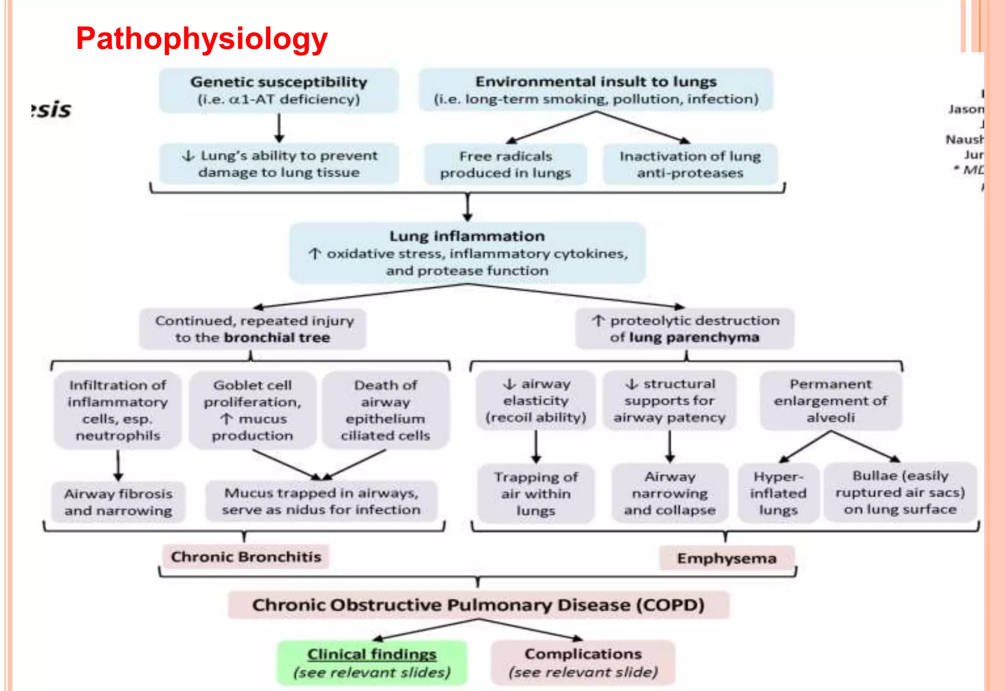
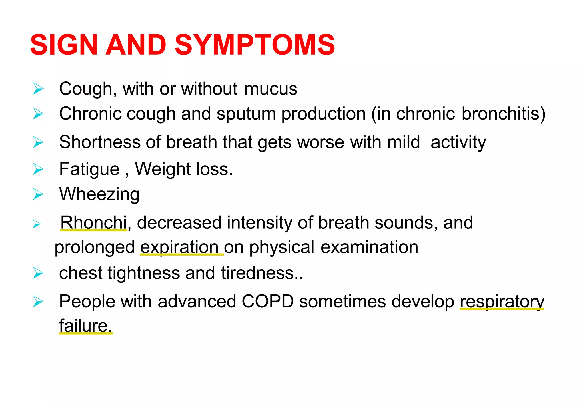
CHRONIC OBSTRUCTIVE
PULMONARY DISEASES
Mr.Veerabhadra B B
Asst Professor
Dept of Medical Surgical Nsg.
INTRODUCTION
 COPD is the progressive and partially reversible disease
of the airway
 Comprises primarily of two related disease-
chronic bronchitis and Emphysema
 Chronic obstruction of the flow of air through the
airway and out of the lungs, progressive obstruction
over time
INCIDENCE
 COPD is the 5th leading cause of death in the
United States for all ages
 Both genders are affected .fifth for men and
fourth for women
 The incidence of COPD increases with age
Chronic bronchitis
 Chronic bronchitis is defined clinically as a daily
cough with production of sputum at least 3 month per
year for 2 or more consecutive year.
 It involves inflammation and swelling of the lining
of the bronchi & trachea (air way) that leads to
narrowing and obstruction of the air way.
 The inflammation also stimulate production of
mucus which can cause further obstruction of the
airway.
EMPHYSEMA
 It is permanent enlargement of the alveoli due to
destruction of the wall between alveoli
which leads to reduce the elasticity of the lungs over all.
 Loss of elasticity leads to collapse of the bronchioles,
obstructing air flow out of the alveoli.
 Air become trapped to the alveoli and reduce the
ability of the lungs to shrink during exhalation .
 Reduce the expansion of the lungs during the
next breath reduce the amount of air that is
inhaled.
 As a result, less air for the exchange of
gasses gets in to the lungs .
 This trapped air also can compress
adjacent less damage lung tissue.
CONTINUED…..
ETIOLOGY AND RISK FACTORS
The specific causes of COPD are not clearly
understood. Some risk factors are tissue.
1.Cigarette smoking. (Primary & Second hand smoke)
2.Air Pollution: Some occupational pollutants such as
cadmium and silica
3.Alpha-1 Antitrypsin (AAT) deficiency- (Genetic diseases)
AAT enzyme is produced by liver and present in normal
lungs. Normal1.5-3.5 g/l. Block the damaging effects of
elastase on elastin.
Pathophysiology
A SAMPLE VIDEO
SIGN AND SYMPTOMS
 Cough, with or without mucus
 Chronic cough and sputum production (in chronic bronchitis)
 Shortness of breath that gets worse with mild activity
 Fatigue , Weight loss.
 Wheezing
 Rhonchi, decreased intensity of breath sounds, and
prolonged expiration on physical examination
 chest tightness and tiredness..
 People with advanced COPD sometimes develop respiratory
failure.
COMMON SIGNS ARE:
 Tachypnea a rapid breathing rate
 Breathing out taking a longer time than breathing in
 Enlargement of the chest, particularly the front- to-back
distance (hyperaeration)
 Breathing through pursed lips
 Increased antero-posterior to lateral ratio of the chest (i.e.
barrel chest)
INVESTIGATION
 Thorough Medical History
 Physical examination finds enlarged chest cavity and
wheezing.
Blood Test
 A hematocrit value of more than 52% in males and
more than 47% in female indicates disease.
 Measure the alpha1-antitrypsin (AAT),theAAT level
is low
 Sputum for culture and microscopic examination
of mucoid sputum
CONTD
• Chest X-ray- Hyper inflated lung, Flat
diaphragm,Tubular heart, Increase broncho vascular
markings
•High Resolution CT scanning(HRCT)
•Pulmonary Function Test
Forced expiratory volume in 1 second (FEV1) is a.
 Mild= FEV1 >80%predicted
 Moderate= FEV1 80-50%predicted
 Sever= FEV1 50-30%predicted
 Very sever = FEV1 <30%predicted
• Arterial blood gas analysis:
(severe hypoxemia and hypercapnia).
Chest X-ray picture of COPD
CT SCAN PICTURE OF COPD
PULMONARY FUNCTION TEST
COMPLICATIONS OF COPD
 Respiratory Infections
 Acute Respiratory Failure
 Spontaneous Pneumothorax due to rupture of
emphysematous bleb.
 Ventilation Perfusion Mismatch
 Severe Hypoxemia & acidosis.
 Corpulmonale
MEDICAL MANAGEMENT
The treatment goal for the client with COPD is
 To improve ventilation
 To facilitate the removal of bronchial secretions
 To prevent complications
 To slow the progression of clinical manifestations
 To promote health maintenance and client
management of disease.
TREATMENT STRATEGIES INCLUDE
• Quitting cigarette smoking
 Taking medications to dilate airways(
bronchodilators)
 Vaccinating against flu influenza and pneumonia
 Regular oxygen supplementation
 Pulmonary rehabilitation
 Bronchodilators:
Beta2 agonists are prescribed. (Albuterol or
Salbutamol, metaproterenol) To relieve bronchospasm
and reduce airway obstruction
 Metered-dose inhaler (MDI)
 Anticholinergic : bronchodilators work by blocking
the cholinergic receptors resulting in
bronchodilatation
MEDICAL MANAGEMENT
CONT…
 Methylxanthines (Theophylline, Aminophylline) are
also used to treat acute exacerbations.
 Nebulization of medication via an air compressor
 Corticosteroids are used in the acute management
of clients with COPD exacerbations ex:
Beclomethasone Diproprionate, Salmeterol And
Fluticasone are used.
Regular oxygen therapy: 2-4 lit/ min.
Antibiotic- Treat with antibiotic therapy for
recurrent bacterial infection.
NURSING MANAGEMENT
Assessment
 history of smoking, family history,
occupational history
 ABG analysis
 Respiratory rate, depth and
characteristics
 sputum amount and type
 anxiety level of the patient
1: Impaired gas exchange related to dyspnoea, mucus
plug and decreased ventilation
 Assess respiratory rate, depth, note use of accessory
muscles, pursed lip breathing, inability to speak.
 Elevate head of bed.
 Encourage deep slow or pursed lip breathing as
individually tolerated.
 Administer low- flow oxygen therapy (1-2lit/min) as
needed via nasal prongs.
 Administer bronchodilators if ordered
 Regularly monitor the client's respiratory rate and
pattern, pulse oximetry, ABG results
2.: Activity intolerance related to inadequate oxygenation
and dyspnea
 Monitor the severity of dyspnea and oxygen
with and following activity
 Keep the patient in semi- flower position.
 Maintain supplemental oxygen therapy (2lit/min)
 Assist the client in scheduling a gradual increase in daily
and exercise
3: Ineffective airwayclearance related to excessive
production of secretions, retained secretions and
ineffective coughing
 Monitor respiratory rate and auscultate breath sounds eg.
wheeze, crackles, rhonchi
 Assist the patient to assume position of comfort eg elevate head
of bed, sitting on edge of bed.
 Keep environmental pollution to minimum eg dust, smoke and
feather pillows according to individual situation
 Encourage/ assist with abdominal or pursed lip breathing
exercises
 Administer medications as indicated such as
bronchodilators
 Perform chest physiotherapy.
 4 : Anxiety related to disease prognosis
 Encourage the use of breathing retraining and
relaxation techniques.
 Explain the patient about disease including cause,
signs and symptoms, medication, procedures,
prevention and follow up care
 Give the opportunity to talk the patient with similar
problem who admitted in the same ward and
almost in recovery phase
THANK YOU

Chronic obstructive pulmonary diseases & Nursing care.

  • 1.
    CHRONIC OBSTRUCTIVE PULMONARY DISEASES Mr.VeerabhadraB B Asst Professor Dept of Medical Surgical Nsg.
  • 2.
    INTRODUCTION  COPD isthe progressive and partially reversible disease of the airway  Comprises primarily of two related disease- chronic bronchitis and Emphysema  Chronic obstruction of the flow of air through the airway and out of the lungs, progressive obstruction over time
  • 4.
    INCIDENCE  COPD isthe 5th leading cause of death in the United States for all ages  Both genders are affected .fifth for men and fourth for women  The incidence of COPD increases with age Chronic bronchitis
  • 5.
     Chronic bronchitisis defined clinically as a daily cough with production of sputum at least 3 month per year for 2 or more consecutive year.  It involves inflammation and swelling of the lining of the bronchi & trachea (air way) that leads to narrowing and obstruction of the air way.  The inflammation also stimulate production of mucus which can cause further obstruction of the airway.
  • 6.
    EMPHYSEMA  It ispermanent enlargement of the alveoli due to destruction of the wall between alveoli which leads to reduce the elasticity of the lungs over all.  Loss of elasticity leads to collapse of the bronchioles, obstructing air flow out of the alveoli.  Air become trapped to the alveoli and reduce the ability of the lungs to shrink during exhalation .
  • 7.
     Reduce theexpansion of the lungs during the next breath reduce the amount of air that is inhaled.  As a result, less air for the exchange of gasses gets in to the lungs .  This trapped air also can compress adjacent less damage lung tissue. CONTINUED…..
  • 8.
    ETIOLOGY AND RISKFACTORS The specific causes of COPD are not clearly understood. Some risk factors are tissue. 1.Cigarette smoking. (Primary & Second hand smoke) 2.Air Pollution: Some occupational pollutants such as cadmium and silica 3.Alpha-1 Antitrypsin (AAT) deficiency- (Genetic diseases) AAT enzyme is produced by liver and present in normal lungs. Normal1.5-3.5 g/l. Block the damaging effects of elastase on elastin.
  • 9.
  • 10.
  • 11.
    SIGN AND SYMPTOMS Cough, with or without mucus  Chronic cough and sputum production (in chronic bronchitis)  Shortness of breath that gets worse with mild activity  Fatigue , Weight loss.  Wheezing  Rhonchi, decreased intensity of breath sounds, and prolonged expiration on physical examination  chest tightness and tiredness..  People with advanced COPD sometimes develop respiratory failure.
  • 12.
    COMMON SIGNS ARE: Tachypnea a rapid breathing rate  Breathing out taking a longer time than breathing in  Enlargement of the chest, particularly the front- to-back distance (hyperaeration)  Breathing through pursed lips  Increased antero-posterior to lateral ratio of the chest (i.e. barrel chest)
  • 13.
    INVESTIGATION  Thorough MedicalHistory  Physical examination finds enlarged chest cavity and wheezing. Blood Test  A hematocrit value of more than 52% in males and more than 47% in female indicates disease.  Measure the alpha1-antitrypsin (AAT),theAAT level is low  Sputum for culture and microscopic examination of mucoid sputum
  • 14.
    CONTD • Chest X-ray-Hyper inflated lung, Flat diaphragm,Tubular heart, Increase broncho vascular markings •High Resolution CT scanning(HRCT) •Pulmonary Function Test Forced expiratory volume in 1 second (FEV1) is a.  Mild= FEV1 >80%predicted  Moderate= FEV1 80-50%predicted  Sever= FEV1 50-30%predicted  Very sever = FEV1 <30%predicted • Arterial blood gas analysis: (severe hypoxemia and hypercapnia).
  • 15.
  • 16.
  • 17.
  • 18.
    COMPLICATIONS OF COPD Respiratory Infections  Acute Respiratory Failure  Spontaneous Pneumothorax due to rupture of emphysematous bleb.  Ventilation Perfusion Mismatch  Severe Hypoxemia & acidosis.  Corpulmonale
  • 19.
    MEDICAL MANAGEMENT The treatmentgoal for the client with COPD is  To improve ventilation  To facilitate the removal of bronchial secretions  To prevent complications  To slow the progression of clinical manifestations  To promote health maintenance and client management of disease.
  • 20.
    TREATMENT STRATEGIES INCLUDE •Quitting cigarette smoking  Taking medications to dilate airways( bronchodilators)  Vaccinating against flu influenza and pneumonia  Regular oxygen supplementation  Pulmonary rehabilitation
  • 21.
     Bronchodilators: Beta2 agonistsare prescribed. (Albuterol or Salbutamol, metaproterenol) To relieve bronchospasm and reduce airway obstruction  Metered-dose inhaler (MDI)  Anticholinergic : bronchodilators work by blocking the cholinergic receptors resulting in bronchodilatation MEDICAL MANAGEMENT
  • 22.
    CONT…  Methylxanthines (Theophylline,Aminophylline) are also used to treat acute exacerbations.  Nebulization of medication via an air compressor  Corticosteroids are used in the acute management of clients with COPD exacerbations ex: Beclomethasone Diproprionate, Salmeterol And Fluticasone are used.
  • 23.
    Regular oxygen therapy:2-4 lit/ min. Antibiotic- Treat with antibiotic therapy for recurrent bacterial infection.
  • 24.
    NURSING MANAGEMENT Assessment  historyof smoking, family history, occupational history  ABG analysis  Respiratory rate, depth and characteristics  sputum amount and type  anxiety level of the patient
  • 25.
    1: Impaired gasexchange related to dyspnoea, mucus plug and decreased ventilation  Assess respiratory rate, depth, note use of accessory muscles, pursed lip breathing, inability to speak.  Elevate head of bed.  Encourage deep slow or pursed lip breathing as individually tolerated.  Administer low- flow oxygen therapy (1-2lit/min) as needed via nasal prongs.  Administer bronchodilators if ordered  Regularly monitor the client's respiratory rate and pattern, pulse oximetry, ABG results
  • 26.
    2.: Activity intolerancerelated to inadequate oxygenation and dyspnea  Monitor the severity of dyspnea and oxygen with and following activity  Keep the patient in semi- flower position.  Maintain supplemental oxygen therapy (2lit/min)  Assist the client in scheduling a gradual increase in daily and exercise
  • 27.
    3: Ineffective airwayclearancerelated to excessive production of secretions, retained secretions and ineffective coughing  Monitor respiratory rate and auscultate breath sounds eg. wheeze, crackles, rhonchi  Assist the patient to assume position of comfort eg elevate head of bed, sitting on edge of bed.  Keep environmental pollution to minimum eg dust, smoke and feather pillows according to individual situation  Encourage/ assist with abdominal or pursed lip breathing exercises  Administer medications as indicated such as bronchodilators  Perform chest physiotherapy.
  • 28.
     4 :Anxiety related to disease prognosis  Encourage the use of breathing retraining and relaxation techniques.  Explain the patient about disease including cause, signs and symptoms, medication, procedures, prevention and follow up care  Give the opportunity to talk the patient with similar problem who admitted in the same ward and almost in recovery phase
  • 29.