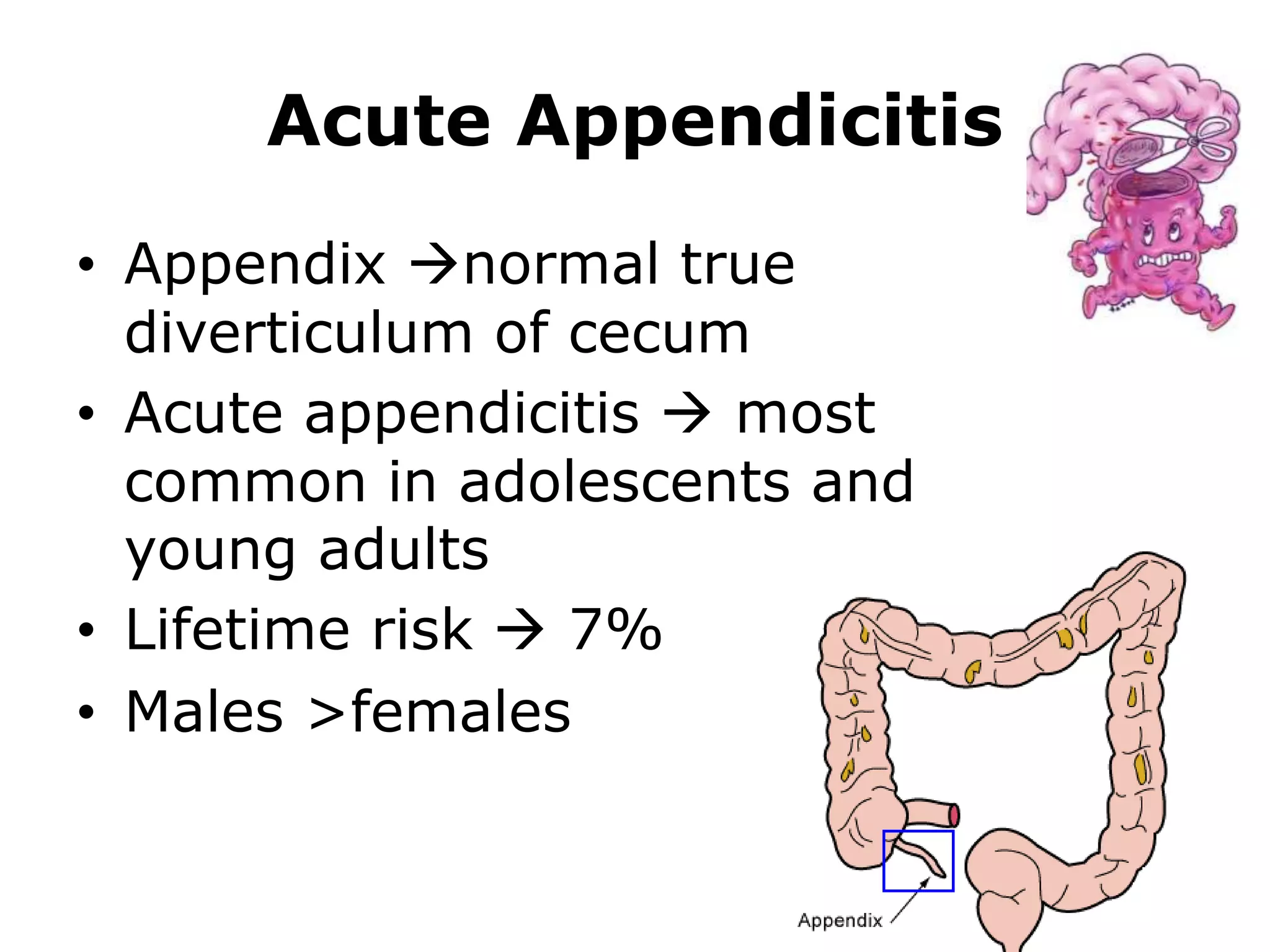
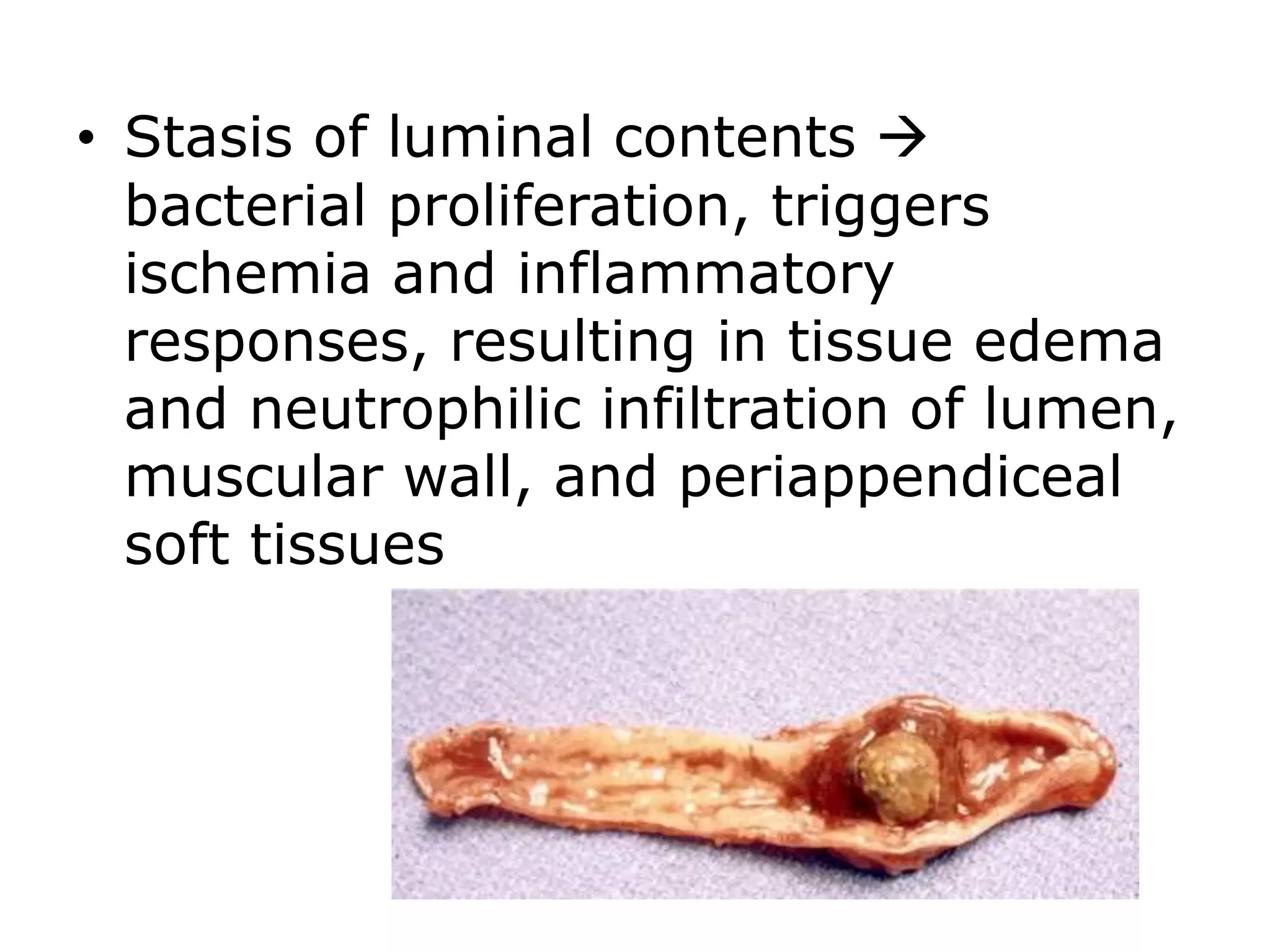
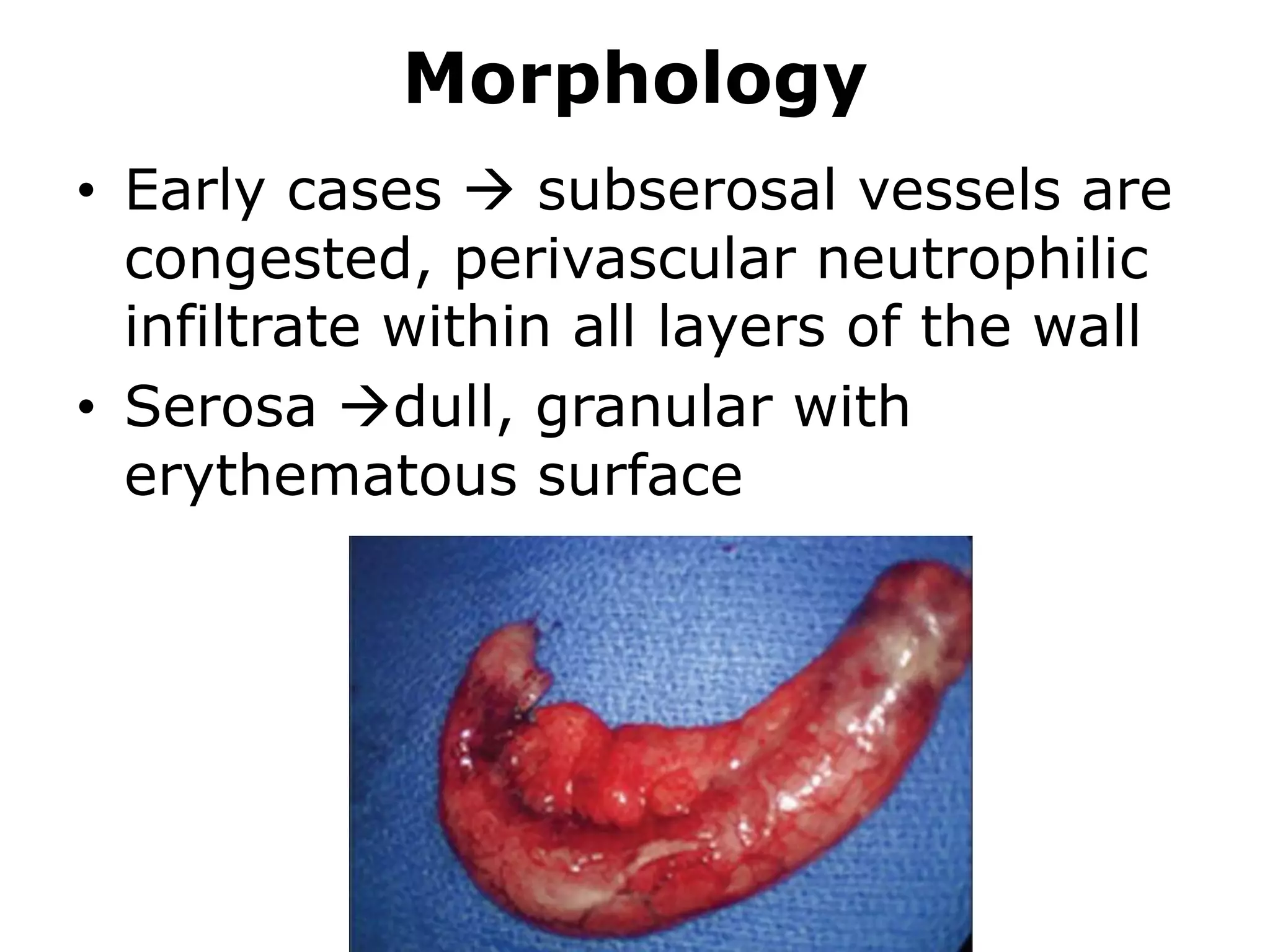
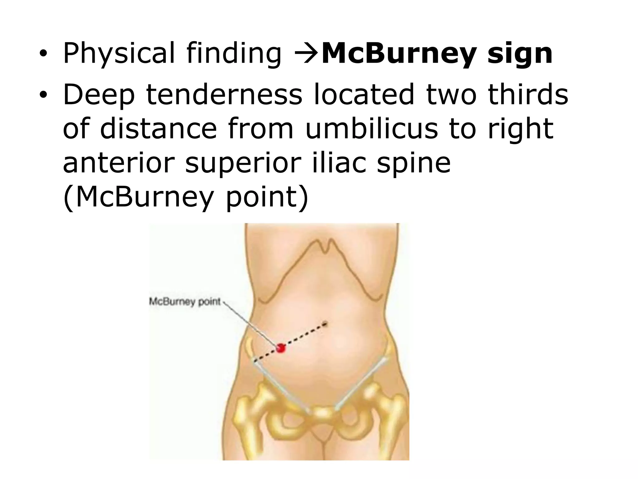
Acute Appendicitis
• Appendix normal true
diverticulum of cecum
• Acute appendicitis  most
common in adolescents and
young adults
• Lifetime risk  7%
• Males >females
Pathogenesis
• Initiated by progressive increase in
intraluminal pressure  compromise
venous outflow
• 50% to 80% of cases associated
with luminal obstruction by small
stone-like mass of stool (fecalith) or
less commonly gallstone, tumor, or
mass of worms (oxyuriasis
vermicularis)
• Stasis of luminal contents 
bacterial proliferation, triggers
ischemia and inflammatory
responses, resulting in tissue edema
and neutrophilic infiltration of lumen,
muscular wall, and periappendiceal
soft tissues
Morphology
• Early cases  subserosal vessels are
congested, perivascular neutrophilic
infiltrate within all layers of the wall
• Serosa dull, granular with
erythematous surface
• Diagnosis requires neutrophilic
infiltration of the muscularis
propria
• Severe cases  prominent
neutrophilic exudate with serosal
fibrinopurulent reaction
• Focal abscesses may form within the
wall (acute suppurative
appendicitis)
• Compromise of appendiceal vessels
leads to large areas of hemorrhagic
ulceration and gangrenous necrosis
extending to serosa creating acute
gangrenous appendicitis followed
by rupture and suppurative
peritonitis
Clinical Features
• Early acute appendicitis
produces periumbilical pain
that ultimately localizes to
right lower quadrant, followed
by:
– nausea
–Vomiting
–low-grade fever
–mildly elevated peripheral
white cell count
• Physical finding McBurney sign
• Deep tenderness located two thirds
of distance from umbilicus to right
anterior superior iliac spine
(McBurney point)
• Retrocecal appendix may generate
right flank or pelvic pain
• Malrotated colon may give rise to
appendicitis in left upper quadrant
• Neutrophilic leukocytosis
Complications
• Appendiceal perforation
• Pyelophlebitis
• Portal venous thrombosis
• Liver abscess
• Bacteremia
Clinical differential diagnosis
• Mesenteric lymphadenitis
• Acute salpingitis
• Ectopic pregnancy
• Mittelschmerz (German: "middle
pain")-pain caused by minor pelvic
bleeding at time of ovulation
• Meckel diverticulitis
Tumors of the Appendix
• Most common tumor of appendix 
welldifferentiated neuroendocrine
(carcinoid) tumor
• Usually discovered incidentally at the
time of surgery or
• examination of resected appendix
• carcinoid  benign tumor, form solid
bulbous swelling at the tip of
appendix
Carcinoid tumor
Gross
Microscopic
• Adenomas or non–mucin-producing
adenocarcinomas also occur in
appendix and may cause obstruction
and enlargement that mimics acute
appendicitis
• Mucocele  dilated appendix filled
with mucin
• Represent obstructed appendix
containing inspissated mucin or
consequence of mucinous
cystadenoma or mucinous
cystadenocarcinoma
• Invasion through appendiceal wall
can lead to intraperitoneal seeding
and spread
• In women resulting peritoneal
implants may be mistaken for
mucinous ovarian tumors
• Most advanced cases  abdomen
fills with tenacious, semisolid mucin,
a condition called pseudomyxoma
peritonei

Appendicitis and tumors of appendix mbbs

  • 1.
    Acute Appendicitis • Appendixnormal true diverticulum of cecum • Acute appendicitis  most common in adolescents and young adults • Lifetime risk  7% • Males >females
  • 2.
    Pathogenesis • Initiated byprogressive increase in intraluminal pressure  compromise venous outflow • 50% to 80% of cases associated with luminal obstruction by small stone-like mass of stool (fecalith) or less commonly gallstone, tumor, or mass of worms (oxyuriasis vermicularis)
  • 3.
    • Stasis ofluminal contents  bacterial proliferation, triggers ischemia and inflammatory responses, resulting in tissue edema and neutrophilic infiltration of lumen, muscular wall, and periappendiceal soft tissues
  • 4.
    Morphology • Early cases subserosal vessels are congested, perivascular neutrophilic infiltrate within all layers of the wall • Serosa dull, granular with erythematous surface
  • 5.
    • Diagnosis requiresneutrophilic infiltration of the muscularis propria • Severe cases  prominent neutrophilic exudate with serosal fibrinopurulent reaction
  • 6.
    • Focal abscessesmay form within the wall (acute suppurative appendicitis) • Compromise of appendiceal vessels leads to large areas of hemorrhagic ulceration and gangrenous necrosis extending to serosa creating acute gangrenous appendicitis followed by rupture and suppurative peritonitis
  • 7.
    Clinical Features • Earlyacute appendicitis produces periumbilical pain that ultimately localizes to right lower quadrant, followed by: – nausea –Vomiting –low-grade fever –mildly elevated peripheral white cell count
  • 8.
    • Physical findingMcBurney sign • Deep tenderness located two thirds of distance from umbilicus to right anterior superior iliac spine (McBurney point)
  • 9.
    • Retrocecal appendixmay generate right flank or pelvic pain • Malrotated colon may give rise to appendicitis in left upper quadrant • Neutrophilic leukocytosis
  • 10.
    Complications • Appendiceal perforation •Pyelophlebitis • Portal venous thrombosis • Liver abscess • Bacteremia
  • 11.
    Clinical differential diagnosis •Mesenteric lymphadenitis • Acute salpingitis • Ectopic pregnancy • Mittelschmerz (German: "middle pain")-pain caused by minor pelvic bleeding at time of ovulation • Meckel diverticulitis
  • 12.
    Tumors of theAppendix • Most common tumor of appendix  welldifferentiated neuroendocrine (carcinoid) tumor • Usually discovered incidentally at the time of surgery or • examination of resected appendix • carcinoid  benign tumor, form solid bulbous swelling at the tip of appendix
  • 13.
  • 14.
    • Adenomas ornon–mucin-producing adenocarcinomas also occur in appendix and may cause obstruction and enlargement that mimics acute appendicitis
  • 15.
    • Mucocele dilated appendix filled with mucin • Represent obstructed appendix containing inspissated mucin or consequence of mucinous cystadenoma or mucinous cystadenocarcinoma
  • 16.
    • Invasion throughappendiceal wall can lead to intraperitoneal seeding and spread • In women resulting peritoneal implants may be mistaken for mucinous ovarian tumors
  • 17.
    • Most advancedcases  abdomen fills with tenacious, semisolid mucin, a condition called pseudomyxoma peritonei