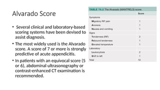
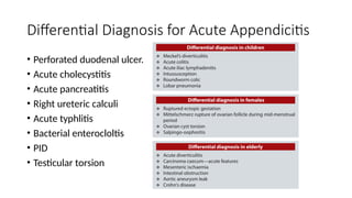
Appendicitis and its
complications
Contents
Anatomy
• Located at the terminal end of the caecum.
• Size: 5 to 10 cm
• Diameter: 3-8 mm
• Mesoappendix: Extention of the mesentery containing
appendicular artery, branch of ileocolic artery.
• Positions of appendix:
• Retrocaecal (74%) – most common
• Pelvic
• Paracaecal
• Preileal
• Subcaecal
• Post ileal (0.5%) – least common
Appendicitis
• Appendicitis is an inflammation of the appendix that develops most
common in adolescents.
• It refers to the acute inflammation of the appendix, and is the most
common cause for acute severe abdominal pain
• The abdomen is most tender at McBurny’s point – 1/3rd
of the
distance from R ASIS to umbilicus, corresponds to the location of the
appendix
Risk Factors
• Faecolith – most common cause
• Infection
• White races
• Lack of fibre rich diet
• Family history: 30%
• Obstuction
• Extremes of age
• Previous abdominal surgery
Causes
• Organism:
• E.Coli (85%)
• enterococci, (30%),
• streptococci,
• Anaerobic
• streptococci,
• Cl. welchii, bacteroides
Pathogenesis
• Acute inflammation of the mucus membrane with secondary infection
without obstruction causes acute nonobstructive appendicitis.
• Luminal obstruction by faecolith, lymphoid hyperplasia, pinworm
(oxyuris vermicularis), other worms, foreign body, carcinoma/Crohn‘s
disease
Types
• Acute nonobstructive appendicitis (catarrhal) (mucosal appendicitis)
• Acute obstructive appendicitis.
• Recurrent appendicitis.
• Subacute appendicitis.
• Stump appendicitis
Clinical Features
• Pain: earliest symptom. Visceral
pain starts around the umbilicus ->
right iliac fossa
• Vomiting: due to reflex
pylorospasm.
• Murphy’s triad
• Pain
• Vomiting
• Febrile
• Constipation is the usual feature
but diarrhoea can occur
• Fever, tachycardia.
• Urinary frequency: bladder
irritation.
• Tenderness and rebound
tenderness at McBurney’s point.
• Rovsing’s sign: On pressing left iliac
fossa, pain occurs in right iliac fossa
• Baldwing’s test is positive in
retrocaecal appendix.
Alvarado Score
• Several clinical and laboratory-based
scoring systems have been devised to
assist diagnosis.
• The most widely used is the Alvarado
score. A score of 7 or more is strongly
predictive of acute appendicitis.
• In patients with an equivocal score (5
or 6), abdominal ultrasonography or
contrast-enhanced CT examination is
recommended.
Differential Diagnosis for Acute Appendicitis
• Perforated duodenal ulcer.
• Acute cholecystitis
• Acute pancreatitis
• Right ureteric calculi
• Acute typhlitis
• Bacterial enterocloltis
• PID
• Testicular torsion
Investigations
• USG:
• Noncompressible appendix of size > 6 mm AP diameter, hyperechoic
thickened appendix wall > 2 mm—target sign.
• Appendicolith.
• Interruption of submucosal continuity.
• Periappendicular fluid.
• CBC (WBC) to correlate clinically.
• CECT: usually in old people
• MRI: useful in pregnancy
Surgery
• Open appendectomy-
• Grid iron incision, Lans incision, Rutherford Morrison’s incision
• Appendix identified and the base of mesoappendix clamped, divided and
ligated.
• Base of the appendix is ligated close to the base, and amputated between
artery forceps and the ligature.
• Appendicualr vessels are then ligated.
Open appendectomy
• Laparoscopic appendicectomy:
• Penumoperitoenum established using infra umbilical approach.
• Umbilical port + 2 working ports.
• The appendix is found using caecel taeniae.
• Appendix elevated and mesoappendix is displayed and is dissected.
• Appendicular vessels are ligated.
• Appendix is free at its base and is ligated.
• The specimen is removed through one of the operating ports.
Laparoscopic appendectomy
Complications
APPENDICULAR MASS
• It is the localisation of infection occurring 3 to 5 days after an attack of
acute appendicitis.
• Inflamed appendix, greater omentum, oedematous caecum, parietal
peritoneum and dilated ileum (ileus) forms a mass in the right iliac
fossa.
• Investigations
• TC is increased.
• Ultrasound confirms the mass.
• Treatment - Ochsner-Sherren Regimen
• Contraindications for
Ochsner-Sherren regimen
• 1. When diagnosis is in
doubt.
• 2. In acute appendicitis in
children and elderly.
• 3. In burst, gangrenous
appendicitis.
• 4. In patients in whom
diffuse peritonitis sets in.
APPENDICULAR ABSCESS
• Suppuration in an acute appendicitis or suppuration in an already formed
appendicular mass.
• commonly occurs in retrocaecal region
• Clinical features
• High fever, tender abdomen, smooth, dull (to percuss),
• soft swelling in right iliac fossa.
• Investigations: Ultrasound confirms the diagnosis.
• Treatment
• Antibiotics are started.
• CT-guided aspiration or catheter drainage is done often as initial
• Drainage of abscess.
Treatment
• Surgery:
• Open appendicectomy
• Laparoscopic appendicectomy
Thank You

Appendicitis and its complications asdpptx

  • 1.
  • 2.
  • 3.
    Anatomy • Located atthe terminal end of the caecum. • Size: 5 to 10 cm • Diameter: 3-8 mm • Mesoappendix: Extention of the mesentery containing appendicular artery, branch of ileocolic artery. • Positions of appendix: • Retrocaecal (74%) – most common • Pelvic • Paracaecal • Preileal • Subcaecal • Post ileal (0.5%) – least common
  • 4.
    Appendicitis • Appendicitis isan inflammation of the appendix that develops most common in adolescents. • It refers to the acute inflammation of the appendix, and is the most common cause for acute severe abdominal pain • The abdomen is most tender at McBurny’s point – 1/3rd of the distance from R ASIS to umbilicus, corresponds to the location of the appendix
  • 5.
    Risk Factors • Faecolith– most common cause • Infection • White races • Lack of fibre rich diet • Family history: 30% • Obstuction • Extremes of age • Previous abdominal surgery
  • 6.
    Causes • Organism: • E.Coli(85%) • enterococci, (30%), • streptococci, • Anaerobic • streptococci, • Cl. welchii, bacteroides
  • 7.
    Pathogenesis • Acute inflammationof the mucus membrane with secondary infection without obstruction causes acute nonobstructive appendicitis. • Luminal obstruction by faecolith, lymphoid hyperplasia, pinworm (oxyuris vermicularis), other worms, foreign body, carcinoma/Crohn‘s disease
  • 8.
    Types • Acute nonobstructiveappendicitis (catarrhal) (mucosal appendicitis) • Acute obstructive appendicitis. • Recurrent appendicitis. • Subacute appendicitis. • Stump appendicitis
  • 9.
    Clinical Features • Pain:earliest symptom. Visceral pain starts around the umbilicus -> right iliac fossa • Vomiting: due to reflex pylorospasm. • Murphy’s triad • Pain • Vomiting • Febrile • Constipation is the usual feature but diarrhoea can occur • Fever, tachycardia. • Urinary frequency: bladder irritation. • Tenderness and rebound tenderness at McBurney’s point. • Rovsing’s sign: On pressing left iliac fossa, pain occurs in right iliac fossa • Baldwing’s test is positive in retrocaecal appendix.
  • 10.
    Alvarado Score • Severalclinical and laboratory-based scoring systems have been devised to assist diagnosis. • The most widely used is the Alvarado score. A score of 7 or more is strongly predictive of acute appendicitis. • In patients with an equivocal score (5 or 6), abdominal ultrasonography or contrast-enhanced CT examination is recommended.
  • 11.
    Differential Diagnosis forAcute Appendicitis • Perforated duodenal ulcer. • Acute cholecystitis • Acute pancreatitis • Right ureteric calculi • Acute typhlitis • Bacterial enterocloltis • PID • Testicular torsion
  • 12.
    Investigations • USG: • Noncompressibleappendix of size > 6 mm AP diameter, hyperechoic thickened appendix wall > 2 mm—target sign. • Appendicolith. • Interruption of submucosal continuity. • Periappendicular fluid. • CBC (WBC) to correlate clinically. • CECT: usually in old people • MRI: useful in pregnancy
  • 13.
    Surgery • Open appendectomy- •Grid iron incision, Lans incision, Rutherford Morrison’s incision • Appendix identified and the base of mesoappendix clamped, divided and ligated. • Base of the appendix is ligated close to the base, and amputated between artery forceps and the ligature. • Appendicualr vessels are then ligated.
  • 14.
  • 15.
    • Laparoscopic appendicectomy: •Penumoperitoenum established using infra umbilical approach. • Umbilical port + 2 working ports. • The appendix is found using caecel taeniae. • Appendix elevated and mesoappendix is displayed and is dissected. • Appendicular vessels are ligated. • Appendix is free at its base and is ligated. • The specimen is removed through one of the operating ports.
  • 16.
  • 17.
  • 18.
    APPENDICULAR MASS • Itis the localisation of infection occurring 3 to 5 days after an attack of acute appendicitis. • Inflamed appendix, greater omentum, oedematous caecum, parietal peritoneum and dilated ileum (ileus) forms a mass in the right iliac fossa. • Investigations • TC is increased. • Ultrasound confirms the mass. • Treatment - Ochsner-Sherren Regimen
  • 19.
    • Contraindications for Ochsner-Sherrenregimen • 1. When diagnosis is in doubt. • 2. In acute appendicitis in children and elderly. • 3. In burst, gangrenous appendicitis. • 4. In patients in whom diffuse peritonitis sets in.
  • 20.
    APPENDICULAR ABSCESS • Suppurationin an acute appendicitis or suppuration in an already formed appendicular mass. • commonly occurs in retrocaecal region • Clinical features • High fever, tender abdomen, smooth, dull (to percuss), • soft swelling in right iliac fossa. • Investigations: Ultrasound confirms the diagnosis. • Treatment • Antibiotics are started. • CT-guided aspiration or catheter drainage is done often as initial • Drainage of abscess.
  • 22.
    Treatment • Surgery: • Openappendicectomy • Laparoscopic appendicectomy
  • 23.