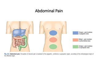
1. Acute appendicitis is a common condition that usually presents with abdominal pain that starts around the navel and shifts to the lower right abdomen. Other common symptoms include anorexia, nausea, and vomiting.
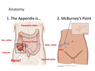
2. A clinical examination often reveals tenderness at McBurney's point with guarding and rebound tenderness. Special tests like Rovsing's sign may also indicate appendicitis. Differential diagnoses include other abdominal conditions.
3. The diagnosis is usually made based on history and clinical exam findings. Laboratory tests are not usually required but may include a CBC and urinalysis. Imaging tests like ultrasound or CT scans can help in unclear cases. Prompt diagnosis and treatment is important to
Information about Management of Appendicular Lump by Dr Dhaval Mangukiya.
Details of Appendicular Lump, Basic to Above the Basics, Incidence, Safe Approach Interval Laparoscopy, Early Surgery etc.
https://drdhavalmangukiya.com/
http://www.youtube.com/c/DrDhavalMangukiyaGastrosurgeonSurat
https://gastrosurgerysurat.blogspot.com/
PERFORATED PEPTIC ULCER
PERFORATION
DEFINITION
It is the terminology used for perforation of duodenal ulcer or gastric ulcer or stomal ulcer.
Otherwise all clinical features and management are similar.
Perforation is common in duodenal ulcer
Mortality is more in gastric ulcer perforation and perforation in elderly
Normally, fistula is defined as an abnormal communication between two epithelized surface.But enterocutaneous fistula is an abnormal communication between the skin with various parts of the gut. The ileum is the most common site of origin of enterocutaneous fistula.
Disclaimer: A lot from this slides were taken also from https://www.slideshare.net/babysurgeon/scrotal-swellings-1 (Dr Selvaraj Balasubramani)
This covers only :
ANATOMY
CAUSES
TORSION OF TESTIS
EPIDIDYMO-ORCHITIS
HYDROCELE
EPIDIDYMAL CYST
VARICOCELE
OPEN INGUINAL HERNIA REPAIR- OPERATIVE SURGERY
#surgicaleducator #operativesurgery #openinguinalherniarepair #usmle #babysurgeon #surgicaltutor
Dear viewers,
• Greetings from “Surgical Educator”
• Because of the popular demand by viewers of the YouTube channel “Surgical Educator”, I have decided to create and upload videos on common surgeries.
• I have already uploaded videos on open and Laparoscopic Appendicectomy, Thyroidectomy, Modified Radical Mastectomy and open and
Laparoscopic Cholecystectomy
• In this video today, I have discussed Open Inguinal Hernia Repair.
• However, these videos are not real surgeries but the theoretical aspect of operative surgery like going through an atlas of operative surgery.
• Along with these videos, I recommend you to watch real operative surgery videos as well and I will give a link for each surgery in the end of the video as end-cards, which I think will be very useful.
• This will give a very good opportunity for the surgical trainees to mentally rehearse various surgical steps in a sequential manner prior to actual surgery. You can watch the video in the following links:
• surgicaleducator.blogspot.com
• youtube.com/c/surgicaleducator
• Thank you for watching the videos.
This presentation describes the Laparoscopic Anatomy of the Inguinal Region and Correlates the differences with Open repairs...eventually changing the perception of the operating surgeon...for a Laparoscopic Transition
URETHRAL INJURY- Trauma Surgery
#surgicaleducator #babysurgeon
Dear viewers,
Greetings from “Surgical Educator”
Today I have uploaded a video on URETHRAL INJURY- one more in Trauma Surgery. I haven’t talked elaborately but have included the essential minimum an undergraduate medical student should know. I have talked about anatomy, classification, etio-pathogenesis, symptoms, signs, investigations, grading, treatment, complications, treatment algorithm and case vignettes of urethral injuries. My aim is, after watching this video all of you should be able to arrive at a correct working diagnosis of the type of urethral injury and should also be able to institute immediate treatment to the patients if there is a need. The definitive urethroplasty should be done by a Urologist. You can watch the video in the following links:
surgicaleducator.blogspot.com
youtube.com/c/surgicaleducator
Thank you for watching the video.
Types of Urethral injury, Mechanism of Urethral injury, Classification of Urethral injury, Symptoms of Urethral injury, Signs of Urethral injury,
Investigations for Urethral injury, Grading of Urethral Injury, Treatment for Urethral injury, Complications of Urethral Injury
acute abdominal pain in pediatrics. include background and approach also there are three cases included, intussusception, Hirschsprung's disease and DKA.
Seminar presentation by 5th year Medical Student under the supervision of a pediatric surgery specialist from HRPZ II. Reference as mentioned in the slide.
Title: Sense of Taste
Presenter: Dr. Faiza, Assistant Professor of Physiology
Qualifications:
MBBS (Best Graduate, AIMC Lahore)
FCPS Physiology
ICMT, CHPE, DHPE (STMU)
MPH (GC University, Faisalabad)
MBA (Virtual University of Pakistan)
Learning Objectives:
Describe the structure and function of taste buds.
Describe the relationship between the taste threshold and taste index of common substances.
Explain the chemical basis and signal transduction of taste perception for each type of primary taste sensation.
Recognize different abnormalities of taste perception and their causes.
Key Topics:
Significance of Taste Sensation:
Differentiation between pleasant and harmful food
Influence on behavior
Selection of food based on metabolic needs
Receptors of Taste:
Taste buds on the tongue
Influence of sense of smell, texture of food, and pain stimulation (e.g., by pepper)
Primary and Secondary Taste Sensations:
Primary taste sensations: Sweet, Sour, Salty, Bitter, Umami
Chemical basis and signal transduction mechanisms for each taste
Taste Threshold and Index:
Taste threshold values for Sweet (sucrose), Salty (NaCl), Sour (HCl), and Bitter (Quinine)
Taste index relationship: Inversely proportional to taste threshold
Taste Blindness:
Inability to taste certain substances, particularly thiourea compounds
Example: Phenylthiocarbamide
Structure and Function of Taste Buds:
Composition: Epithelial cells, Sustentacular/Supporting cells, Taste cells, Basal cells
Features: Taste pores, Taste hairs/microvilli, and Taste nerve fibers
Location of Taste Buds:
Found in papillae of the tongue (Fungiform, Circumvallate, Foliate)
Also present on the palate, tonsillar pillars, epiglottis, and proximal esophagus
Mechanism of Taste Stimulation:
Interaction of taste substances with receptors on microvilli
Signal transduction pathways for Umami, Sweet, Bitter, Sour, and Salty tastes
Taste Sensitivity and Adaptation:
Decrease in sensitivity with age
Rapid adaptation of taste sensation
Role of Saliva in Taste:
Dissolution of tastants to reach receptors
Washing away the stimulus
Taste Preferences and Aversions:
Mechanisms behind taste preference and aversion
Influence of receptors and neural pathways
Impact of Sensory Nerve Damage:
Degeneration of taste buds if the sensory nerve fiber is cut
Abnormalities of Taste Detection:
Conditions: Ageusia, Hypogeusia, Dysgeusia (parageusia)
Causes: Nerve damage, neurological disorders, infections, poor oral hygiene, adverse drug effects, deficiencies, aging, tobacco use, altered neurotransmitter levels
Neurotransmitters and Taste Threshold:
Effects of serotonin (5-HT) and norepinephrine (NE) on taste sensitivity
Supertasters:
25% of the population with heightened sensitivity to taste, especially bitterness
Increased number of fungiform papillae
Pulmonary Thromboembolism - etilogy, types, medical- Surgical and nursing man...VarunMahajani
Disruption of blood supply to lung alveoli due to blockage of one or more pulmonary blood vessels is called as Pulmonary thromboembolism. In this presentation we will discuss its causes, types and its management in depth.
Explore natural remedies for syphilis treatment in Singapore. Discover alternative therapies, herbal remedies, and lifestyle changes that may complement conventional treatments. Learn about holistic approaches to managing syphilis symptoms and supporting overall health.
These lecture slides, by Dr Sidra Arshad, offer a quick overview of physiological basis of a normal electrocardiogram.
Learning objectives:
1. Define an electrocardiogram (ECG) and electrocardiography
2. Describe how dipoles generated by the heart produce the waveforms of the ECG
3. Describe the components of a normal electrocardiogram of a typical bipolar leads (limb II)
4. Differentiate between intervals and segments
5. Enlist some common indications for obtaining an ECG
Study Resources:
1. Chapter 11, Guyton and Hall Textbook of Medical Physiology, 14th edition
2. Chapter 9, Human Physiology - From Cells to Systems, Lauralee Sherwood, 9th edition
3. Chapter 29, Ganong’s Review of Medical Physiology, 26th edition
4. Electrocardiogram, StatPearls - https://www.ncbi.nlm.nih.gov/books/NBK549803/
5. ECG in Medical Practice by ABM Abdullah, 4th edition
6. ECG Basics, http://www.nataliescasebook.com/tag/e-c-g-basics
New Drug Discovery and Development .....NEHA GUPTA
The "New Drug Discovery and Development" process involves the identification, design, testing, and manufacturing of novel pharmaceutical compounds with the aim of introducing new and improved treatments for various medical conditions. This comprehensive endeavor encompasses various stages, including target identification, preclinical studies, clinical trials, regulatory approval, and post-market surveillance. It involves multidisciplinary collaboration among scientists, researchers, clinicians, regulatory experts, and pharmaceutical companies to bring innovative therapies to market and address unmet medical needs.
Report Back from SGO 2024: What’s the Latest in Cervical Cancer?bkling
Are you curious about what’s new in cervical cancer research or unsure what the findings mean? Join Dr. Emily Ko, a gynecologic oncologist at Penn Medicine, to learn about the latest updates from the Society of Gynecologic Oncology (SGO) 2024 Annual Meeting on Women’s Cancer. Dr. Ko will discuss what the research presented at the conference means for you and answer your questions about the new developments.
Tom Selleck Health: A Comprehensive Look at the Iconic Actor’s Wellness Journeygreendigital
Tom Selleck, an enduring figure in Hollywood. has captivated audiences for decades with his rugged charm, iconic moustache. and memorable roles in television and film. From his breakout role as Thomas Magnum in Magnum P.I. to his current portrayal of Frank Reagan in Blue Bloods. Selleck's career has spanned over 50 years. But beyond his professional achievements. fans have often been curious about Tom Selleck Health. especially as he has aged in the public eye.
Follow us on: Pinterest
Introduction
Many have been interested in Tom Selleck health. not only because of his enduring presence on screen but also because of the challenges. and lifestyle choices he has faced and made over the years. This article delves into the various aspects of Tom Selleck health. exploring his fitness regimen, diet, mental health. and the challenges he has encountered as he ages. We'll look at how he maintains his well-being. the health issues he has faced, and his approach to ageing .
Early Life and Career
Childhood and Athletic Beginnings
Tom Selleck was born on January 29, 1945, in Detroit, Michigan, and grew up in Sherman Oaks, California. From an early age, he was involved in sports, particularly basketball. which played a significant role in his physical development. His athletic pursuits continued into college. where he attended the University of Southern California (USC) on a basketball scholarship. This early involvement in sports laid a strong foundation for his physical health and disciplined lifestyle.
Transition to Acting
Selleck's transition from an athlete to an actor came with its physical demands. His first significant role in "Magnum P.I." required him to perform various stunts and maintain a fit appearance. This role, which he played from 1980 to 1988. necessitated a rigorous fitness routine to meet the show's demands. setting the stage for his long-term commitment to health and wellness.
Fitness Regimen
Workout Routine
Tom Selleck health and fitness regimen has evolved. adapting to his changing roles and age. During his "Magnum, P.I." days. Selleck's workouts were intense and focused on building and maintaining muscle mass. His routine included weightlifting, cardiovascular exercises. and specific training for the stunts he performed on the show.
Selleck adjusted his fitness routine as he aged to suit his body's needs. Today, his workouts focus on maintaining flexibility, strength, and cardiovascular health. He incorporates low-impact exercises such as swimming, walking, and light weightlifting. This balanced approach helps him stay fit without putting undue strain on his joints and muscles.
Importance of Flexibility and Mobility
In recent years, Selleck has emphasized the importance of flexibility and mobility in his fitness regimen. Understanding the natural decline in muscle mass and joint flexibility with age. he includes stretching and yoga in his routine. These practices help prevent injuries, improve posture, and maintain mobilit
The prostate is an exocrine gland of the male mammalian reproductive system
It is a walnut-sized gland that forms part of the male reproductive system and is located in front of the rectum and just below the urinary bladder
Function is to store and secrete a clear, slightly alkaline fluid that constitutes 10-30% of the volume of the seminal fluid that along with the spermatozoa, constitutes semen
A healthy human prostate measures (4cm-vertical, by 3cm-horizontal, 2cm ant-post ).
It surrounds the urethra just below the urinary bladder. It has anterior, median, posterior and two lateral lobes
It’s work is regulated by androgens which are responsible for male sex characteristics
Generalised disease of the prostate due to hormonal derangement which leads to non malignant enlargement of the gland (increase in the number of epithelial cells and stromal tissue)to cause compression of the urethra leading to symptoms (LUTS
Acute scrotum is a general term referring to an emergency condition affecting the contents or the wall of the scrotum.
There are a number of conditions that present acutely, predominantly with pain and/or swelling
A careful and detailed history and examination, and in some cases, investigations allow differentiation between these diagnoses. A prompt diagnosis is essential as the patient may require urgent surgical intervention
Testicular torsion refers to twisting of the spermatic cord, causing ischaemia of the testicle.
Testicular torsion results from inadequate fixation of the testis to the tunica vaginalis producing ischemia from reduced arterial inflow and venous outflow obstruction.
The prevalence of testicular torsion in adult patients hospitalized with acute scrotal pain is approximately 25 to 50 percent
Ethanol (CH3CH2OH), or beverage alcohol, is a two-carbon alcohol
that is rapidly distributed in the body and brain. Ethanol alters many
neurochemical systems and has rewarding and addictive properties. It
is the oldest recreational drug and likely contributes to more morbidity,
mortality, and public health costs than all illicit drugs combined. The
5th edition of the Diagnostic and Statistical Manual of Mental Disorders
(DSM-5) integrates alcohol abuse and alcohol dependence into a single
disorder called alcohol use disorder (AUD), with mild, moderate,
and severe subclassifications (American Psychiatric Association, 2013).
In the DSM-5, all types of substance abuse and dependence have been
combined into a single substance use disorder (SUD) on a continuum
from mild to severe. A diagnosis of AUD requires that at least two of
the 11 DSM-5 behaviors be present within a 12-month period (mild
AUD: 2–3 criteria; moderate AUD: 4–5 criteria; severe AUD: 6–11 criteria).
The four main behavioral effects of AUD are impaired control over
drinking, negative social consequences, risky use, and altered physiological
effects (tolerance, withdrawal). This chapter presents an overview
of the prevalence and harmful consequences of AUD in the U.S.,
the systemic nature of the disease, neurocircuitry and stages of AUD,
comorbidities, fetal alcohol spectrum disorders, genetic risk factors, and
pharmacotherapies for AUD.
4. Acute Appendicitis
• Appendicitis is common- 7-9% lifetime risk
• Mostly young people but can present at any age.
• Delay in diagnosis/management causes significant morbidity- can be
a surgical emergency
• Usually clinical diagnosis- not reliant on imaging
• Has classic presentation but often presents atypically- it is a common
pitfall!
Appendix role :
1- part of immunity
2- stabilize the ureter
7. ACUTE APPENDICITIS
Incidence:
- rare in infants, increasingly common in childhood and early
adult life, peak incidence in the teens and early 20s.
- before puberty males = females.
- In teenagers and young adults the male–female ratio 3:2
- Thereafter, the greater incidence in males declines.
8. Aetiology
No definite cause
1-Decreased dietary fibre and increased consumption of refined carbohydrates
(low fiber diet)
2-Bacterial proliferation within the appendix,
no single organism is responsible(mixed growth of aerobic and anaerobic
organisms ).
3- Obstruction of the appendix lumen
- Lymphoid hyperplasia
- faecolith (composed of inspissated faecal material,
calcium phosphates, bacteria and epithelial debris
- stricture
- foreign body(rare)
- tumour, particularly carcinoma of the caecum(middle age and elderly)
- Intestinal parasites, particularly Oxyuris vermicularis
9. Pathology
- Lymphoid hyperplasia narrows the lumen luminal obstruction.
- Continued mucus secretion and inflammatory exudation increase
intraluminal pressure obstructing lymphatic drainageOedema
- Mucosal ulceration
- Bacterial translocation to the submucosa.
10. Resolution may occur (spontaneously/antibiotic therapy).
or
Condition progresses,more distension venous obstruction & wall
ischaemia bacterial invasion through the muscularis propria and
submucosa, producing acute appendicitis.
Ischaemic necrosis gangrenous appendicitisfree bacterial
contamination of the peritoneal cavity(peritonitis).
11. • Alternatively, the greater omentum and loops of small bowel become
adherent to the inflamed appendix, walling off the spread of peritoneal
contamination, and resulting in a phlegmonous mass or paracaecal
abscess.
Rarely, appendiceal inflammation resolves, leaving a distended mucus-filled
organ termed a ‘mucocoele’ of the appendix .
12. Diffuse peritonitis is the great threat of acute appendicitis, it occurs as a result
of:
- free migration of bacteria through an ischaemic appendicular wall
- frank perforation of a gangrenous appendix
- delayed perforation of an appendix abscess.
Factors that promote this process include:
- extremes of age ( child : immature omentum , elderly : atrophic omentum )
- immunosuppression pregnant , steroid taker
- diabetes mellitus
- faecolith obstruction of the appendix lumen
- free-lying pelvic appendix
- previous abdominal surgery
In these situations, a rapidly deteriorating clinical course is accompanied by
signs of diffuse peritonitis and systemic sepsis syndrome.
13. Clinical diagnosis
History
PAIN
-poorly localised, colicky abdominal pain(midgut visceral discomfort in
response to appendiceal inflammation and obstruction).
-The pain in periumbilical region.
- Central abdominal pain + anorexia, nausea 20% and usually one or two
episodes of vomiting 75% that follow the onset of pain.
- If the oral contrast get inside the appendix under CT , never appendicitis
14. With progressive inflammation of the appendix:
Shifting of pain: from central abdominal pain to right iliac fossa(irritation
of RIF parietal peritoneum) visceral to somatic pain which is :
- more intense
- constant
- localised to right iliac fossa
Typically, coughing or sudden movement exacerbates the right iliac fossa
pain.
15. ANOREXIA
-constant clinical feature, particularly in children.
Family history
1/3 of children with appendicitis have a first-degree relative with a similar
history .
One half of acute appendicitis classic visceral–somatic sequence of pain
.
Signs
Temperature and pulse rate:
- After 6 hours, slight pyrexia (37.2–37.7°C) with a corresponding
increase in the pulse rate to 80 or 90 is usual.
-Changes of greater magnitude may indicate complications.
16. - Unwell patient with low-grade pyrexia
- Inspection of the abdomen limitation of respiratory movement in the
lower abdomen.
- localised abdominal tenderness maximum at McBurney’s point.
- Muscle guarding
- Rebound tenderness
Asking the patient to cough or gentle percussion over the site of
maximum tenderness will elicit rebound tenderness .
- Pointing sign: The patient is asked to point to where the pain began and
where it moved .
17. - Rovsing’s sign: Deep palpation of the left iliac
fossa may cause pain in the right iliac fossa.
- Psoas sign : passive extension of hip or active
flexion of hip against resistance pain. The
patient will lie with the right hip flexed for pain
relief .
- Obturator sign : hip flexion and internal
rotation cause pain in the hypogastrium (the
obturator test).
-Cutaneous hyperaesthesia may be
demonstrable
in the right iliac fossa
18. Two clinical syndromes of acute appendicitis:
1- acute catarrhal (non-obstructive) appendicitis
2- acute obstructive appendicitis:
-more acute course(abrupt)
-generalised abdominal pain from the start.
- Temperature normal
- vomiting common.
19. Special features, according to position of the
appendix
1-Retrocaecal appendicitis
- Rigidity is often absent
- Application of deep pressure may fail to
elicit tenderness (silent appendix)
-Deep tenderness is often present in the loin,
and rigidity of the quadratus lumborum.
-Psoas spasm leading to flexion of the hip
joint. Hyperextension of the hip joint may
induce abdominal pain (Psoas sign +ve).
20. 2-Pelvic appendicitis
- More common in children
- Early diarrhoea (inflamed appendix in contact with the rectum).
- Complete absence of abdominal rigidity
- Tenderness over McBurney’s point is also lacking.
- Deep tenderness can be made out just above and to the right of the
symphysis pubis.
- Rectal examination reveals tenderness in the rectovesical pouch or
the pouch of Douglas, especially on the right
side.
- +ve psoas and obturator signs.
- Frequency of micturition (inflamed appendix in contact with the
bladder).
21. 3-Postileal appendicitis
The inflamed appendix lies behind the terminal
Ileum(difficult to diagnose)
- Pain may not shift
- Diarrhea is a feature
- Marked retching
- Tenderness, if any, is ill defined, although it may be
present immediately to the right of the umbilicus.
22. Special features, according to age
Infants
- Rare in infants under 36 months of age.
- The patient is unable to give a history. Diagnosis is
often delayed.
- Higher incidence of perforation and postoperative
morbidity than older children.
- Diffuse peritonitis can develop rapidly because of the
underdeveloped greater omentum.
Children
- Vomiting is more common.
- complete aversion to food.
23. The elderly
- Gangrene and perforation occur much more frequently .
- The abdominal clinical picture is not obvious even in the presence of
gangrenous appendicitis (lax abdominal walls or obesity)
- Clinical picture may simulate subacute intestinal obstruction.
- Higher mortality
(coincident medical conditions plus the previous factors)
The obese
- Obesity can obscure and diminish all the local signs of acute
appendicitis.
- Midline abdominal incision
(Delay in diagnosis + technical difficulty of operating in the obese)
- Laparoscopy is particularly useful in the obese.
24. Pregnancy
Appendicitis is the most common extrauterine acute abdominal condition in
pregnancy.
- Delay in presentation (early non-specific symptoms are often attributed
to the pregnancy).
- The physiologic leukocytosis of pregnancy (high as 16,000 cells/mm3).
Obstetric teaching has been that the caecum and appendix are progressively
pushed to the right upper quadrant of the abdomen as pregnancy develops
during the second and
third trimesters.
- Pain in the right lower quadrant of the abdomen remains the cardinal
feature of appendicitis in pregnancy.
- Fetal loss occurs in 3–5 per cent of cases, increasing to 20 per cent if
perforation is found at operation.
25. Differential diagnosis
Children
1- Acute gastroenteritis and mesenteric lymphadenitis:
the pain is diffuse, and tenderness is not as sharply localized and cervical
lymph nodes may be enlarged.
2- Meckel’s diverticulitis:
signs may be central or left sided.
history of antecedent abdominal pain or intermittent lower
gastrointestinal bleeding.
3- Intussusception:
Appendicitis is uncommon before the age of two years, whereas the
median age for intussusception is 18 months.
A mass may be palpable in the right lower quadrant,
26. 5-Lobar pneumonia and pleurisy, especially at the right
base
Abdominal tenderness is minimal,
pyrexia is marked
+ve pleural friction rub or altered breath
sounds on auscultation.
A chest radiograph is diagnostic.
4- Henoch–Schönlein purpura
preceded by a sore throat or respiratory
infection.
Abdominal pain can be severe.
ecchymotic rash, affecting the extensor
surfaces of the limbs and on the buttocks.
The face is usually spared.
Microscopic haematuria is common.
27. Adults
1- Terminal ileitis
- a doughy mass of inflamed ileum may be felt.
- history of abdominal cramping, weight loss and diarrhoea.
The ileitis may be non-specific, due to Crohn’s disease or Yersinia
infection.
- serum antibody titres are diagnostic, and treatment with
intravenous tetracycline is appropriate.
2- Ureteric colic:
Urinalysis should always be performed, and the presence of red cells
supine abdominal radiograph. Renal ultrasound or intravenous urogram
is diagnostic.
3- Right-sided acute pyelonephritis
increased frequency of micturition.
tenderness confined to the loin, fever (temperature 39°C) and possibly
rigors and pyuria.
28. 4- perforated peptic ulcer
the duodenal contents pass along the paracolic gutter to the right iliac fossa.
- history of dyspepsia
- very sudden onset of pain that starts in the epigastrium and passes down
the right paracolic gutter.
- rigidity is usually greater in the right hypochondrium.
- rigidity and tenderness in the right iliac fossa
- erect chest radiograph gas under the diaphragm in 70 %.
- abdominal (CT) examination in difficult cases.
5- Testicular torsion (teenage or young adult male)
Pain can be referred to the right iliac fossa, and shyness on the part of the
patient may lead the unwary to suspect appendicitis unless the scrotum is
examined in all cases.
29. 7- Rectus sheath haematoma
- relatively rare
- acute pain and localised tenderness in the RIF
- after an episode of strenuous physical exercise.
- Localised pain without gastrointestinal upset is the rule. Occasionally, in
an elderly patient, particularly one taking anticoagulant therapy, a rectus
sheath haematoma may present as a mass and tenderness in the right
iliac fossa after minor trauma.
6-Acute pancreatitis
should be considered in the
differential diagnosis of all adults
suspected of having acute
appendicitis and, when appropriate,
should be excluded by serum or
urinary amylase measurement.
30. Adult female
pelvic disease in women of childbearing age most often mimics acute
appendicitis.
A careful gynaecological history should be taken in all women with
suspected appendicitis, concentrating on:
- menstrual cycle
- vaginal discharge
- possible pregnancy .
The most common diagnostic mimics are
pelvic inflammatory disease (PID), Mittelschmerz, torsion or
haemorrhage of an ovarian cyst and ectopic pregnancy.
31. 1-Pelvic inflammatory disease
(salpingitis, endometritis and tubo-ovarian sepsis)
- The pain is lower than in appendicitis and is bilateral.
- history of vaginal discharge, dysmenorrhoea and dysurea .
- vaginal examination adnexal and cervical tenderness .
- High vaginal swab & culture Chlamydia trachomatis and Neisseria
gonorrhoeae.
- gynaecologist opinion should be obtained.
- Transvaginal ultrasound
- diagnostic laparoscopy
32. 2- Mittelschmerz
Midcycle rupture of a follicular cyst with bleeding produces lower
abdominal and pelvic pain, typically midcycle.
- No systemic upset
- Pregnancy test is negative
- Symptoms usually subside within hours.
Occasionally, diagnostic laparoscopy is required. Retrograde
menstruation may cause similar symptoms.
33. 3- Torsion/haemorrhage of an ovarian cyst
(difficult differential diagnosis)
pelvic ultrasound and a gynaecological opinion should be sought.
If encountered at operation untwisting of the involved adnexa and
ovarian cystectomy should be performed.
34. Right-sided unruptured tubal pregnancy :
- pain commences on the right side and stays there.
- pain is severe.
- history of a missed menstrual period
- Signs of intraperitoneal bleeding with referred pain in the
shoulder.
- cervical excitation test positive
- urinary pregnancy test may be positive.
- Pelvic ultrasonography.
4- Ectopic pregnancy
- Ectopic preg. signs of
haemoperitoneum
-right-sided tubal abortion or
right-sided unruptured tubal pregnancy.
35. Elderly
1- Diverticulitis
Abdominal CT scanning is particularly useful and should be considered in
the management of all patients over the age of 60 years.
Right-sided diverticulitis is unusual and may be clinically
indistinguishable from appendicitis.
36. 2-Intestinal obstruction
In elderly, occasionally, it may be difficult to differentiate IO from acute
appendicitis
3-Carcinoma of the caecum
-Carcinoma of the caecum when obstructed or locally perforated, may
mimic or cause obstructive appendicitis in adults.
-history of antecedent discomfort, altered bowel habit or unexplained
anaemia.
- mass may be palpable and an abdominal CT scan
diagnostic.
37. Rare differential diagnoses
1-Preherpetic pain of the right 10th and 11th dorsal nerves.
- no shift of pain
- marked hyperaesthesia.
- no intestinal upset, no rigidity.
The herpetic eruption may be delayed for 3–8 hours.
2-Diabetic crises.
3-Spinal conditions include tuberculosis of the spine, metastatic
carcinoma, osteoporotic vertebral collapse and multiple myeloma.
4-porphyria and diabetes mellitus
5-Typhlitis or leukaemic ileocaecal syndrome
38. Investigation
The diagnosis of acute appendicitis is essentially clinical
• Routine
Full blood count WBC > 10,000
Urinalysis hematuria/ pyuria due to irritation of nearby ureter/ urinary
bladder
• Selective
Pregnancy test
Urea and electrolytes
Supine abdominal radiograph
Ultrasound of the abdomen/pelvis
Contrast-enhanced abdomen and pelvic computed tomography scan
39. Diagnostic Scoring
•Diagnosis is essentially clinical;
•A number of clinical and laboratory-based
scoring systems have been devised to assist
diagnosis.
•The most widely used is Alvarado score.
Source: Bailey & Loves Short Practice of
Surgery 25th ed
40. The Alvarado (MANTRELS) Score
Score
Symptoms
• Migratory RIF pain
• Anorexia
• Nausea and vomiting
1
1
1
Signs
• Tenderness (RIF)
• Rebound tenderness
• Elevated temperature
2
1
1
Laboratory
• Leucocytosis
• Shift to the left (segmented
neutrophils)
2
1
TOTAL 10
• < 5 is strongly against a diagnosis of appendicitis
• 7 or more is strongly predictive of acute appendicitis
• In patients with an equivocal score of 5 or 6, abdominal
USG or contrast-enhanced CT scan is used to further
reduce the rate of negative appendicectomy
1-4: Very
unlikely
5-6: Possible
7-8: Very
probable
9-10:
Definite
41. Abdominal ultrasound is useful in:
- children and thin adults
- if gynaecological pathology is suspected, with a diagnostic
accuracy >90 %.
Contrast-enhanced CT scan is most useful in:
- diagnostic uncertainty+older patients
- acute diverticulitis, intestinal obstruction and neoplasm suspected.
42. Appendicectomy
- no unnecessary delay
- short period of intensive preoperative preparation:
1- NPO
2- Intravenous fluids
(catheterisation is needed only in the very ill)
3- IV antibiotics
In the absence of purulant peritonitis give single preoperative dose of
antibiotics .
When peritonitis is suspected, therapeutic intravenous antibiotics to cover
Gram negative bacilli, as well as anaerobic cocci, should be given.
43. Appendicectomy
- general anaesthetic
- patient supine on the operating table.
- When a laparoscopic technique is to be used, the bladder must be empty
(ensure that the patient has voided before leaving the ward).
palpated for a mass prior to preparing the entire abdomen with an
antiseptic solution.(mass conservative)
Draping of the abdomen
Incisions used:
1- The gridiron incision(most common)
2- Rutherford Morison incision(muscle cutting)
3- Transverse skin crease (Lanz) incision
4- lower midline abdominal incision( doubtful diagnosis)
44. 1-The caecum is identified by the presence of taeniae
coli and is withdrawn.
2- Appendix may be felt at the base
of the caecum.
3- Inflammatory adhesions must be gently broken
with a finger, the appendix delivered into the wound.
4- Mesoappendix devided between ligature, the base
of appendix ligated and divided.
5- Purse-string to invaginates the stump of the
appendix(?????)
45. Laparoscopic appendicectomy
Laparoscopy is diagnostic tool(esp. in female) and therapeutic.
Used more in:
• Female (child bearing age)
• in obese patients
• early pregnancy
Advantages:
• less postoperative pain
• Early discharge from hospital
• Early return to daily activities
47. Problems encountered during appendicectomy
1-A normal appendix is found
- exclude other possible diagnoses, particularly terminal ileitis,
Meckel’s diverticulitis and tubal or ovarian causes in women.
- remove the appendix (avoid future diagnostic difficulties)
2-The appendix cannot be found.
The caecum should be mobilised, and the taeniae coli should be traced
to their confluence on the caecum before the diagnosis of ‘absent
appendix’ is made.
48. 3-An appendicular tumour is found.
Small tumours (under 2.0 cm in diameter) can be removed by
appendicectomy.
Larger: right hemicolectomy
4- An appendix abscess is found and the appendix cannot be removed
easily.
Preoperatively diagnosed abscessPercutaneous drainage of the abscess and
intravenous antibiotic .
At operationdraine the abscess +intravenous antibiotics.
Frankly necrotic appendix(very rarely) caecectomy or partial right
hemicolectomy .
49. Appendix abscess
Failure of resolution of an appendix mass or continued spiking pyrexia
usually indicates that there is pus within the phlegmonous appendix
mass.
Treatment :
Ultrasound or abdominal CT scan percutaneous drain.
If unsuccessful laparotomy through a midline incision is indicated.
50. Pelvic abscess
- an occasional complication of acute appendicitis.
- Presented with spiking pyrexia several days after appendicitis
- Pelvic pressure or discomfort
- loose stool or tenesmus is common.
- Rectal examination mass in the pelvis.
Pelvic ultrasound or CT scan confirm.
Treatment:
- Radiologically guided percutaneous drainage
- Transrectal drainage under general anaesthetic.
51. Postoperative complications
1- Wound infection
most common postoperative complication
usually presents with pain and erythema of the wound on
the 4th or 5th postoperative day.
Treatment: wound drainage + antibiotics.
52. 2- Intra-abdominal abscess
presented with spiking fever, malaise and anorexia developing 5–7 days
after operation .
Sites of collection:
Interloop, paracolic, pelvic and subphrenic
Abdominal ultrasonography and CT scanning diagnostic and allow
percutaneous drainage.
Laparotomy in intra-abdominal sepsis without localised collection.
53. 3- slipped ligature
internal bleeding, hypotension, tacchycardia reoperate
4- Ileus
Ileus persisting for more than 4 or 5 days + fevercontinuing intra-
abdominal sepsis investigation.
5- Respiratory
- rare
- Adequate postoperative analgesia and physiotherapy reduce the
incidence.
6- Venous thrombosis and embolism
- rare after appendicectomy
- more in elderly and in women taking the oral contraceptive pill and
prophylactic measures should be considered.
54. 7- Portal pyaemia (pylephlebitis)
- rare
- complication of gangrenous appendicitis
- high fever, rigors and jaundice.
- caused by septicaemia in the portal venous system intrahepatic
abscesses (multiple).
Treatment: systemic antibiotics + percutaneous drainage of hepatic
abscesses.
screen for underlying thrombophilia
55. 8- Faecal fistula
- Rare
- leakage from appendicular stump.
- More in appendicectomy in Crohn’s disease.
Treatment: Conservative management with low-residue enteral nutrition.
9- Adhesive intestinal obstruction
This is the most common late complication of appendicectomy.
56. Management of an appendix mass
Conservative Ochsner–Sherren regimen;
A nonoperative programme but to be prepared to operate should clinical
deterioration occur . This includes:
Careful recording of the patient’s condition
1- Temperature and pulse rate should be recorded 4-hourly
2- Fluid balance record
3- The abdomen regularly reexamined.
4- The extent of the mass should be made (mark the limits of the mass on
the abdominal wall using a skin pencil)
5- A contrast-enhanced CT examination of the abdomen
6- Antibiotic
7- An abscess, if present, should be drained radiologically.
57. Criteria for stopping conservative treatment of
an appendix mass
_ A rising pulse rate
_ Increasing or spreading abdominal pain
_ Increasing size of the mass
Clinical deterioration or evidence of peritonitis is an indication for early
laparotomy.
If the mass resolve, Patients over the age of 40 should have colonoscopy and
follow-up imaging to ensure resolution and exclude appendicular or colonic
malignancy.
58. Recurrent acute appendicitis
- not uncommon
- attacks vary in intensity and may occur every few months.
- majority of cases ends in severe acute appendicitis.
The appendix in these cases shows fibrosis indicative of previous
inflammation.
60. 1-Carcinoid tumours
- arise in argentaffin tissue (Kulchitsky cells of the crypts of
Lieberkühn)
- most common in the vermiform appendix.
- appendix removed because of symptoms of subacute or
recurrent appendicitis.
-frequently in the distal third of the
appendix.
-moderately hard , yellow tumour
-the intact mucosa .
-Microscopicallycharacteristic
pattern using immunohistochemical
stain for chromogranin B .
-rarely gives rise to metastases.
61. Treatment:
- Appendectomy .
- Right hemicolectomy is indicated if:
- caecal wall involvement .
- 2 or more in size.
- Involved lymph nodes.
2- Goblet cell carcinoid tumour
3- Primary adenocarcinoma of the appendix
extremely rare, presented as appendicitis
treated by right hemicolectomy
62. Mucinous cystadenoma
- A mucin-secreting adenoma of the appendix
- rupture into the peritoneal cavity , seeding it with mucussecreting cells.
- delayed presentation with gross abdominal distension as a result of
pseudomyxoma peritoneii, which may mimic ascites .
Treatment
radical resection of all involved parietal peritoneal
surfaces and aggressive intraperitoneal chemotherapy.