Downloaded 1,777 times


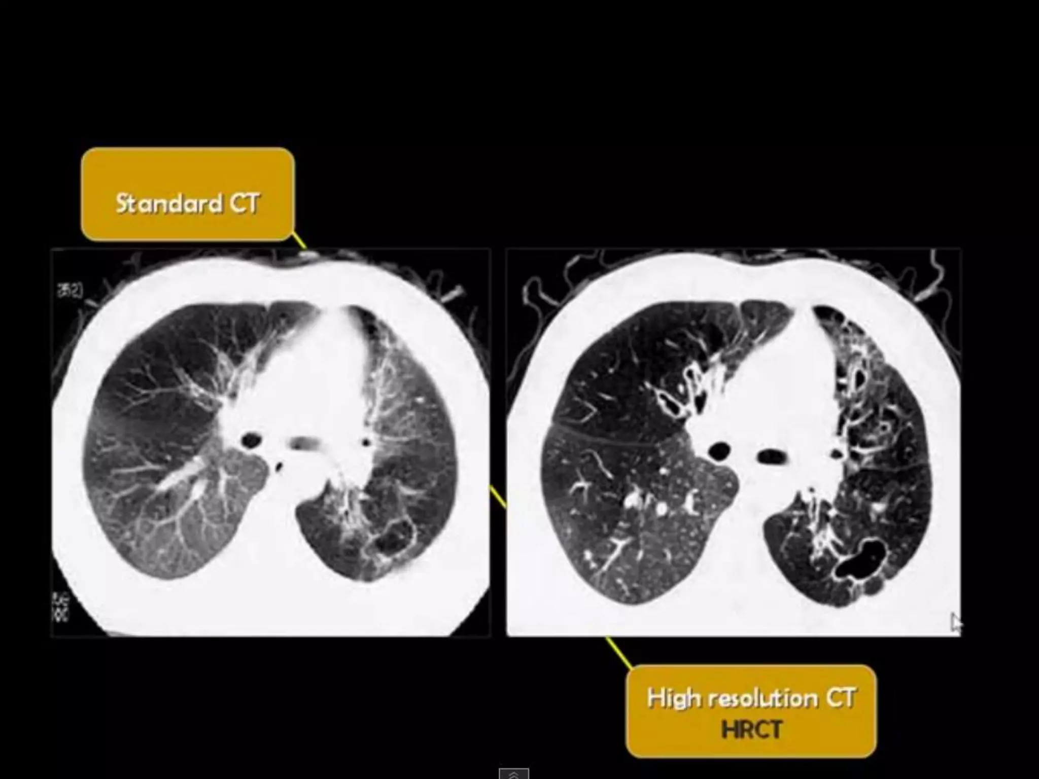
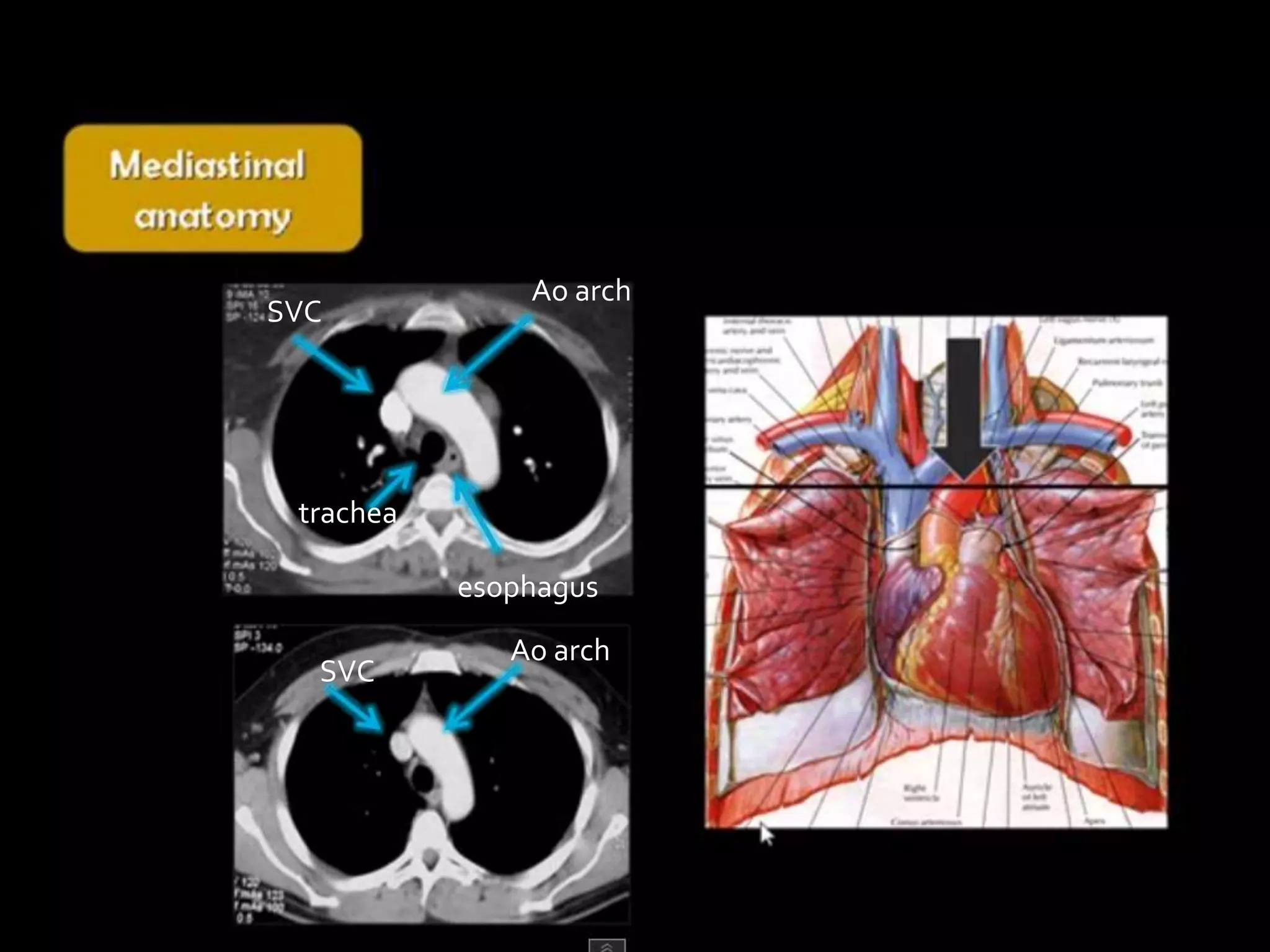


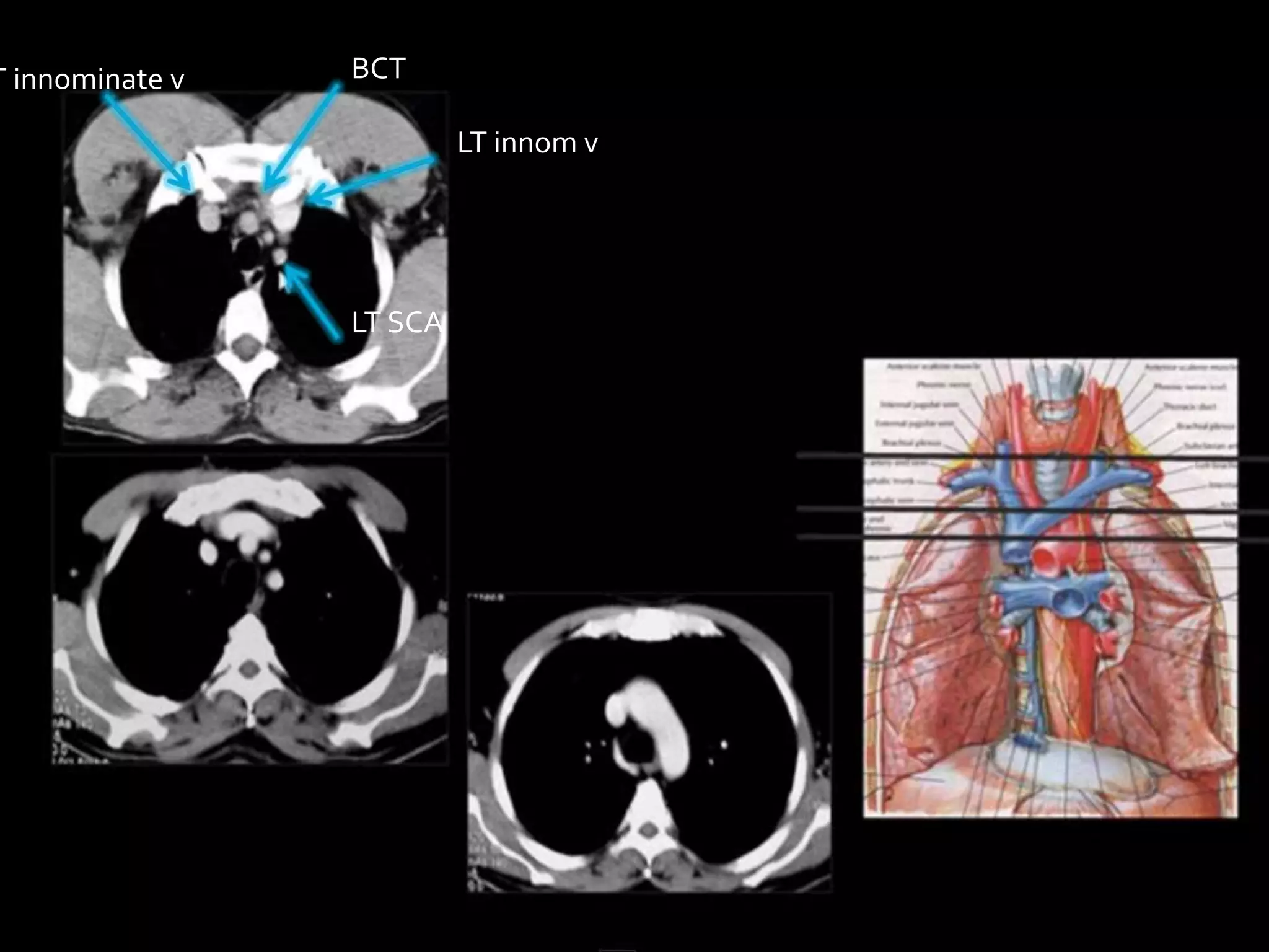


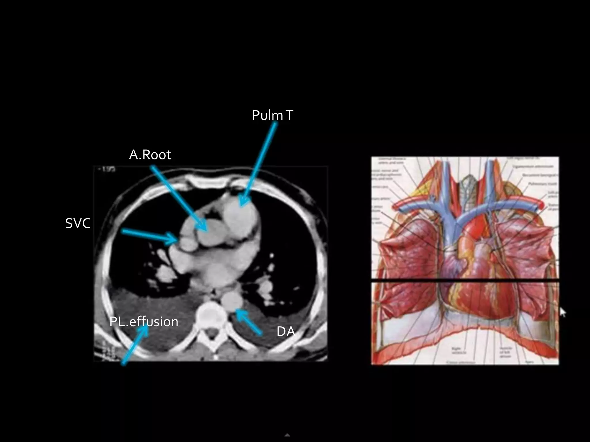

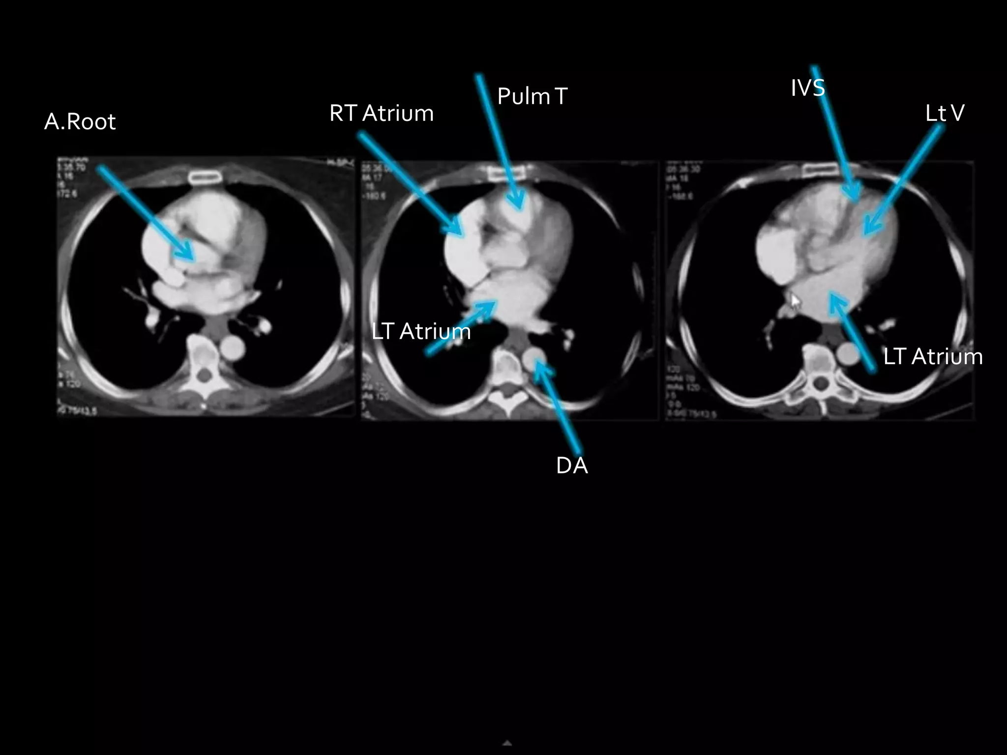


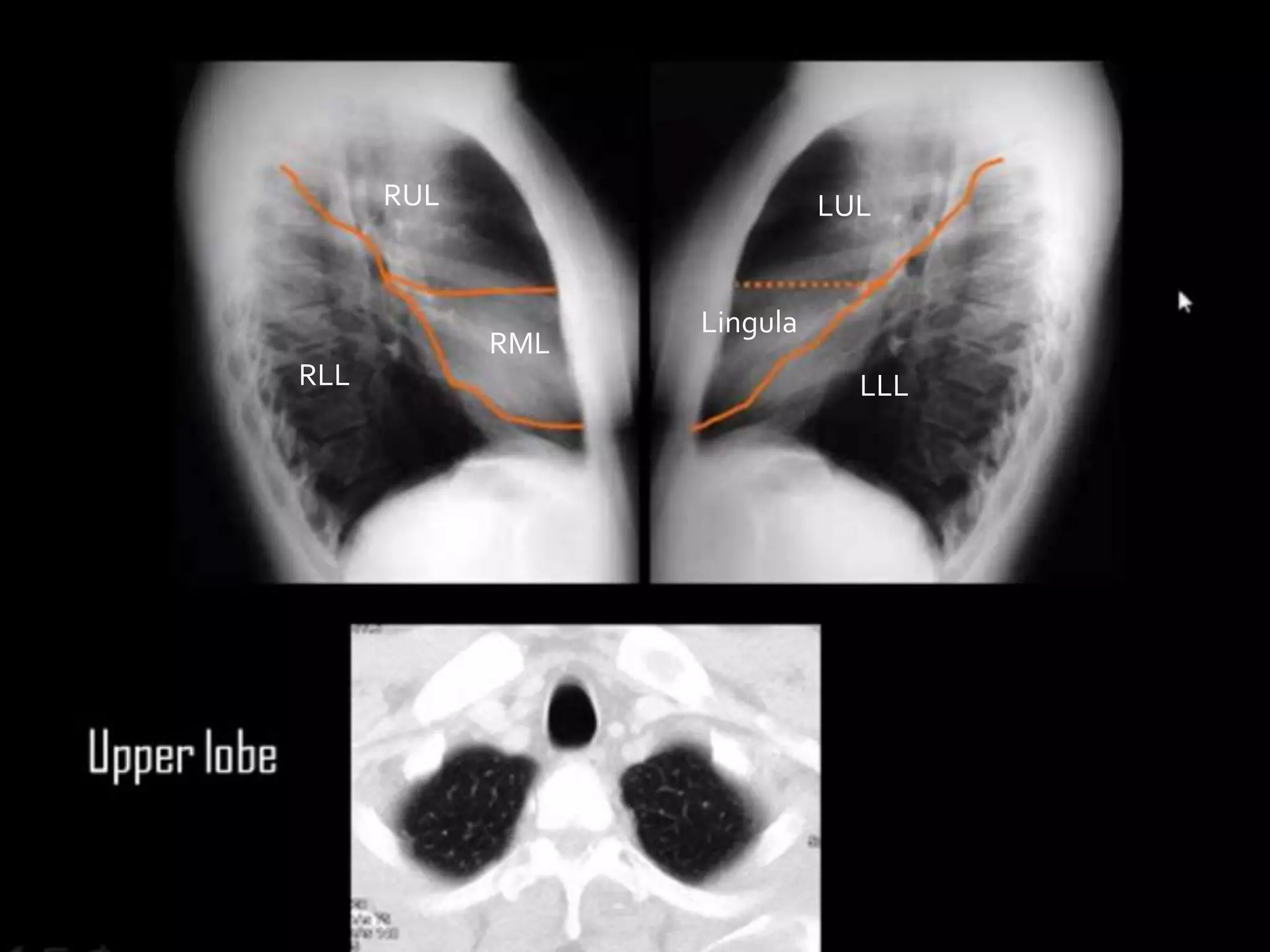

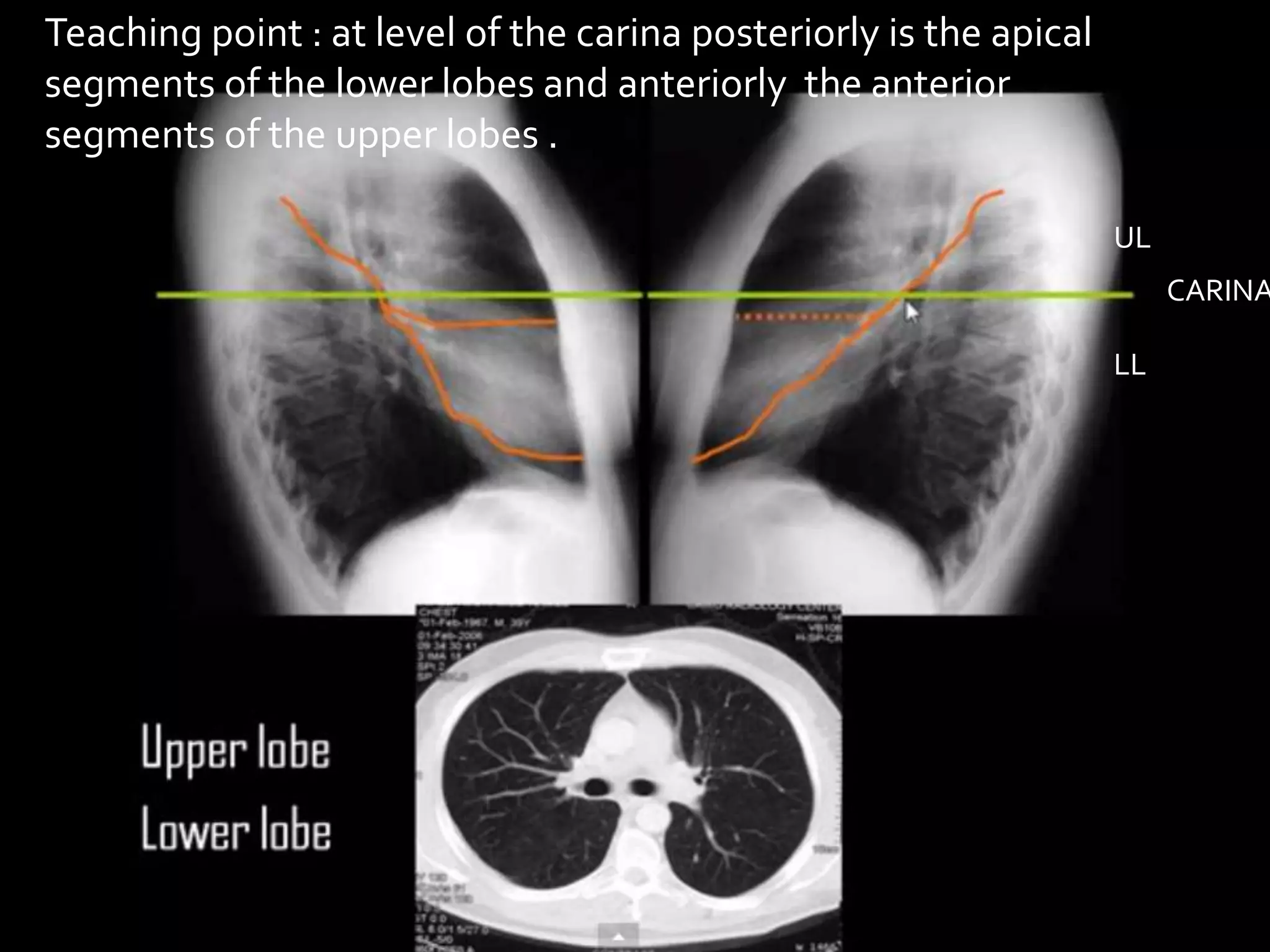

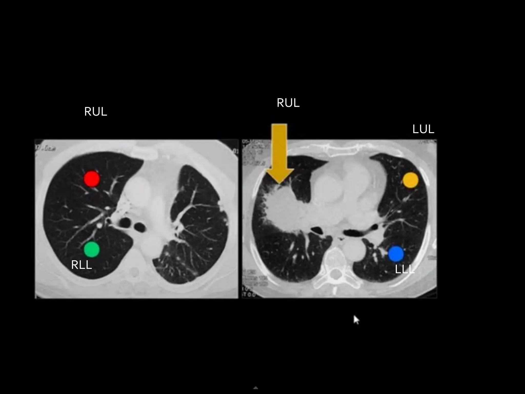

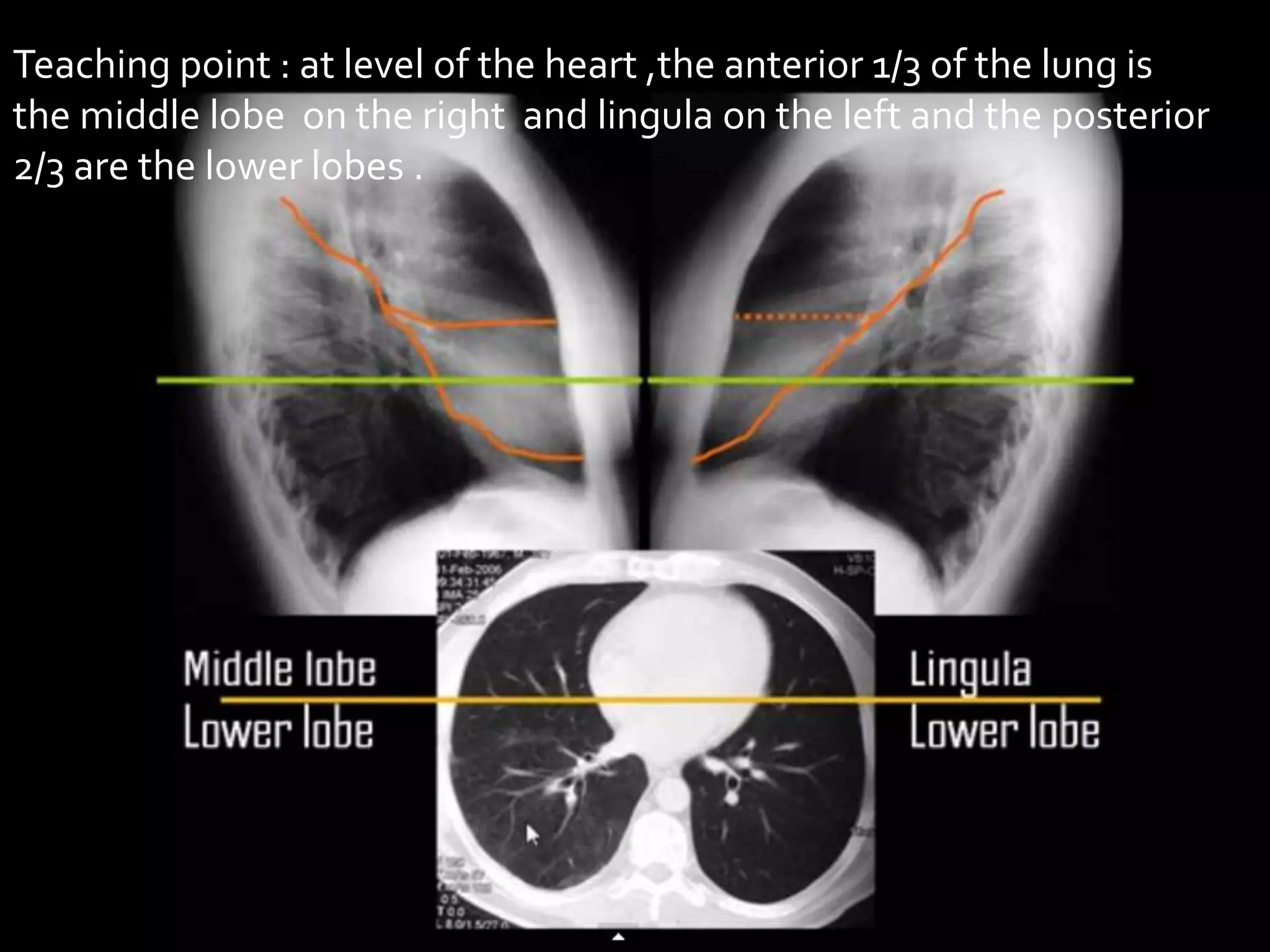
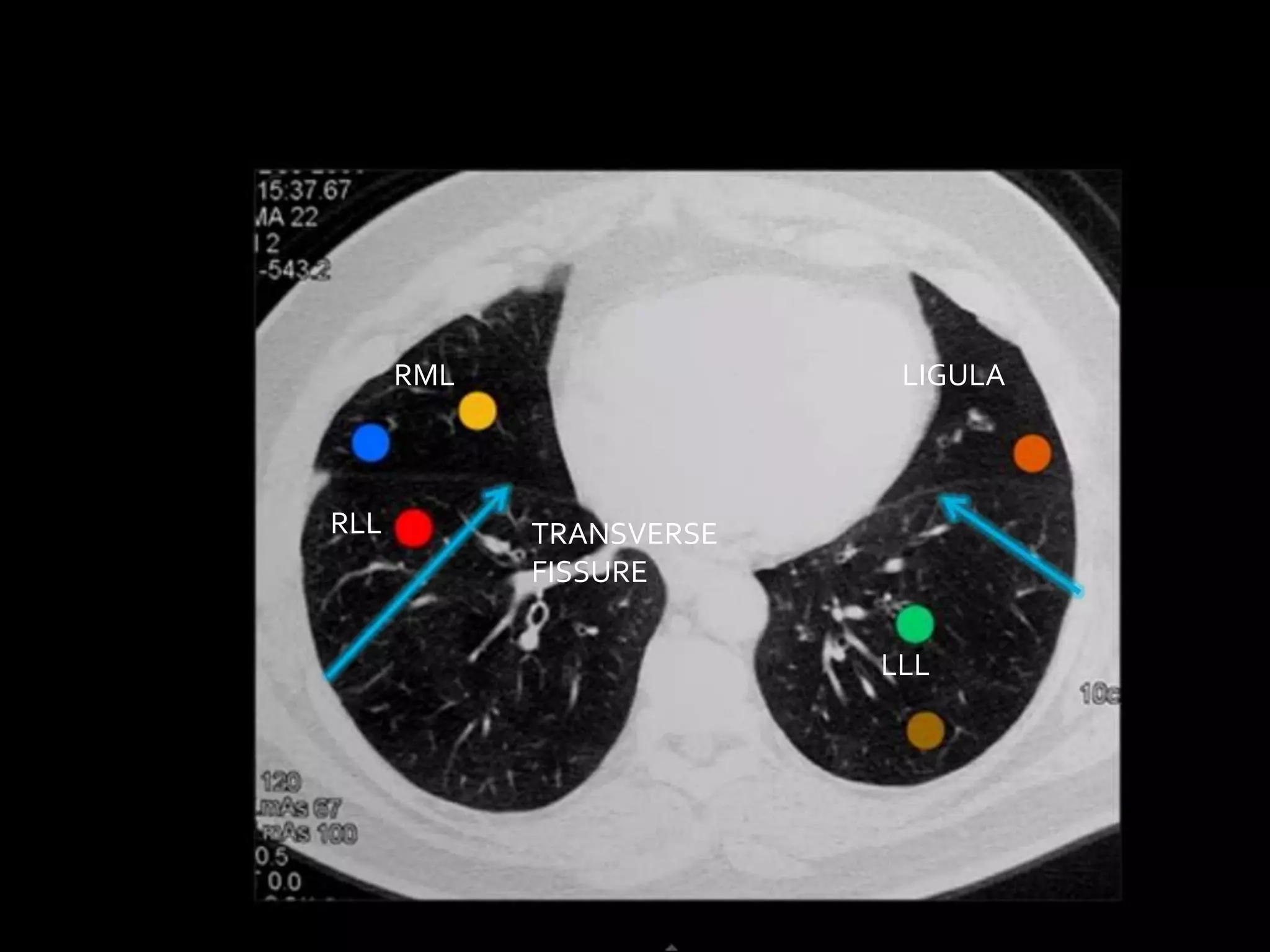


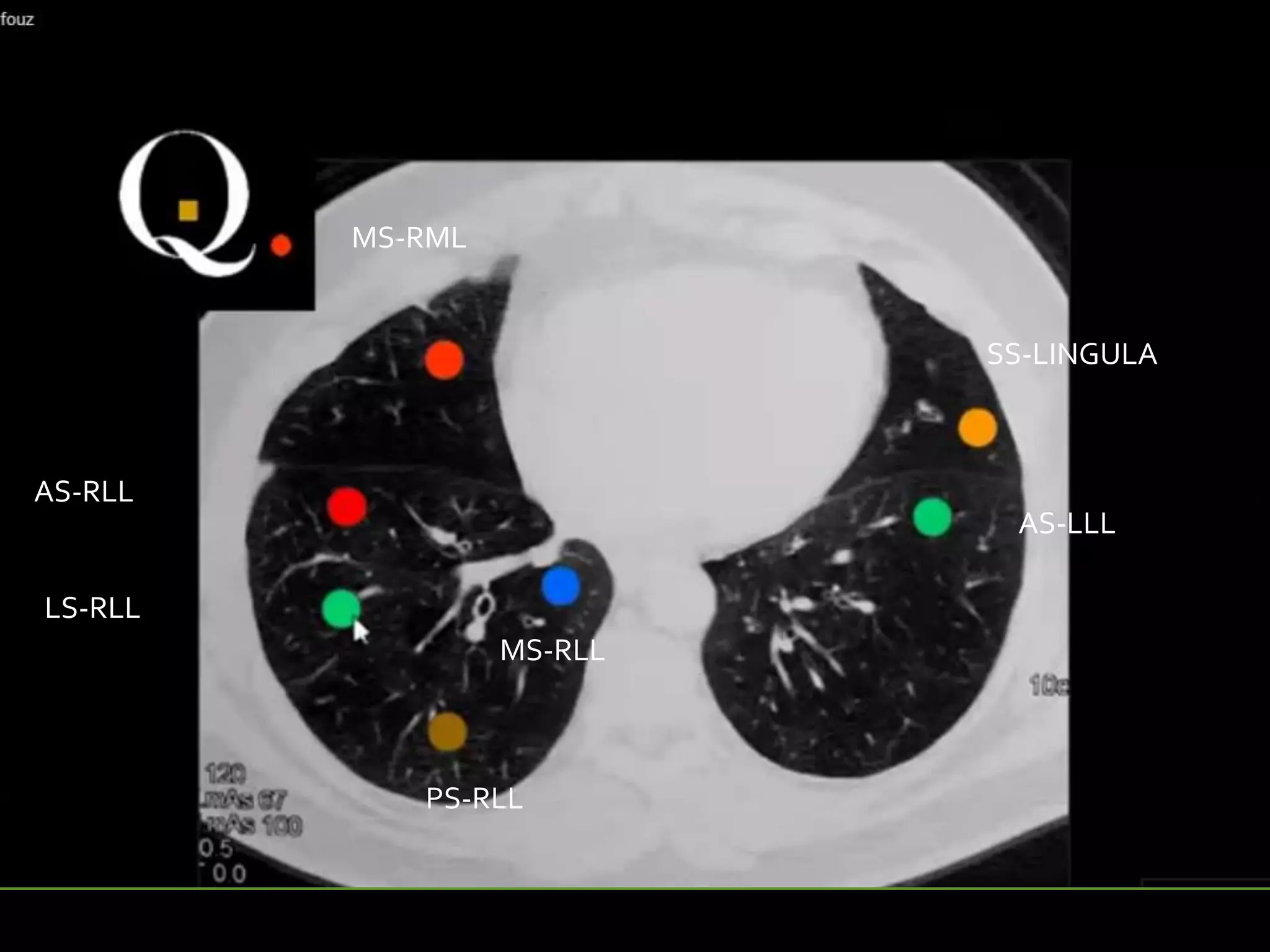
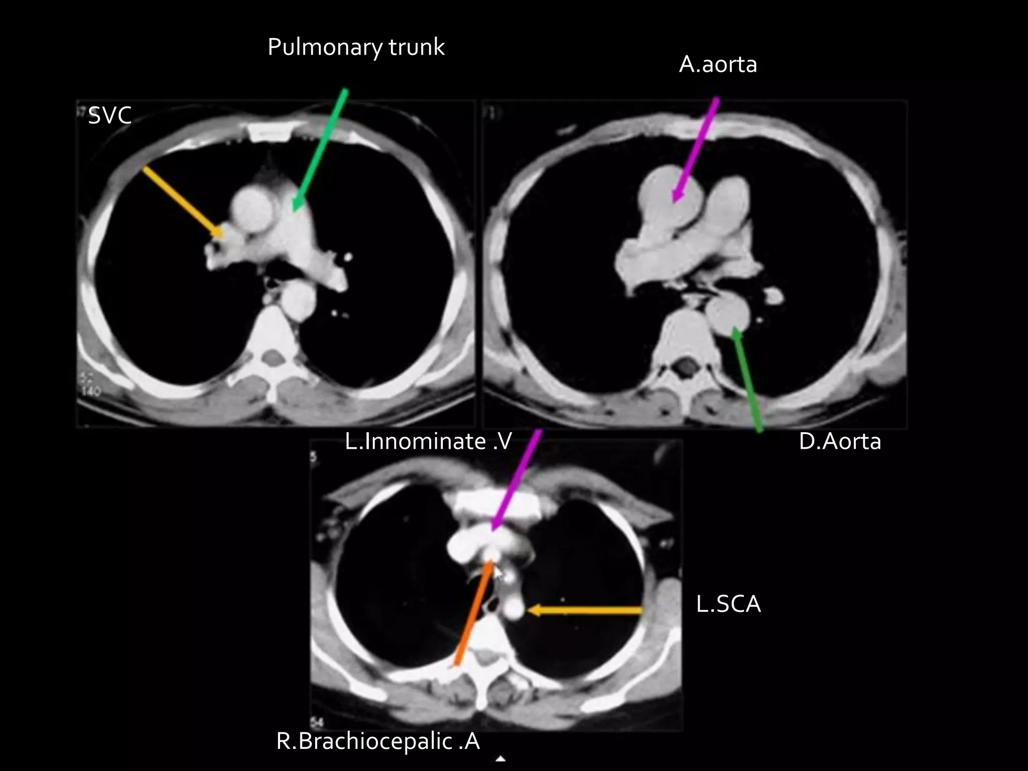
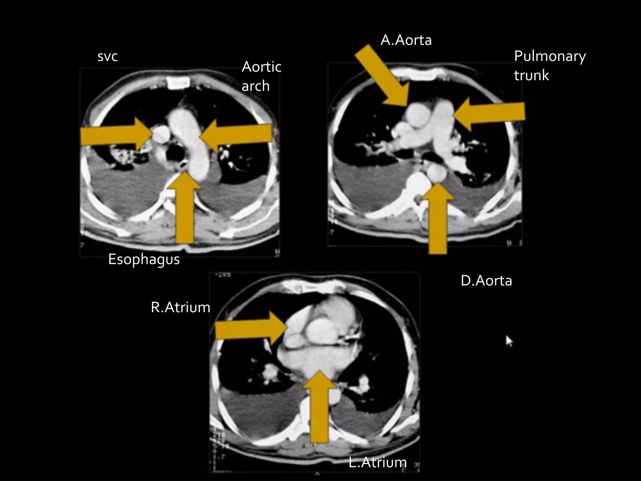

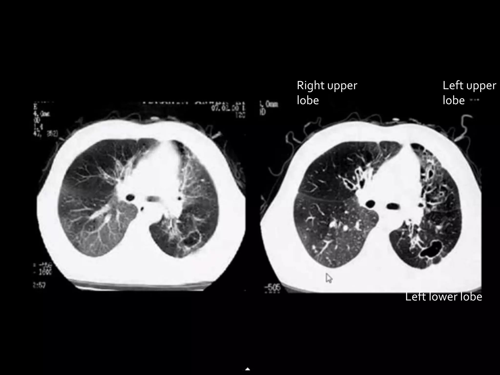
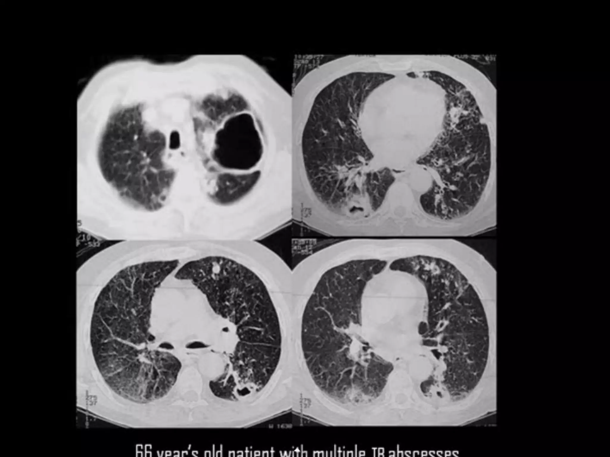
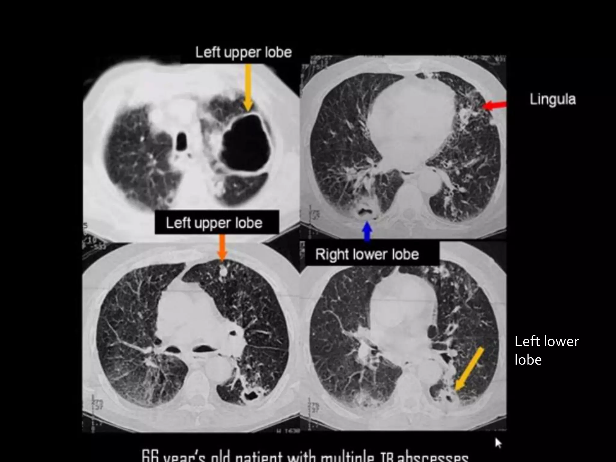


The document discusses the use of high-resolution computed tomography (HRCT) for examining the chest anatomy, particularly the pulmonary parenchyma and airways. It outlines key anatomical landmarks and their relevance in interpreting CT images, including the roles of the trachea and levels of the heart in lung segment identification. Additionally, it emphasizes certain teaching points related to lung subdivisions and their clinical implications.


