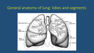
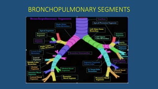
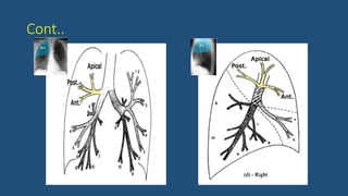
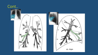
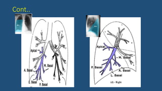
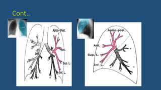
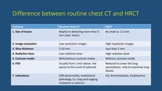
CT IN PULMONOLOGY
Dr. Ajay Kumar Yadav
PGY3, Internal Medicine
2018/12/31
LAYOUT
• Anatomy
• Bronchopulmonary segments
• Mediastinum
• Introduction to CT chest
• Utility of CT chest in diagnosis of lung pathology
General anatomy of lung: lobes and segments
BRONCHOPULMONARY SEGMENTS
Cont..
Cont..
Cont..
Cont..
Cont..
Anatomy Of Mediastinum
 3 almost equal divisions:
• Supra-aortic mediastinum: from thoracic inlet to the top of the aortic arch.
• Sub-aortic mediastinum: Aortic arch to the superior aspect of the heart.
• Para-cardiac mediastinum: Superior aspect of the heart to the diaphragm.
 In adults, each of these division is made up of about 15 contiguous 5 mm
slices.
Cont..
Sub-divisions of Thorax
Sub-divisions of mediastinum
1- superior mediastinum
2- anterior mediastinum
3- middle mediastinum
4- posterior mediastinum
Sub-divisions of mediastinum
1- anterior mediastinum
2- middle mediastinum
3- posterior mediastinum
D/D of mediastinal mass
CT
CHEST
Introduction to CT chest
• The most sensitive & advanced way of evaluating the chest & lungs.
• Images are far more detailed than a conventional chest radiograph.
• Thorax contains tissues with HU ranging from -1000 for air through lung
parenchyma, fat & soft tissue to cortical bone with HU of over 1500.
• Useful to show different types of tissues (lungs, heart, bones, soft tissues,
muscles & blood vessels.) in single examination.
Appearance on CT scan
Indications of CT chest
 Pulmonary diseases
 Intra-thoracic assessment of tumor and follow up
 Pleural diseases
 Diaphragmatic abnormalities
 Congenital abnormalities
 Cardio-vascular abnormalities
 To plan radiation therapy & assess the response to treatment
 To plan Surgical treatment / post-operative assessment
 To guide tissue biopsy
 Traumatic/emergency conditions
Contra-indications to CT
• Pregnancy
• Allergy to contrast
• Renal dysfunction
CT preparation
• CECT: 6 hours NPO
• Blood urea & Serum creatinine
report (done within a month) is
usually required within normal
limit.
• Management of life saving drugs
& life support facilities.
• Rule out pregnancy.
CT chest: General principles
Variables Characteristics
1. Scan level Lung apices to the posterior costophrenic angles (25-30 cm)
2. Patient position Supine; prone scans sometimes used for diagnosis of lung disease or pleural
fluid collections
3. Lung volume Full inspiration, single breath hold; expiratory scans sometimes used to
diagnose air trapping
4. Gantry rotation time Rapid (to reduce scan time); approximately 0.5 s in most instances
5. Scan duration Approximately 2.5 s (single breath hold) for the thorax using MDCT and fast
scanning
6. Slice thickness and pitch If data are recorded using 0.625- mm detectors, slices can be reconstructed
at any thickness from 0.625 to 5 mm.
7. Reconstruction algorithm High-resolution algorithm used for most studies; standard or soft-tissue
algorithm usually used for vascular studies
8. Two- or three- dimensional
reconstructions
Not routine; occasionally useful for lung, airway, or vascular studies
9. Contrast agents IV contrast in CECT
Window Settings
• Lung window
• Typically have a window mean of approx. −600 to −700 HU and a window
width of 1000 to 1500 HU
• Best demonstrate lung anatomy and pathology, contrasting soft-tissue
structures with surrounding air-filled lung parenchyma.
• Mediastinal or soft-tissue window
• Typically have window mean of 20 to 40 HU and width of 450 to 500 HU
• Best demonstrate soft-tissue anatomy in the mediastinum and in other areas
of the thorax, allowing the differentiation of fat, fluid, tissue, calcium, and
contrast-opacified vessels.
Cont..
• Bone window
• Typically have a window mean of approx. 300 to 500 HU and a window width
of 2000 HU.
• Best demonstrate skeletal structures or very dense objects.
Types of CT
• Standard (Helical) CT/Contrast Enhanced CT (CECT)
• High Resolution CT (HRCT)
• Low Dose CT (LDCT)
• CT Angiogram (CTA)
Difference between routine chest CT and HRCT
Features Routine chest CT HRCT
1. Size of lesion Helpful in detecting more than 5
mm sized lesion.
As small as 1-2 mm
2. Image resolution Low resolution images High resolution images
3. Slice thickness 5-10 mm Less than 2 mm
4. Radiation dose Low radiation dose High radiation dose
5. Contrast media With/without contrast media Without contrast media
6. FOV Usually from 1 inch above the
apices to the Level of adrenals
Reduced to cover the lung
parenchyma only to maximize lung
details
7. indications CXR abnormality, mediastinal
pathology, Ca. lung and staging,
empyema vs abscess
ILD, Broncheictasis, Emphysema
HRCT
• Compared to helical CT, HRCT uses narrow beam collimation to take thin slice
images of lung parenchyma
• High resolution reconstruction algorithm
• High definition images of lung alveoli, airways, interstitium
• Expiratory images may assist in identifying air trapping
• Scans performed with patient supine and prone. Prone scans are used to detect
subtle posterior lung abnormalities and posterior pleural fluid collections.
Indications of HRCT
• ILD: disease activity, patterns (UIP VS NSIP), optimal biopsy site, prognosis, response to
treatment
• Bronchiectasis
• Emphysema
• Normal or equivocal CXR: suspected lung diseases and pattern clarification
• Normal CXR with abnormal PFT
• Evaluation of hemoptysis
Drawbacks of HRCT
• Does not Image whole lungs: unsuitable for assessment of Ca lung
and localized diseases.
• Unsuitable for soft tissue and mediastinum (thin section and high
resolution)
• Unsuitable for assessment of blood vessels
Low Dose CT (LDCT)
• Lower dose radiation will not reduce diagnostic functionality of scan
• Detail is decreased
• Uses:
• Screening of lung cancer
• F/U: Infection, post lung transplant, metastasis
CT Angiography (CTA)
• Indications
• Pulmonary embolism
• Aortic aneurysm
• Aortic dissection
• Risk
• HSR
• Contrast induced nephropathy (CIN)
Radiation Exposure
Shall we begin the CT interpretation of lung
disease ?
SECONDARY LOBULE
• Basic anatomic unit of pulmonary structure and function.
• Smallest lung unit that is surrounded by connective tissue septa.
• Interpretation of ILD is based on the type of involvement of the
secondary lobule.
• Measures about 1-2 cm and is made up of 5-15 pulmonary acini, that
contain the alveoli for gas exchange.
• The secondary lobule is supplied
by a small bronchiole (terminal
bronchiole) in the center, that is
parallelled by the centrilobular
artery.
Pulmonary veins and lymphatics
run in the periphery of the lobule
within the interlobular septa.
• Centrilobular area
• Central part of the secundary lobule.
• It is usually the site of diseases, that enter the lung through the airways ( i.e.
hypersensitivity pneumonitis, respiratory bronchiolitis, centrilobular
emphysema).
• Perilymphatic area
• Peripheral part of the secundary lobule.
• It is usually the site of diseases, that are located in the lymphatics of in the
interlobular septa ( i.e. sarcoid, lymphangitic carcinomatosis, pulmonary
edema).
Basic interpretation
A structured approach to interpretation of HRCT involves the following questions:
• What is the dominant HR-pattern ?
• reticular
• nodular
• high attenuation (ground-glass, consolidation)
• low attenuation (emphysema, cystic)
• Where is it located within the secondary lobule (centrilobular, perilymphatic or random)?
• Is there an upper versus lower zone or a central versus peripheral predominance ?
• Are there additional findings (pleural fluid, lymphadenopathy, traction bronchiectasis) ?
• Reticular pattern
• There are too many lines, either as a result of thickening of the interlobular
septa or as a result of fibrosis as in honeycombing.
Thickening of interlobular septa
Nodular Pattern
• Peri-lymphatic distribution
• Nodules are seen in relation to pleural surfaces, interlobular septa and the peri-broncho-
vascular interstitium.
• Nodules are almost always visible in a sub-pleural location, particularly in relation to the
fissures
Satellite nodules in Sarcoidosis
Aka “Galaxy sign”
• Centrilobular distribution
• Nodules are limited to the centrilobular region.
• Spare the pleural surfaces.
• The most peripheral nodules are centered 5-10 mm from fissures or the pleural surface
• Random distribution
• Nodules are randomly distributed relative to structures of the lung and secondary lobule.
• Nodules can usually be seen to involve the pleural surfaces and fissures, but lack the sub-
pleural predominance often seen in pts with a perilymphatic distribution.
Pulmonary metastasis with Feeding vessel sign
Approach to nodules
Nodule calcification
• Indicates that that the nodule is benign.
• Soft-tissue window settings are best for proving the presence of
calcium.
• Types of calcification
• Diffuse calcification: typical of granuloma
• Dense central (i.e., bull’s-eye) calcification: most typical of histoplasmosis
• Central and popcorn calcification: typical of hamartoma
• Concentric rings of calcification (target calcification): typical of histoplasmosis.
Diffuse calcification
Bull’s eye calcification Concentric calcification
Tree-in-bud appearance
• Tree-in-bud describes the appearance of an irregular and often nodular branching
structure, most easily identified in the lung periphery.
• It represents dilated and impacted (mucus or pus-filled) centrilobular
bronchioles.
Tree-in-bud appearance
Causes of tree-in-bud appearance
High attenuation pattern
• Ground-glass-opacity (GGO)
• Hazy increase in lung opacity without obscuration of underlying vessels
• Consolidation
• Increase in lung opacity obscures the vessels.
• In both, increase in lung density is the result of replacement of air in the alveoli by fluid,
cells or fibrosis.
• In GGO the density of the intra-bronchial air appears darker as the air in the surrounding
alveoli : 'dark bronchus' sign
• In consolidation, there is exclusively air left intra-bronchial :'air bronchogram'.
• Ground-glass opacity (GGO) represents:
• Filling of the alveolar spaces with pus, edema, hemorrhage, inflammation or tumor cells.
• Thickening of the interstitium or alveolar walls below the spatial resolution of the HRCT as
seen in fibrosis.
• So GGO may either be the result of air space disease (filling of the alveoli) or
interstitial lung disease (i.e. fibrosis).
• The location of the abnormalities in ground glass pattern can be helpfull:
• Upper zone predominance: Respiratory bronchiolitis, PCP.
• Lower zone predominance: UIP, NSIP, DIP.
• Centrilobular distribution: Hypersensitivity pneumonitis, Respiratory bronchiolitis
Causes of GGO
Treatable or not treatable?
• 60-80% of patients with GGO on HRCT have an active and potentially treatable lung disease.
• Remaining 20-40% of the cases is not treatable and the GGO is the result of fibrosis.
• Usually associated HRCT findings of fibrosis, such as traction bronchiectasis and
honeycombing.
Mosaic attenuation
• Used to describe density differences between affected and non-affected lung
areas.
• There are patchy areas of black and white lung.
• The role of the radiologist is to determine which part is abnormal: the black or
the white lung.
• When ground glass opacity presents as mosaic attenuation consider:
• Infiltrative process adjacent to normal lung
• Normal lung appearing relatively dense adjacent to lung with air-trapping
• Hyperperfused lung adjacent to oligemic lung due to chronic thromboembolic disease
Cont..
• There are two diagnostic hints for further differentiation:
• Look at expiratory scans for air trapping
• Look at the vessels
• If the vesses are difficult to see in the 'black' lung as compared to the
'white' lung, than it is likely that the 'black' lung is abnormal.
• Then there are two possibilities: obstructive bronchiolitis or chronic PE.
• Sometimes these can be differentiated with an expiratory scan.
• If the vessels are the same in the 'black' lung and 'white' lung, then we are
looking at a patient with infiltrative lung disease e.g. pulmonary h’ge.
Crazy pavement phenomenon
• Crazy Paving is a combination of ground glass opacity with superimposed septal
thickening.
• It was first thought to be specific for alveolar proteinosis, but later was also seen
in other diseases.
• Crazy Paving can also be seen in:
• Sarcoidosis
• NSIP
• Organizing pneumonia (COP/BOOP)
• Infection (PCP, viral, Mycoplasma, bacterial)
• Neoplasm (Bronchoalveolar carcinoma (BAC)
• Pulmonary hemorrhage
• Edema (heart failure, ARDS, AIP)
Consolidation
• Consolidation is synonymous with airspace disease.
• Acute consolidation is seen in:
• Pneumonias (bacterial, mycoplasma, PCP)
• Pulmonary edema due to heart failure or ARDS
• Hemorrhage
• Acute eosinophilic pneumonia
• Chronic consolidation is seen in:
• Organizing Pneumonia
• Chronic eosinophilic pneumonia
• Fibrosis in UIP and NSIP
• Bronchoalveolar carcinoma or lymphoma
Causes of consolidation
• Organizing pneumonia (OP)
• Represents an inflammatory process in which the healing process is
characterized by organization and cicatrization of the exudate rather than
by resolution and resorption.
• Aka 'unresolved pneumonia'.
• If no cause can be identified it is called cryptogenic organizing pneumonia
(COP).
• It was described in earlier years as Bronchiolitis-obliterans-organizing
pneumonia (BOOP).
Low attenuation pattern
• Decreased lung attenuation or air-filled lesions.
• These include:
• Emphysema
• Lung cysts (LAM, LIP, Langerhans cell histiocytosis)
• Bronchiectasis
• Honeycombing
Emphysema
• Emphysema typically presents as areas of low attenuation without
visible walls.
• Centrilobular emphysema
• Most common type
• Irreversible destruction of alveolar walls in the centrilobular portion of the
lobule
• Upper lobe predominance
• Strongly associated with smoking.
• Panlobular emphysema
• Affects the whole secondary lobule
• Lower lobe predominance
• In alpha-1-antitrypsin deficiency + smokers with advanced emphysema
• Paraseptal emphysema
• Adjacent to the pleura and interlobar fissures
• In older patients  a/w centrilobular emphysema
• In young adults  spontaneous pneumothorax
Cystic lung disease
• CYSTS
• Radiolucent areas with a wall
thickness of less than 4mm
• Causes
• LCH
• LAM
• Pneumatocele
• Lymphocytic interstitial pneumonia
• CAVITY
• Radiolucent areas with a wall
thickness of more than 4mm
• Causes
• Infection (TB, Staph, fungal, hydatid)
• Septic emboli
• Squamous and large cell carcinoma
• Wegener's granulomatosis (GPA)
Bronchiectasis
• Bronchiectasis is defined as localized bronchial dilatation.
• The diagnosis of bronchiectasis is usually based on a combination of
the following findings:
• Bronchial dilatation (signet-ring sign, tram track sign)
• Bronchial wall thickening
• Lack of normal tapering with visibility of airways in the peripheral lung
• Mucus retention in the broncial lumen
• Associated atelectasis and sometimes air trapping
Types of bronchiectasis
Bronchiectasis changes in
specimen of lung
Traction bronchiectasis
Cylinderical and cystic
bronchiectasis
• Predominant upper lobe involvement
• Cystic fibrosis
• Tb sequale
• Central bronchiectasis
• ABPA
• Predominant middle lobe involvement
• MAC infection
• Predominant lower lobe involvement
• Immune deficiency
• Ciliary dysmotility (e.g. kartagenar syndrome)
Honeycombing
• Presence of small cystic spaces with irregularly thickened walls
composed of fibrous tissue.
• Honeycomb cysts often predominate in the peripheral and sub-
pleural lung regions regardless of their cause.
• Irreversible – no t/t required
Atelactasis
• Atelectasis most commonly occurs because of
 Bronchial obstruction (obstructive atelectasis),
 Pleural effusion or other pleural processes that allow the lung to
collapse (passive or relaxation atelectasis), or
 Lung fibrosis (cicatrization atelectasis)
Rounded atelactasis
• Represents focal, collapsed, and often folded lung.
• Appear as mass or mass-like consolidation and are usually several cms in
diameter.
• 4 findings must be present to make absolute diagnosis
• Ipsilateral pleural thickening or effusion
• Significant contact between the lung lesion and the abnormal pleural surface
• Comet tail sign: Bending or bowing of adjacent bronchi and arteries toward the
edge of the area of rounded atelectasis.
• Volume loss in the lobe in which there is opacity.
Comet tail sign
Halo sign and angioinvasive aspergillosis
• The term halo sign is used to
describe a nodule with a soft-
tissue attenuation center
surrounded by a less dense halo
of GGO.
• In patients with angioinvasive
aspergillosis, the halo sign reflects
the presence of a septic infarction
(the dense center) surrounded by
hemorrhage (the halo).
Halo sign
Air crescent sign and mycetoma
• Presence of a lung mass capped by a crescent of air.
• Usually indicates the presence of a mass within a cavity.
• Most typical of mycetoma.
• On CT, a round or oval mass (the fungus ball) can be seen in a
dependent location within the cavity and is typically mobile. The mass
is capped by a crescent of air.
Lung abscess and cavities
• The hallmark of a lung abscess is necrosis or cavitation within an area
of pneumonia or dense consolidation.
• Commonly visible on CECT as one or more areas of low attenuation
within opacified lung.
• Cavitation is said to be present if air is visible within the lesion
Lung abscess with air-fluid level
ILD
• UIP pattern on HRCT is characterized by:
 Subpleural and basal predominance
 Reticulation (with traction bronchiectasis)
 Honeycombing
 Absence of inconsistent features, including the following:
 Upper, mid-lung, or peribronchial predominance
 Extensive GGO ( > reticulation in extent)
 Profuse micronodules (bilateral, upper lobe)
 Discrete cysts not representing honeycombing
 Mosaic perfusion or air trapping (bilateral, ≥ three lobes)
 Segmental or lobar consolidation
• NSIP pattern on HRCT
• Predominance in the peripheral, posterior, and basal lung regions, with a
concentric distribution.
• Sparing of the immediate subpleural lung (seen in 50% of cases), a finding
that is highly predictive of NSIP
• GGO in cellular NSIP
• Reticulation
• Traction bronchiectasis (usually but not always in fibrotic NSIP)
• Rare honeycombing, which, if present, is of limited extent
Hypersensitivity pneumonitis
• HRCT finding in sub-acute HP
• Patchy or geographic GGO (80%)
• Poorly defined centrilobular GGO nodules
• Upper or mid-lung predominance, with involvement of the entire cross-
section of the lung (i.e., there is no subpleural predominance)
• Mosaic perfusion caused by bronchiolar obstruction
• Air trapping (commonly present on expiratory Scans)
• A combination of patchy GGO and patchy mosaic perfusion (termed the
headcheese sign) : typical of HP
• HRCT in chronic or fibrotic stage
• Patchy or geographic reticulation and traction bronchiectasis
• Honeycombing in some cases
• Upper or mid-lung predominance, with involvement of the entire cross-
section of the lung
• Mosaic perfusion caused by bronchiolar obstruction
• Air trapping (commonly present on expiratory scans)
• Never lie to ur physician – we are looking right through u…..
• Thank you!!!

Ct in pulmonology

  • 1.
    CT IN PULMONOLOGY Dr.Ajay Kumar Yadav PGY3, Internal Medicine 2018/12/31
  • 2.
    LAYOUT • Anatomy • Bronchopulmonarysegments • Mediastinum • Introduction to CT chest • Utility of CT chest in diagnosis of lung pathology
  • 3.
    General anatomy oflung: lobes and segments
  • 4.
  • 5.
  • 6.
  • 7.
  • 8.
  • 9.
  • 10.
    Anatomy Of Mediastinum 3 almost equal divisions: • Supra-aortic mediastinum: from thoracic inlet to the top of the aortic arch. • Sub-aortic mediastinum: Aortic arch to the superior aspect of the heart. • Para-cardiac mediastinum: Superior aspect of the heart to the diaphragm.  In adults, each of these division is made up of about 15 contiguous 5 mm slices.
  • 11.
    Cont.. Sub-divisions of Thorax Sub-divisionsof mediastinum 1- superior mediastinum 2- anterior mediastinum 3- middle mediastinum 4- posterior mediastinum Sub-divisions of mediastinum 1- anterior mediastinum 2- middle mediastinum 3- posterior mediastinum
  • 12.
  • 13.
  • 14.
    Introduction to CTchest • The most sensitive & advanced way of evaluating the chest & lungs. • Images are far more detailed than a conventional chest radiograph. • Thorax contains tissues with HU ranging from -1000 for air through lung parenchyma, fat & soft tissue to cortical bone with HU of over 1500. • Useful to show different types of tissues (lungs, heart, bones, soft tissues, muscles & blood vessels.) in single examination.
  • 15.
  • 16.
    Indications of CTchest  Pulmonary diseases  Intra-thoracic assessment of tumor and follow up  Pleural diseases  Diaphragmatic abnormalities  Congenital abnormalities  Cardio-vascular abnormalities  To plan radiation therapy & assess the response to treatment  To plan Surgical treatment / post-operative assessment  To guide tissue biopsy  Traumatic/emergency conditions
  • 17.
    Contra-indications to CT •Pregnancy • Allergy to contrast • Renal dysfunction
  • 18.
    CT preparation • CECT:6 hours NPO • Blood urea & Serum creatinine report (done within a month) is usually required within normal limit. • Management of life saving drugs & life support facilities. • Rule out pregnancy.
  • 19.
    CT chest: Generalprinciples Variables Characteristics 1. Scan level Lung apices to the posterior costophrenic angles (25-30 cm) 2. Patient position Supine; prone scans sometimes used for diagnosis of lung disease or pleural fluid collections 3. Lung volume Full inspiration, single breath hold; expiratory scans sometimes used to diagnose air trapping 4. Gantry rotation time Rapid (to reduce scan time); approximately 0.5 s in most instances 5. Scan duration Approximately 2.5 s (single breath hold) for the thorax using MDCT and fast scanning 6. Slice thickness and pitch If data are recorded using 0.625- mm detectors, slices can be reconstructed at any thickness from 0.625 to 5 mm. 7. Reconstruction algorithm High-resolution algorithm used for most studies; standard or soft-tissue algorithm usually used for vascular studies 8. Two- or three- dimensional reconstructions Not routine; occasionally useful for lung, airway, or vascular studies 9. Contrast agents IV contrast in CECT
  • 20.
    Window Settings • Lungwindow • Typically have a window mean of approx. −600 to −700 HU and a window width of 1000 to 1500 HU • Best demonstrate lung anatomy and pathology, contrasting soft-tissue structures with surrounding air-filled lung parenchyma. • Mediastinal or soft-tissue window • Typically have window mean of 20 to 40 HU and width of 450 to 500 HU • Best demonstrate soft-tissue anatomy in the mediastinum and in other areas of the thorax, allowing the differentiation of fat, fluid, tissue, calcium, and contrast-opacified vessels.
  • 21.
    Cont.. • Bone window •Typically have a window mean of approx. 300 to 500 HU and a window width of 2000 HU. • Best demonstrate skeletal structures or very dense objects.
  • 22.
    Types of CT •Standard (Helical) CT/Contrast Enhanced CT (CECT) • High Resolution CT (HRCT) • Low Dose CT (LDCT) • CT Angiogram (CTA)
  • 23.
    Difference between routinechest CT and HRCT Features Routine chest CT HRCT 1. Size of lesion Helpful in detecting more than 5 mm sized lesion. As small as 1-2 mm 2. Image resolution Low resolution images High resolution images 3. Slice thickness 5-10 mm Less than 2 mm 4. Radiation dose Low radiation dose High radiation dose 5. Contrast media With/without contrast media Without contrast media 6. FOV Usually from 1 inch above the apices to the Level of adrenals Reduced to cover the lung parenchyma only to maximize lung details 7. indications CXR abnormality, mediastinal pathology, Ca. lung and staging, empyema vs abscess ILD, Broncheictasis, Emphysema
  • 24.
    HRCT • Compared tohelical CT, HRCT uses narrow beam collimation to take thin slice images of lung parenchyma • High resolution reconstruction algorithm • High definition images of lung alveoli, airways, interstitium • Expiratory images may assist in identifying air trapping • Scans performed with patient supine and prone. Prone scans are used to detect subtle posterior lung abnormalities and posterior pleural fluid collections.
  • 25.
    Indications of HRCT •ILD: disease activity, patterns (UIP VS NSIP), optimal biopsy site, prognosis, response to treatment • Bronchiectasis • Emphysema • Normal or equivocal CXR: suspected lung diseases and pattern clarification • Normal CXR with abnormal PFT • Evaluation of hemoptysis
  • 26.
    Drawbacks of HRCT •Does not Image whole lungs: unsuitable for assessment of Ca lung and localized diseases. • Unsuitable for soft tissue and mediastinum (thin section and high resolution) • Unsuitable for assessment of blood vessels
  • 27.
    Low Dose CT(LDCT) • Lower dose radiation will not reduce diagnostic functionality of scan • Detail is decreased • Uses: • Screening of lung cancer • F/U: Infection, post lung transplant, metastasis
  • 28.
    CT Angiography (CTA) •Indications • Pulmonary embolism • Aortic aneurysm • Aortic dissection • Risk • HSR • Contrast induced nephropathy (CIN)
  • 29.
  • 30.
    Shall we beginthe CT interpretation of lung disease ?
  • 31.
    SECONDARY LOBULE • Basicanatomic unit of pulmonary structure and function. • Smallest lung unit that is surrounded by connective tissue septa. • Interpretation of ILD is based on the type of involvement of the secondary lobule. • Measures about 1-2 cm and is made up of 5-15 pulmonary acini, that contain the alveoli for gas exchange.
  • 32.
    • The secondarylobule is supplied by a small bronchiole (terminal bronchiole) in the center, that is parallelled by the centrilobular artery. Pulmonary veins and lymphatics run in the periphery of the lobule within the interlobular septa.
  • 33.
    • Centrilobular area •Central part of the secundary lobule. • It is usually the site of diseases, that enter the lung through the airways ( i.e. hypersensitivity pneumonitis, respiratory bronchiolitis, centrilobular emphysema). • Perilymphatic area • Peripheral part of the secundary lobule. • It is usually the site of diseases, that are located in the lymphatics of in the interlobular septa ( i.e. sarcoid, lymphangitic carcinomatosis, pulmonary edema).
  • 34.
    Basic interpretation A structuredapproach to interpretation of HRCT involves the following questions: • What is the dominant HR-pattern ? • reticular • nodular • high attenuation (ground-glass, consolidation) • low attenuation (emphysema, cystic) • Where is it located within the secondary lobule (centrilobular, perilymphatic or random)? • Is there an upper versus lower zone or a central versus peripheral predominance ? • Are there additional findings (pleural fluid, lymphadenopathy, traction bronchiectasis) ?
  • 35.
    • Reticular pattern •There are too many lines, either as a result of thickening of the interlobular septa or as a result of fibrosis as in honeycombing.
  • 37.
  • 38.
    Nodular Pattern • Peri-lymphaticdistribution • Nodules are seen in relation to pleural surfaces, interlobular septa and the peri-broncho- vascular interstitium. • Nodules are almost always visible in a sub-pleural location, particularly in relation to the fissures
  • 40.
    Satellite nodules inSarcoidosis Aka “Galaxy sign”
  • 41.
    • Centrilobular distribution •Nodules are limited to the centrilobular region. • Spare the pleural surfaces. • The most peripheral nodules are centered 5-10 mm from fissures or the pleural surface
  • 43.
    • Random distribution •Nodules are randomly distributed relative to structures of the lung and secondary lobule. • Nodules can usually be seen to involve the pleural surfaces and fissures, but lack the sub- pleural predominance often seen in pts with a perilymphatic distribution.
  • 45.
    Pulmonary metastasis withFeeding vessel sign
  • 46.
  • 47.
    Nodule calcification • Indicatesthat that the nodule is benign. • Soft-tissue window settings are best for proving the presence of calcium. • Types of calcification • Diffuse calcification: typical of granuloma • Dense central (i.e., bull’s-eye) calcification: most typical of histoplasmosis • Central and popcorn calcification: typical of hamartoma • Concentric rings of calcification (target calcification): typical of histoplasmosis.
  • 48.
  • 49.
    Bull’s eye calcificationConcentric calcification
  • 50.
    Tree-in-bud appearance • Tree-in-buddescribes the appearance of an irregular and often nodular branching structure, most easily identified in the lung periphery. • It represents dilated and impacted (mucus or pus-filled) centrilobular bronchioles.
  • 51.
  • 52.
  • 53.
    High attenuation pattern •Ground-glass-opacity (GGO) • Hazy increase in lung opacity without obscuration of underlying vessels • Consolidation • Increase in lung opacity obscures the vessels. • In both, increase in lung density is the result of replacement of air in the alveoli by fluid, cells or fibrosis. • In GGO the density of the intra-bronchial air appears darker as the air in the surrounding alveoli : 'dark bronchus' sign • In consolidation, there is exclusively air left intra-bronchial :'air bronchogram'.
  • 55.
    • Ground-glass opacity(GGO) represents: • Filling of the alveolar spaces with pus, edema, hemorrhage, inflammation or tumor cells. • Thickening of the interstitium or alveolar walls below the spatial resolution of the HRCT as seen in fibrosis. • So GGO may either be the result of air space disease (filling of the alveoli) or interstitial lung disease (i.e. fibrosis). • The location of the abnormalities in ground glass pattern can be helpfull: • Upper zone predominance: Respiratory bronchiolitis, PCP. • Lower zone predominance: UIP, NSIP, DIP. • Centrilobular distribution: Hypersensitivity pneumonitis, Respiratory bronchiolitis
  • 56.
  • 57.
    Treatable or nottreatable? • 60-80% of patients with GGO on HRCT have an active and potentially treatable lung disease. • Remaining 20-40% of the cases is not treatable and the GGO is the result of fibrosis. • Usually associated HRCT findings of fibrosis, such as traction bronchiectasis and honeycombing.
  • 58.
    Mosaic attenuation • Usedto describe density differences between affected and non-affected lung areas. • There are patchy areas of black and white lung. • The role of the radiologist is to determine which part is abnormal: the black or the white lung. • When ground glass opacity presents as mosaic attenuation consider: • Infiltrative process adjacent to normal lung • Normal lung appearing relatively dense adjacent to lung with air-trapping • Hyperperfused lung adjacent to oligemic lung due to chronic thromboembolic disease
  • 59.
    Cont.. • There aretwo diagnostic hints for further differentiation: • Look at expiratory scans for air trapping • Look at the vessels • If the vesses are difficult to see in the 'black' lung as compared to the 'white' lung, than it is likely that the 'black' lung is abnormal. • Then there are two possibilities: obstructive bronchiolitis or chronic PE. • Sometimes these can be differentiated with an expiratory scan. • If the vessels are the same in the 'black' lung and 'white' lung, then we are looking at a patient with infiltrative lung disease e.g. pulmonary h’ge.
  • 62.
    Crazy pavement phenomenon •Crazy Paving is a combination of ground glass opacity with superimposed septal thickening. • It was first thought to be specific for alveolar proteinosis, but later was also seen in other diseases. • Crazy Paving can also be seen in: • Sarcoidosis • NSIP • Organizing pneumonia (COP/BOOP) • Infection (PCP, viral, Mycoplasma, bacterial) • Neoplasm (Bronchoalveolar carcinoma (BAC) • Pulmonary hemorrhage • Edema (heart failure, ARDS, AIP)
  • 64.
    Consolidation • Consolidation issynonymous with airspace disease. • Acute consolidation is seen in: • Pneumonias (bacterial, mycoplasma, PCP) • Pulmonary edema due to heart failure or ARDS • Hemorrhage • Acute eosinophilic pneumonia • Chronic consolidation is seen in: • Organizing Pneumonia • Chronic eosinophilic pneumonia • Fibrosis in UIP and NSIP • Bronchoalveolar carcinoma or lymphoma
  • 65.
  • 67.
    • Organizing pneumonia(OP) • Represents an inflammatory process in which the healing process is characterized by organization and cicatrization of the exudate rather than by resolution and resorption. • Aka 'unresolved pneumonia'. • If no cause can be identified it is called cryptogenic organizing pneumonia (COP). • It was described in earlier years as Bronchiolitis-obliterans-organizing pneumonia (BOOP).
  • 68.
    Low attenuation pattern •Decreased lung attenuation or air-filled lesions. • These include: • Emphysema • Lung cysts (LAM, LIP, Langerhans cell histiocytosis) • Bronchiectasis • Honeycombing
  • 70.
    Emphysema • Emphysema typicallypresents as areas of low attenuation without visible walls. • Centrilobular emphysema • Most common type • Irreversible destruction of alveolar walls in the centrilobular portion of the lobule • Upper lobe predominance • Strongly associated with smoking.
  • 72.
    • Panlobular emphysema •Affects the whole secondary lobule • Lower lobe predominance • In alpha-1-antitrypsin deficiency + smokers with advanced emphysema • Paraseptal emphysema • Adjacent to the pleura and interlobar fissures • In older patients  a/w centrilobular emphysema • In young adults  spontaneous pneumothorax
  • 74.
    Cystic lung disease •CYSTS • Radiolucent areas with a wall thickness of less than 4mm • Causes • LCH • LAM • Pneumatocele • Lymphocytic interstitial pneumonia • CAVITY • Radiolucent areas with a wall thickness of more than 4mm • Causes • Infection (TB, Staph, fungal, hydatid) • Septic emboli • Squamous and large cell carcinoma • Wegener's granulomatosis (GPA)
  • 76.
    Bronchiectasis • Bronchiectasis isdefined as localized bronchial dilatation. • The diagnosis of bronchiectasis is usually based on a combination of the following findings: • Bronchial dilatation (signet-ring sign, tram track sign) • Bronchial wall thickening • Lack of normal tapering with visibility of airways in the peripheral lung • Mucus retention in the broncial lumen • Associated atelectasis and sometimes air trapping
  • 77.
    Types of bronchiectasis Bronchiectasischanges in specimen of lung
  • 78.
  • 80.
    • Predominant upperlobe involvement • Cystic fibrosis • Tb sequale • Central bronchiectasis • ABPA • Predominant middle lobe involvement • MAC infection • Predominant lower lobe involvement • Immune deficiency • Ciliary dysmotility (e.g. kartagenar syndrome)
  • 81.
    Honeycombing • Presence ofsmall cystic spaces with irregularly thickened walls composed of fibrous tissue. • Honeycomb cysts often predominate in the peripheral and sub- pleural lung regions regardless of their cause. • Irreversible – no t/t required
  • 84.
    Atelactasis • Atelectasis mostcommonly occurs because of  Bronchial obstruction (obstructive atelectasis),  Pleural effusion or other pleural processes that allow the lung to collapse (passive or relaxation atelectasis), or  Lung fibrosis (cicatrization atelectasis)
  • 86.
    Rounded atelactasis • Representsfocal, collapsed, and often folded lung. • Appear as mass or mass-like consolidation and are usually several cms in diameter. • 4 findings must be present to make absolute diagnosis • Ipsilateral pleural thickening or effusion • Significant contact between the lung lesion and the abnormal pleural surface • Comet tail sign: Bending or bowing of adjacent bronchi and arteries toward the edge of the area of rounded atelectasis. • Volume loss in the lobe in which there is opacity.
  • 87.
  • 88.
    Halo sign andangioinvasive aspergillosis • The term halo sign is used to describe a nodule with a soft- tissue attenuation center surrounded by a less dense halo of GGO. • In patients with angioinvasive aspergillosis, the halo sign reflects the presence of a septic infarction (the dense center) surrounded by hemorrhage (the halo). Halo sign
  • 89.
    Air crescent signand mycetoma • Presence of a lung mass capped by a crescent of air. • Usually indicates the presence of a mass within a cavity. • Most typical of mycetoma. • On CT, a round or oval mass (the fungus ball) can be seen in a dependent location within the cavity and is typically mobile. The mass is capped by a crescent of air.
  • 91.
    Lung abscess andcavities • The hallmark of a lung abscess is necrosis or cavitation within an area of pneumonia or dense consolidation. • Commonly visible on CECT as one or more areas of low attenuation within opacified lung. • Cavitation is said to be present if air is visible within the lesion
  • 92.
    Lung abscess withair-fluid level
  • 93.
    ILD • UIP patternon HRCT is characterized by:  Subpleural and basal predominance  Reticulation (with traction bronchiectasis)  Honeycombing  Absence of inconsistent features, including the following:  Upper, mid-lung, or peribronchial predominance  Extensive GGO ( > reticulation in extent)  Profuse micronodules (bilateral, upper lobe)  Discrete cysts not representing honeycombing  Mosaic perfusion or air trapping (bilateral, ≥ three lobes)  Segmental or lobar consolidation
  • 94.
    • NSIP patternon HRCT • Predominance in the peripheral, posterior, and basal lung regions, with a concentric distribution. • Sparing of the immediate subpleural lung (seen in 50% of cases), a finding that is highly predictive of NSIP • GGO in cellular NSIP • Reticulation • Traction bronchiectasis (usually but not always in fibrotic NSIP) • Rare honeycombing, which, if present, is of limited extent
  • 96.
    Hypersensitivity pneumonitis • HRCTfinding in sub-acute HP • Patchy or geographic GGO (80%) • Poorly defined centrilobular GGO nodules • Upper or mid-lung predominance, with involvement of the entire cross- section of the lung (i.e., there is no subpleural predominance) • Mosaic perfusion caused by bronchiolar obstruction • Air trapping (commonly present on expiratory Scans) • A combination of patchy GGO and patchy mosaic perfusion (termed the headcheese sign) : typical of HP
  • 98.
    • HRCT inchronic or fibrotic stage • Patchy or geographic reticulation and traction bronchiectasis • Honeycombing in some cases • Upper or mid-lung predominance, with involvement of the entire cross- section of the lung • Mosaic perfusion caused by bronchiolar obstruction • Air trapping (commonly present on expiratory scans)
  • 99.
    • Never lieto ur physician – we are looking right through u….. • Thank you!!!