Downloaded 1,353 times
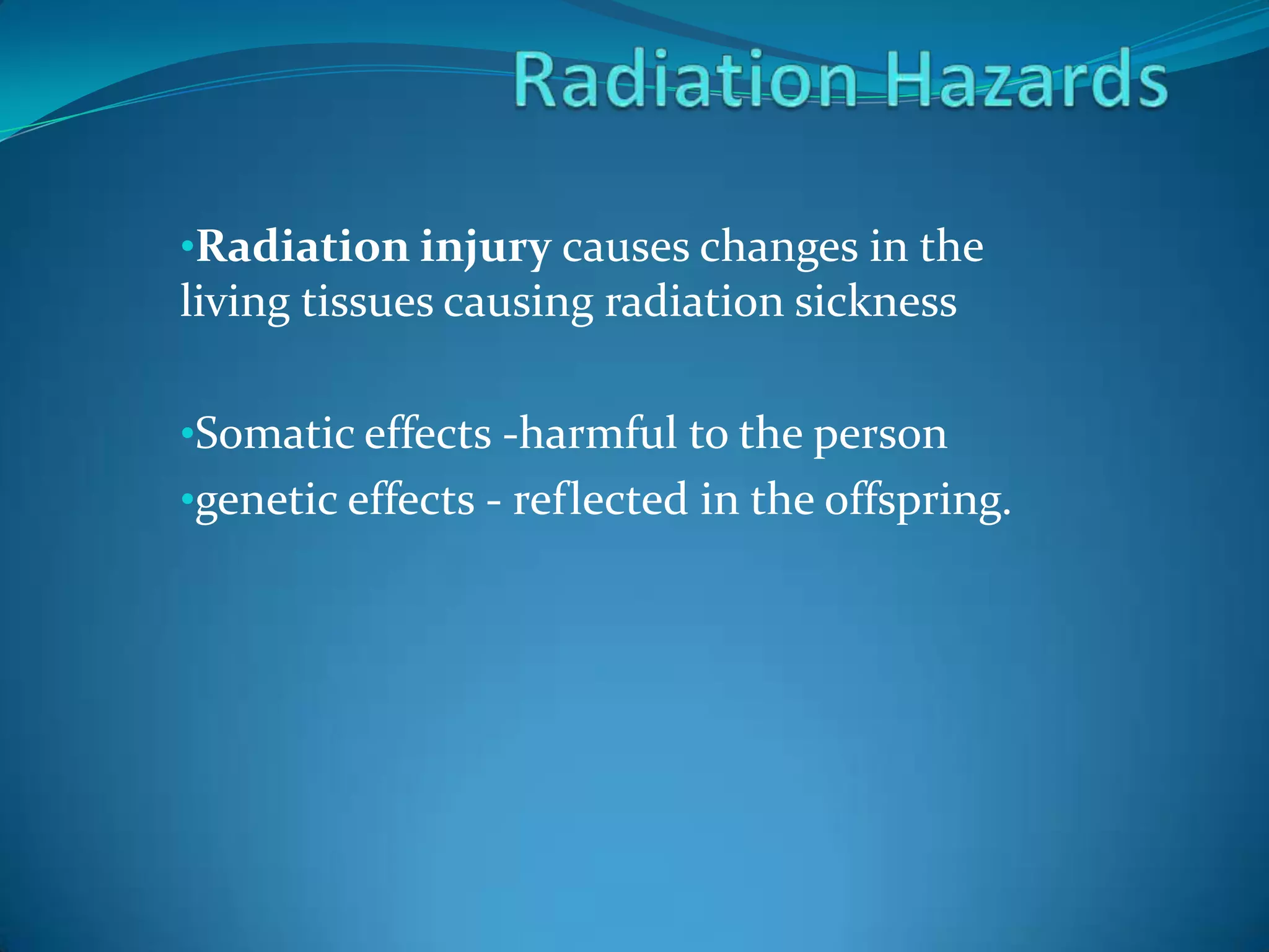

The document discusses the health effects of radiation exposure, including radiation sickness caused by changes to living tissues, as well as somatic and genetic effects. It describes the mechanisms by which ionizing radiation interacts with and damages biological molecules and cells, leading to both acute and long-term health consequences like cancer and genetic mutations. Guidelines are provided for radiation safety and protection measures to minimize exposure when working with radiation sources.