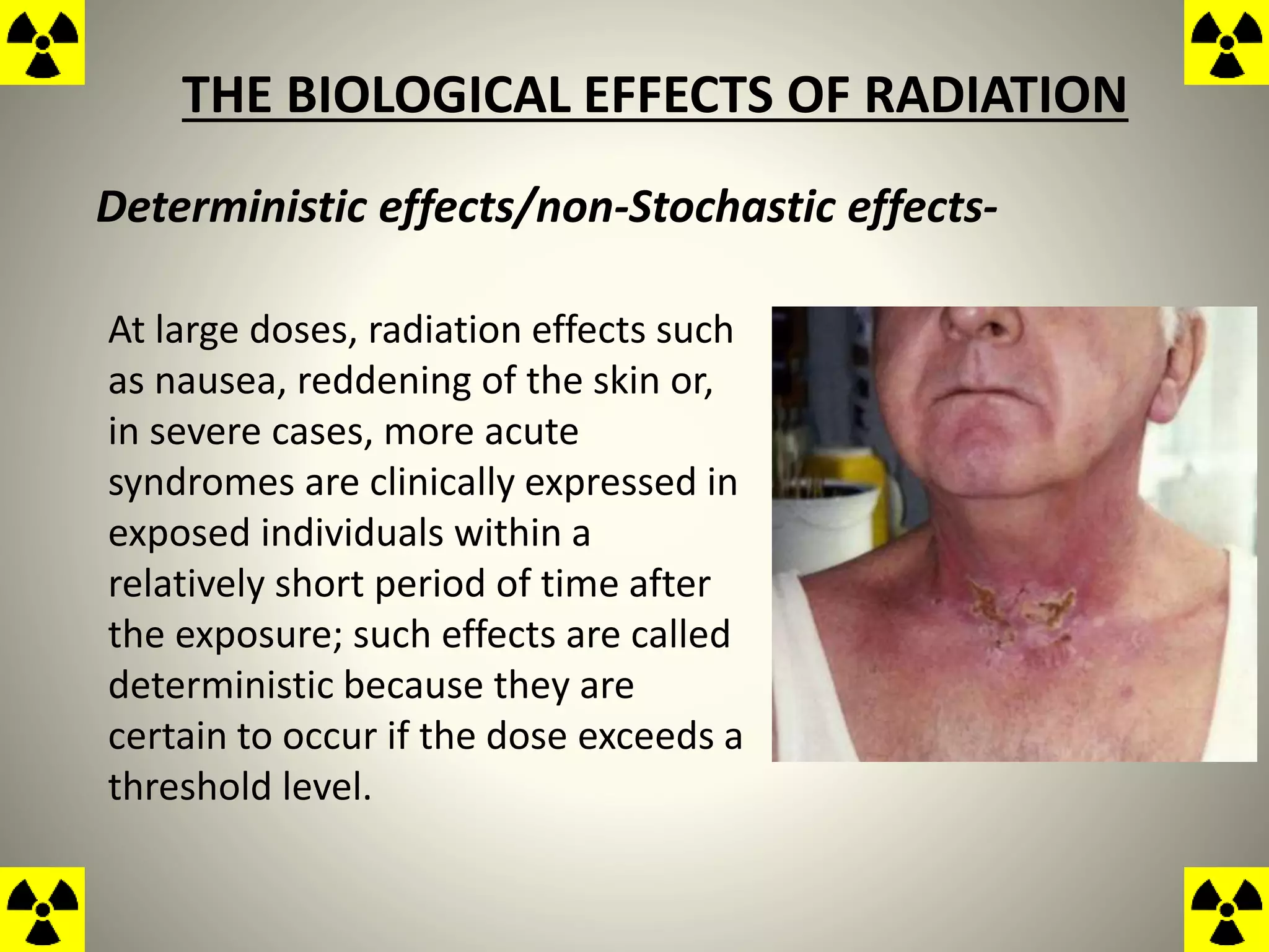
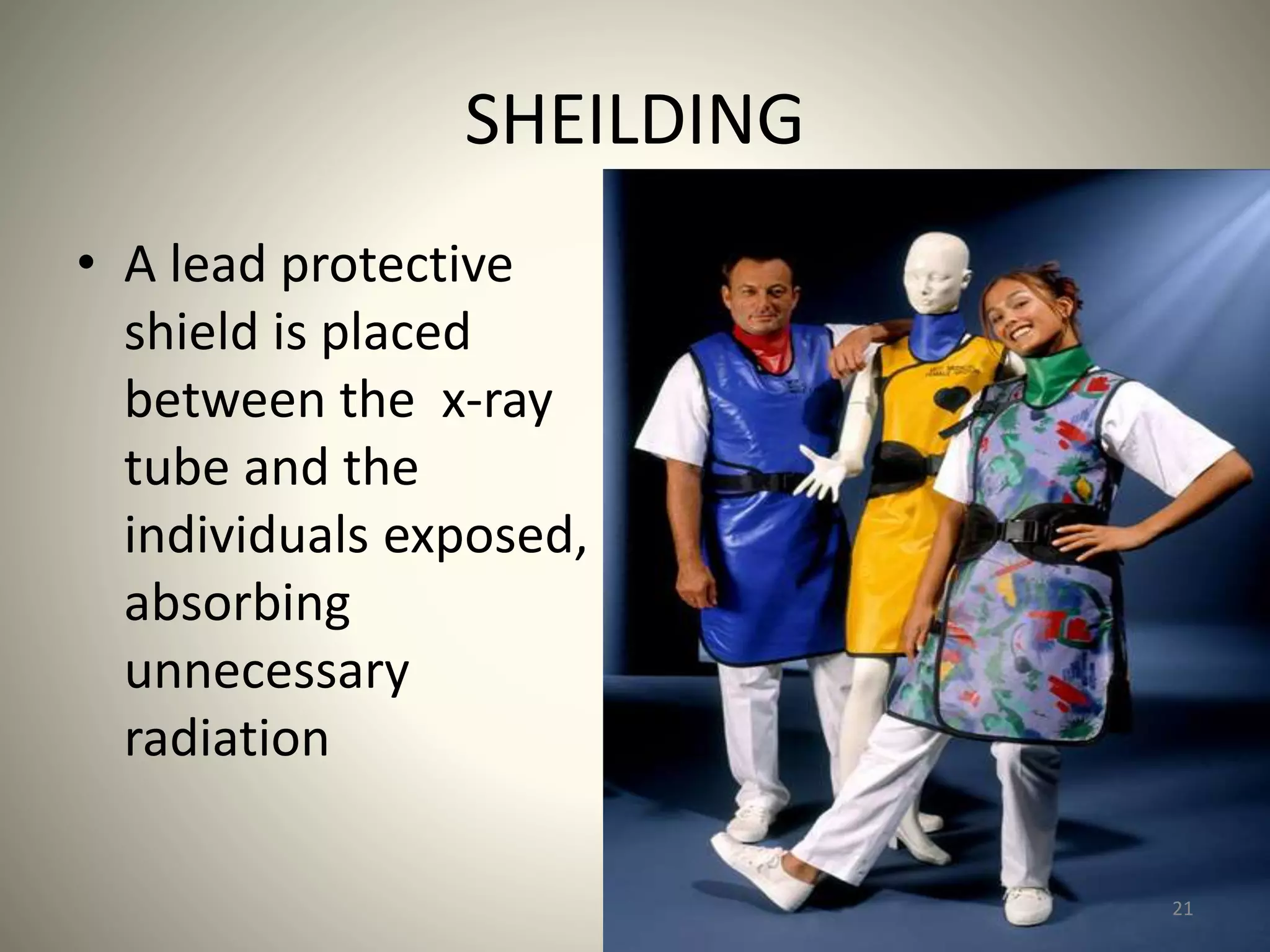
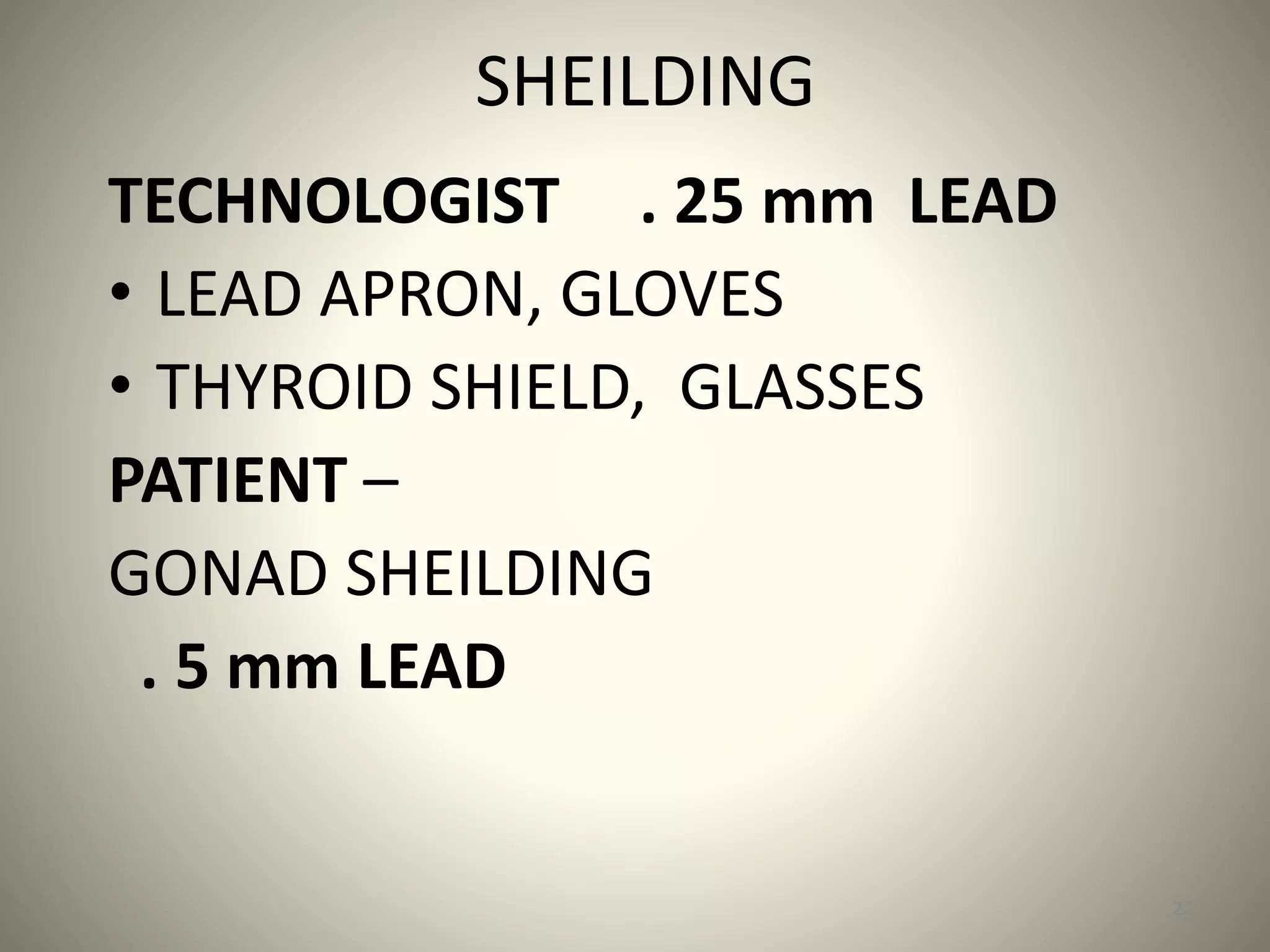
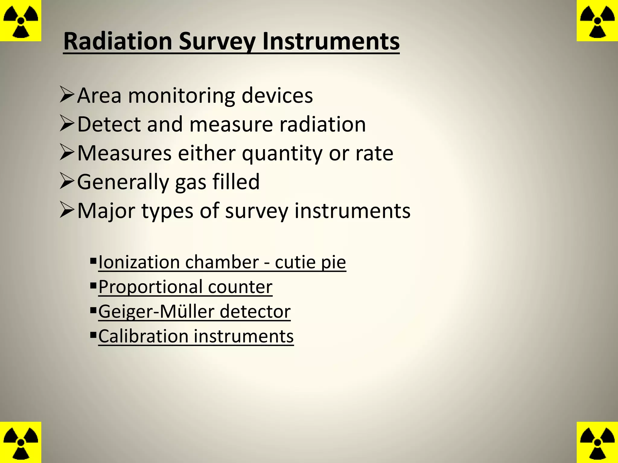
RADIATION PROTECTION
&
Personal Monitoring
Dr Jyotiman Nath
The medical use of ionizing radiations, whether
for diagnosis or therapy, not only results in the
irradiation of the patient but may also result in
some degree of exposure of radiologists,
radiographers, other workers of the department.
All these people are, therefore, subject to some degree
of radiation hazard and it is the object, of what is
usually called 'Radiation Protection‘ , to ensure
that the doses received are as small as possible, so that
the consequent damage never constitutes a significant
hazard to the health of the irradiated person.
The protection of people and the
environment from the harmful effects of
ionizing radiation, includes both particle
radiation and high energy
electromagnetic radiation
THE BIOLOGICAL EFFECTS OF RADIATION
Deterministic effects/non-Stochastic effects-
At large doses, radiation effects such
as nausea, reddening of the skin or,
in severe cases, more acute
syndromes are clinically expressed in
exposed individuals within a
relatively short period of time after
the exposure; such effects are called
deterministic because they are
certain to occur if the dose exceeds a
threshold level.
Deterministic effects/non-Stochastic effects-
Increases in severity with increasing absorbed dose in
affected individuals, owing to damage to increasing number
of cells and tissues.”
Examples : organ atrophy, fibrosis, lens opacification, blood
changes, and decrease in sperm count.
Stochastic effects-
Radiation exposure can also induce delayed effects
such as malignancies, which are expressed after a
latency period and may be epidemiologically
detectable in a population; this induction is assumed
to take place over the entire range of doses, without a
threshold level.
The probability of occurrence increases with increasing
absorbed dose but the severity in affected individuals does
not depend on the magnitude of the absorbed dose
OBJECTIVE OF RADIATION PROTECTION
 To prevent clinically significant radiation-induced
deterministic effects by adhering to dose limits that
are below the apparent or practical threshold,
To limit the risk of stochastic effects (cancer and
hereditary effects) to a reasonable level in relation to
societal needs, values, and benefits gained.
R.B.E. , Dose Equivalent, and Rem
Because the biological effectiveness of one radiation may be
different from that of another, equal absorbed doses (rad) of
different radiations do not necessarily produce biological
effects of the same magnitude.
Radiations with a high L.E.T. have a greater biological
effectiveness than those with a low L.E.T.
The difference is usually expressed by the R.B.E.
Protection regulations must assume the safer aspect
Therefore for each radiation a quality factor (Q.F.) is laid
down.
This is essentially the upper limit of the R.B.E. for the
particular radiation compared with Co-60 gamma rays,
for the most important biological effect produced
The sum of the products of the absorbed dose in rads
and the quality factor for each radiation is called the
dose equivalent, the unit of which is the rem.
Dose equivalent in rem = (Dose in rad X Q.F.)
The rem is, therefore, the quantity of any radiation
which will produce the same biological effect as 1 rad of
Co-60 gamma rays
Maximum Permissible Doses
The maximum permissible doses which are of particular interest to
radiological workers are.—
When the whole body is fairly uniformly irradiated—5 rem in a
year.
For the skin, thyroid gland, or bone—30 rem in a year.
For the hands and forearms, feet and ankles—75 rem in a year.
The dose equivalent to the lens of the eye must not exceed 15
rem (0.15 Sv) per year.
14
Reduction of Occupational
Radiation Exposure
• Radiation Therapy as a profession is very safe
.. if the ALARA rules are followed
• Most technologist exposure occurs from
fluoroscopy exams and mobile exams
– During all fluoroscopy and mobile exams
technologists should wear a protective apron
– The primary beam should never be pointed at the
tech or other staff… primary at the patient!
15
ALARA
• ALWAYS KEEP RADIATION EXPOSURES AS LOW
AS REASONABLY ACHIEVABLE
• What are the ways to do this?
16
CARDINAL RULES
OF RADIATION PROTECTION
•TIME
•DISTANCE
•SHIELDING
17
18
TIME
• The exposure is to be
kept as short as
possible because the
exposure is directly
proportional to time.
• Megavoltage>>>Kilovoltage
19
DISTANCE
• Distance from the
radiation source
should be kept as
great as possible
• Physical Law:
–Inverse Square
Law
SHEILDING
• A lead protective
shield is placed
between the x-ray
tube and the
individuals exposed,
absorbing
unnecessary
radiation
21
SHEILDING
TECHNOLOGIST . 25 mm LEAD
• LEAD APRON, GLOVES
• THYROID SHIELD, GLASSES
PATIENT –
GONAD SHEILDING
. 5 mm LEAD
22
GONAD SHIELDING
• MUST BE . 5 MM OF LEAD
• MUST BE USED WHEN GONADS WILL LIE
WITHIN 5 CM OF THE COLLIMATED AREA
23
Door Shielding
Require a motor drive as well as a means of manual operation
in case of emergency
With a proper maze design, the door is exposed mainly to the
multiply scattered radiation of significantly reduced intensity
and energy
•In most cases, the required shielding turns out to be less
than 6 mm of lead.
Barrier of Radiation protection
Primary and Secondary Barriers………………..
The amount of radiation reaching any place depends not only on
the distance of that place from the radiation source, the nature
and thickness of any interposed barrier,
but also upon the quantity and quality of radiation leaving the
source
Barriers to provide protection against the primary beam are
usually called Primary Barriers and must be incorporated in any
part of the floor, walls, and ceiling of the X-ray room at which
the primary beam can be fired.
Any surfaces at which the primary beam cannot be fired, but
which may receive scattered radiation or leakage radiation,
need only Secondary Barriers.
Lead is the most commonly used protective material, having the
double advantage of high density and high atomic number, which
means that it has a higher linear attenuation coefficient at all
radiation energies than any other commonly available
material.
In many cases lead is not a suitable material for protection
purposes and alternatives have to be sought. For example, it
cannot be used in a viewing window (!), neither is it suitable for
the protective gloves and aprons worn by those who may
have to work in radiation beams.
Ionising Radiations Regulations(IRR99)
Designation of areas
Controlled areas
Supervised Area
Uncontrolled Area
•For protection calculations, the dose-equivalent limit is assumed
to be
0.1 rem/week for the controlled areas (5 rem/year )and
0.01 rem/week for the noncontrolled areas. (0.5rem/year )
Controlled areas
Areas where a person is likely to receive an effective whole body
dose of more than 6mSv per year or where there is significant
risk of spreading contamination outside the work area.
Must be physically demarcated
Must have suitable signage
Local rules should be drawn up
Radiation Protection Supervisor appointed
Environmental and personal monitoring should take place
Uncontrolled Areas
•All other areas in the hospital or clinic and the surrounding
environment
• Uncontrolled areas are those occupied by individuals such
as patients, visitors to the facility, and employees who do
not work routinely with or around radiation sources.
• Areas adjacent to but not part of the x-ray facility is also
uncontrolled areas
Supervised areas
• Any area where the conditions need to be kept under
review
• Any person is likely to receive an effective dose
>1mSv/y or > than 1/10 of any other dose limit.
• It does not automatically follow that outside every
controlled area there will be a supervised area.
.
DEPARTMENTAL SURVEYS
The Exposure rates of the radiation leaking through barriers,
or through cracks will usually be very small (of the order of mill
roentgens per hour),
So ionization chambers of quite large volume are usually used
for their measurement.
Radiation Survey Instruments
Area monitoring devices
Detect and measure radiation
Measures either quantity or rate
Generally gas filled
Major types of survey instruments
Ionization chamber - cutie pie
Proportional counter
Geiger-Müller detector
Calibration instruments
Ionization Chamber (Cutie Pie)
• Measures x or gamma radiation generally - can be
equipped to measure beta
• Measures intensity from 1mR/hr to several thousand
R/hr
• Most commonly used to measure patients receiving
brachytherapy or diagnostic isotopes
Proportional Counter
• Generally used in laboratories to measure
beta or alpha radiation
• Can discriminate between these particles
• Operator must hold the counter close to the
object being surveyed to obtain accurate
reading
Geiger-Müller Detector
• Generally used for nuclear medicine facilities
• Unit is sensitive enough to detect individual particles
• Can be used to locate a lost radioactive source
• Has an audible sound system
• Alerts to presence of radiation
• Meter readings are generally displayed in mR/hr
GAMMA AREA MONITOR
primarily meant to serve as a Gamma Zone Monitor to indicate
dose rates and alarm status (visual and aural), once the dose
rates exceed the preset level fixed by the user.
The purpose of monitoring and exposure assessment is to gather
and provide information on the actual exposure of workers and
to confirm good working practices contributing to reassurance
and motivation.
Radiation oncologists, Radiotherapy physicists,
Radiation protection officers, Radiotherapy technologists, source handlers,
maintenance staff and any nursing or other staff who must spend time with
patients who contain radioactive sources are the ones who need
personal monitorin
Monitoring includes not just measuring and determining the
equivalent dose;
It includes interpretation and assessment.
Employers… and licensees should make arrangements
for appropriate health surveillance in accordance with
the rules established by the Regulatory Authority.”
Personnel Dosimeters
Desirable characteristics
Should be lightweight, durable, and reliable
Should be inexpensive
Types of personnel dosimeters
Film badge
Pocket ionization chambers
Thermo luminescent dosimeters (TLD)
Film Badge
Most widely used and most economical
Consists of three parts:
Plastic film holder
Metal filters
Film packet
Can read x, gamma, and beta
radiation
Accurate from 10mrem - 500rem
Developed and read by densitometer
A certain density value equals a
certain level of radiation
Read with a control badge
Results generally sent as a printout
Film Badge characteristics
Lightweight, durable, portable
Cost efficient
Permanent legal record
Can differentiate between scatter and primary beam
Can discriminate between x, gamma, and beta radiation
Can indicate direction from where radiation came from
Control badge can indicate if exposed in transit
Advantages Of The Film Badge
Only records exposure when it’s worn
Not effective if not worn
Can be affected by heat and humidity
Sensitivity is decreased above and
below 50 keV
Exposure cannot be determined on
day of exposure
Accuracy limited to + or - 20%
Disadvantages Of The Film Badge
Pocket Dosimeter
The most sensitive personnel
dosimeter
Two types
Self-reading
Non self-reading
Can only be read once
Detects gamma or x-radiation
Advantages And Disadvantages Of The Pocket Dosimeter
Small, compact, easy to use
Reasonably accurate and
sensitive
Provides immediate reading
Expensive
Readings can be lost
Must be read each day
No permanent record
Susceptible to false
readout if dropped
Thermo luminescent Dosimeters (TLD)
Looks like a film badge
Contains a lithium fluoride crystal
Responds to radiation similarly to skin
Measured by a TLD analyzer
Crystal will luminescence if exposed to
radiation, then heated
More accurate than a film badge
TLD badge consists of a set of TLD chips enclosed in a plastic
holder with filters.
The most frequently used TLD material is Lithium
TLD
Energy absorbed from the incident radiation
excites and ionizes the molecules of the
thermoluminescent material.
Some of the energy is trapped by impurities or
deformations in the material, and remains trapped
until the material is heated to a high temperature.
How TLD Works ????
Once heated, the trapped energy is released as an
emission of light.
The amount of light emitted is proportional to the
energy absorbed within the thermoluminescent
material, which is proportional to the radiation dose
absorbed.
The emitted light is measured with a photomultiplier
tube, the output of which is applied to a readout
instrument.
Small in size and chemically inert.
Almost tissue-equivalent.
Small change in sensitivity with radiation quality.
Usable over a wide range of radiation qualities.
Usable over a wide range of dose values (1 mR-1000 R).
Advantages of TLD
Sensitivity independent of dose rate.
Read-out system consistent and suitable for automation.
Read-out simple and quick (less than 1 minute per sample).
Apart from initial fading can store dose over long periods of
time.
Advantages of TLD
The initial cost is greater than that of
a film badge
Can only be read once
Records exposure only where worn
Fading of the stored signal can occur,
which is dependent on the time
interval between exposure and
development
Disadvantages of TLD
TLD’s do not give as much information about the energy of
the incident radiation as do film and OSL dosimeters
Optically stimulated luminescent (OSL) dosimeter
OSL dosimeters are currently the most common type of
personnel dosimeter used in the United States
The basic principle of operation is similar to that of the TLD
However, green laser light, rather than heat(as in TLD), is used
to stimulate release of the stored energy
OSL dosimeters have several advantages over TLD’s
They are more sensitive to a wider range of photon and beta
particle energies, and provide more information about the energy
of the incident radiation
This information is used to provide estimates of deep dose, dose
to the lens of the eye, and shallow dose.
The good resolution of the detector also allows analysis of
whether the exposure was dynamic or static
i.e., whether the badge was exposed while moving or from many
different angles, as would be expected if worn for an extended
period of time, or exposed without being moved, such as in the
case of an accidental exposure.
OSL dosimeters are also relatively unaffected by environmental
conditions such as heat, light, and humidity
Declared Pregnant Worker
• Must declare pregnancy – 2 badges
provided
• 1 worn at collar (Mother’s exposure)
• 1 worn inside apron at waist level
Under 5 rad – negligible risk
Risk increases above 15 rad
Recommend abortion (spontaneous)
25 rad
58
Radiation protection

Radiation protection

  • 1.
  • 2.
    The medical useof ionizing radiations, whether for diagnosis or therapy, not only results in the irradiation of the patient but may also result in some degree of exposure of radiologists, radiographers, other workers of the department.
  • 3.
    All these peopleare, therefore, subject to some degree of radiation hazard and it is the object, of what is usually called 'Radiation Protection‘ , to ensure that the doses received are as small as possible, so that the consequent damage never constitutes a significant hazard to the health of the irradiated person.
  • 4.
    The protection ofpeople and the environment from the harmful effects of ionizing radiation, includes both particle radiation and high energy electromagnetic radiation
  • 5.
    THE BIOLOGICAL EFFECTSOF RADIATION Deterministic effects/non-Stochastic effects- At large doses, radiation effects such as nausea, reddening of the skin or, in severe cases, more acute syndromes are clinically expressed in exposed individuals within a relatively short period of time after the exposure; such effects are called deterministic because they are certain to occur if the dose exceeds a threshold level.
  • 6.
    Deterministic effects/non-Stochastic effects- Increasesin severity with increasing absorbed dose in affected individuals, owing to damage to increasing number of cells and tissues.” Examples : organ atrophy, fibrosis, lens opacification, blood changes, and decrease in sperm count.
  • 7.
    Stochastic effects- Radiation exposurecan also induce delayed effects such as malignancies, which are expressed after a latency period and may be epidemiologically detectable in a population; this induction is assumed to take place over the entire range of doses, without a threshold level.
  • 8.
    The probability ofoccurrence increases with increasing absorbed dose but the severity in affected individuals does not depend on the magnitude of the absorbed dose
  • 9.
    OBJECTIVE OF RADIATIONPROTECTION  To prevent clinically significant radiation-induced deterministic effects by adhering to dose limits that are below the apparent or practical threshold, To limit the risk of stochastic effects (cancer and hereditary effects) to a reasonable level in relation to societal needs, values, and benefits gained.
  • 10.
    R.B.E. , DoseEquivalent, and Rem Because the biological effectiveness of one radiation may be different from that of another, equal absorbed doses (rad) of different radiations do not necessarily produce biological effects of the same magnitude. Radiations with a high L.E.T. have a greater biological effectiveness than those with a low L.E.T. The difference is usually expressed by the R.B.E.
  • 11.
    Protection regulations mustassume the safer aspect Therefore for each radiation a quality factor (Q.F.) is laid down. This is essentially the upper limit of the R.B.E. for the particular radiation compared with Co-60 gamma rays, for the most important biological effect produced
  • 12.
    The sum ofthe products of the absorbed dose in rads and the quality factor for each radiation is called the dose equivalent, the unit of which is the rem. Dose equivalent in rem = (Dose in rad X Q.F.) The rem is, therefore, the quantity of any radiation which will produce the same biological effect as 1 rad of Co-60 gamma rays
  • 13.
    Maximum Permissible Doses Themaximum permissible doses which are of particular interest to radiological workers are.— When the whole body is fairly uniformly irradiated—5 rem in a year. For the skin, thyroid gland, or bone—30 rem in a year. For the hands and forearms, feet and ankles—75 rem in a year. The dose equivalent to the lens of the eye must not exceed 15 rem (0.15 Sv) per year.
  • 14.
  • 15.
    Reduction of Occupational RadiationExposure • Radiation Therapy as a profession is very safe .. if the ALARA rules are followed • Most technologist exposure occurs from fluoroscopy exams and mobile exams – During all fluoroscopy and mobile exams technologists should wear a protective apron – The primary beam should never be pointed at the tech or other staff… primary at the patient! 15
  • 16.
    ALARA • ALWAYS KEEPRADIATION EXPOSURES AS LOW AS REASONABLY ACHIEVABLE • What are the ways to do this? 16
  • 17.
    CARDINAL RULES OF RADIATIONPROTECTION •TIME •DISTANCE •SHIELDING 17
  • 18.
  • 19.
    TIME • The exposureis to be kept as short as possible because the exposure is directly proportional to time. • Megavoltage>>>Kilovoltage 19
  • 20.
    DISTANCE • Distance fromthe radiation source should be kept as great as possible • Physical Law: –Inverse Square Law
  • 21.
    SHEILDING • A leadprotective shield is placed between the x-ray tube and the individuals exposed, absorbing unnecessary radiation 21
  • 22.
    SHEILDING TECHNOLOGIST . 25mm LEAD • LEAD APRON, GLOVES • THYROID SHIELD, GLASSES PATIENT – GONAD SHEILDING . 5 mm LEAD 22
  • 23.
    GONAD SHIELDING • MUSTBE . 5 MM OF LEAD • MUST BE USED WHEN GONADS WILL LIE WITHIN 5 CM OF THE COLLIMATED AREA 23
  • 24.
    Door Shielding Require amotor drive as well as a means of manual operation in case of emergency With a proper maze design, the door is exposed mainly to the multiply scattered radiation of significantly reduced intensity and energy •In most cases, the required shielding turns out to be less than 6 mm of lead.
  • 25.
    Barrier of Radiationprotection Primary and Secondary Barriers……………….. The amount of radiation reaching any place depends not only on the distance of that place from the radiation source, the nature and thickness of any interposed barrier, but also upon the quantity and quality of radiation leaving the source
  • 26.
    Barriers to provideprotection against the primary beam are usually called Primary Barriers and must be incorporated in any part of the floor, walls, and ceiling of the X-ray room at which the primary beam can be fired. Any surfaces at which the primary beam cannot be fired, but which may receive scattered radiation or leakage radiation, need only Secondary Barriers.
  • 27.
    Lead is themost commonly used protective material, having the double advantage of high density and high atomic number, which means that it has a higher linear attenuation coefficient at all radiation energies than any other commonly available material. In many cases lead is not a suitable material for protection purposes and alternatives have to be sought. For example, it cannot be used in a viewing window (!), neither is it suitable for the protective gloves and aprons worn by those who may have to work in radiation beams.
  • 28.
    Ionising Radiations Regulations(IRR99) Designationof areas Controlled areas Supervised Area Uncontrolled Area •For protection calculations, the dose-equivalent limit is assumed to be 0.1 rem/week for the controlled areas (5 rem/year )and 0.01 rem/week for the noncontrolled areas. (0.5rem/year )
  • 29.
    Controlled areas Areas wherea person is likely to receive an effective whole body dose of more than 6mSv per year or where there is significant risk of spreading contamination outside the work area. Must be physically demarcated Must have suitable signage Local rules should be drawn up Radiation Protection Supervisor appointed Environmental and personal monitoring should take place
  • 30.
    Uncontrolled Areas •All otherareas in the hospital or clinic and the surrounding environment • Uncontrolled areas are those occupied by individuals such as patients, visitors to the facility, and employees who do not work routinely with or around radiation sources. • Areas adjacent to but not part of the x-ray facility is also uncontrolled areas
  • 31.
    Supervised areas • Anyarea where the conditions need to be kept under review • Any person is likely to receive an effective dose >1mSv/y or > than 1/10 of any other dose limit. • It does not automatically follow that outside every controlled area there will be a supervised area. .
  • 33.
    DEPARTMENTAL SURVEYS The Exposurerates of the radiation leaking through barriers, or through cracks will usually be very small (of the order of mill roentgens per hour), So ionization chambers of quite large volume are usually used for their measurement.
  • 34.
    Radiation Survey Instruments Areamonitoring devices Detect and measure radiation Measures either quantity or rate Generally gas filled Major types of survey instruments Ionization chamber - cutie pie Proportional counter Geiger-Müller detector Calibration instruments
  • 35.
    Ionization Chamber (CutiePie) • Measures x or gamma radiation generally - can be equipped to measure beta • Measures intensity from 1mR/hr to several thousand R/hr • Most commonly used to measure patients receiving brachytherapy or diagnostic isotopes
  • 36.
    Proportional Counter • Generallyused in laboratories to measure beta or alpha radiation • Can discriminate between these particles • Operator must hold the counter close to the object being surveyed to obtain accurate reading
  • 37.
    Geiger-Müller Detector • Generallyused for nuclear medicine facilities • Unit is sensitive enough to detect individual particles • Can be used to locate a lost radioactive source • Has an audible sound system • Alerts to presence of radiation • Meter readings are generally displayed in mR/hr
  • 38.
    GAMMA AREA MONITOR primarilymeant to serve as a Gamma Zone Monitor to indicate dose rates and alarm status (visual and aural), once the dose rates exceed the preset level fixed by the user.
  • 39.
    The purpose ofmonitoring and exposure assessment is to gather and provide information on the actual exposure of workers and to confirm good working practices contributing to reassurance and motivation. Radiation oncologists, Radiotherapy physicists, Radiation protection officers, Radiotherapy technologists, source handlers, maintenance staff and any nursing or other staff who must spend time with patients who contain radioactive sources are the ones who need personal monitorin
  • 40.
    Monitoring includes notjust measuring and determining the equivalent dose; It includes interpretation and assessment. Employers… and licensees should make arrangements for appropriate health surveillance in accordance with the rules established by the Regulatory Authority.”
  • 41.
    Personnel Dosimeters Desirable characteristics Shouldbe lightweight, durable, and reliable Should be inexpensive Types of personnel dosimeters Film badge Pocket ionization chambers Thermo luminescent dosimeters (TLD)
  • 42.
    Film Badge Most widelyused and most economical Consists of three parts: Plastic film holder Metal filters Film packet
  • 43.
    Can read x,gamma, and beta radiation Accurate from 10mrem - 500rem Developed and read by densitometer A certain density value equals a certain level of radiation Read with a control badge Results generally sent as a printout Film Badge characteristics
  • 44.
    Lightweight, durable, portable Costefficient Permanent legal record Can differentiate between scatter and primary beam Can discriminate between x, gamma, and beta radiation Can indicate direction from where radiation came from Control badge can indicate if exposed in transit Advantages Of The Film Badge
  • 45.
    Only records exposurewhen it’s worn Not effective if not worn Can be affected by heat and humidity Sensitivity is decreased above and below 50 keV Exposure cannot be determined on day of exposure Accuracy limited to + or - 20% Disadvantages Of The Film Badge
  • 46.
    Pocket Dosimeter The mostsensitive personnel dosimeter Two types Self-reading Non self-reading Can only be read once Detects gamma or x-radiation
  • 47.
    Advantages And DisadvantagesOf The Pocket Dosimeter Small, compact, easy to use Reasonably accurate and sensitive Provides immediate reading Expensive Readings can be lost Must be read each day No permanent record Susceptible to false readout if dropped
  • 48.
    Thermo luminescent Dosimeters(TLD) Looks like a film badge Contains a lithium fluoride crystal Responds to radiation similarly to skin Measured by a TLD analyzer Crystal will luminescence if exposed to radiation, then heated More accurate than a film badge
  • 49.
    TLD badge consistsof a set of TLD chips enclosed in a plastic holder with filters. The most frequently used TLD material is Lithium TLD
  • 50.
    Energy absorbed fromthe incident radiation excites and ionizes the molecules of the thermoluminescent material. Some of the energy is trapped by impurities or deformations in the material, and remains trapped until the material is heated to a high temperature. How TLD Works ????
  • 51.
    Once heated, thetrapped energy is released as an emission of light. The amount of light emitted is proportional to the energy absorbed within the thermoluminescent material, which is proportional to the radiation dose absorbed. The emitted light is measured with a photomultiplier tube, the output of which is applied to a readout instrument.
  • 52.
    Small in sizeand chemically inert. Almost tissue-equivalent. Small change in sensitivity with radiation quality. Usable over a wide range of radiation qualities. Usable over a wide range of dose values (1 mR-1000 R). Advantages of TLD
  • 53.
    Sensitivity independent ofdose rate. Read-out system consistent and suitable for automation. Read-out simple and quick (less than 1 minute per sample). Apart from initial fading can store dose over long periods of time. Advantages of TLD
  • 54.
    The initial costis greater than that of a film badge Can only be read once Records exposure only where worn Fading of the stored signal can occur, which is dependent on the time interval between exposure and development Disadvantages of TLD TLD’s do not give as much information about the energy of the incident radiation as do film and OSL dosimeters
  • 55.
    Optically stimulated luminescent(OSL) dosimeter OSL dosimeters are currently the most common type of personnel dosimeter used in the United States The basic principle of operation is similar to that of the TLD However, green laser light, rather than heat(as in TLD), is used to stimulate release of the stored energy
  • 56.
    OSL dosimeters haveseveral advantages over TLD’s They are more sensitive to a wider range of photon and beta particle energies, and provide more information about the energy of the incident radiation This information is used to provide estimates of deep dose, dose to the lens of the eye, and shallow dose. The good resolution of the detector also allows analysis of whether the exposure was dynamic or static
  • 57.
    i.e., whether thebadge was exposed while moving or from many different angles, as would be expected if worn for an extended period of time, or exposed without being moved, such as in the case of an accidental exposure. OSL dosimeters are also relatively unaffected by environmental conditions such as heat, light, and humidity
  • 58.
    Declared Pregnant Worker •Must declare pregnancy – 2 badges provided • 1 worn at collar (Mother’s exposure) • 1 worn inside apron at waist level Under 5 rad – negligible risk Risk increases above 15 rad Recommend abortion (spontaneous) 25 rad 58