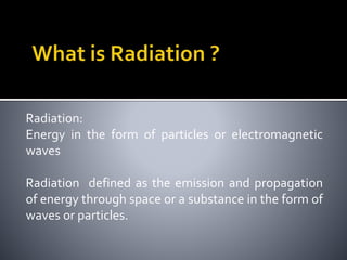
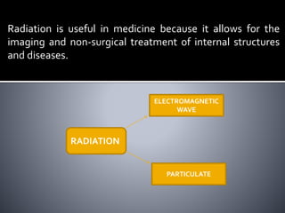
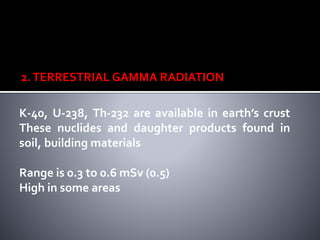
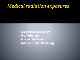
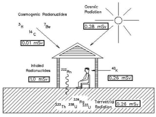
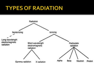
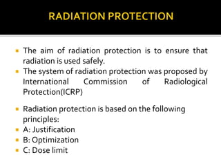
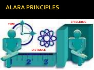
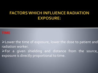
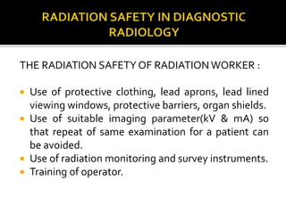
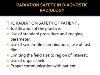
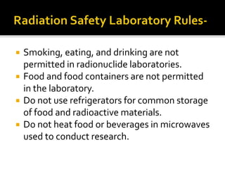
PRESENTING BY
ASIF KT
4TH SEMESTER
MBA HCM
JHON MATTHAI CENTER,THRISSURE
Radiation:
Energy in the form of particles or electromagnetic
waves
Radiation defined as the emission and propagation
of energy through space or a substance in the form of
waves or particles.
Radiation is useful in medicine because it allows for the
imaging and non-surgical treatment of internal structures
and diseases.
RADIATION
ELECTROMAGNETIC
WAVE
PARTICULATE
1. COSMIC RADIATION
Extremely energetic particles (protons) On interaction
in atmosphere produces low energy particles Mainly
muons, gamma, neutrons, electrons at sea level
Range is 0.3 to 1 mSv (0.4 mSv)
(1.0 mSv)
EXTERNAL EXPOSURE
2.TERRESTRIAL GAMMA RADIATION
K-40, U-238, Th-232 are available in earth’s crust
These nuclides and daughter products found in
soil, building materials
Range is 0.3 to 0.6 mSv (0.5)
High in some areas
• Diagnostic Radiology
• Radiotherapy
• Nuclear Medicine
• Interventional Radiology
 The higher frequencies of EM radiation, consisting
of x-rays and gamma rays, are types of ionizing
radiation.
 Lower frequency radiation, consisting of ultraviolet
(UV), infrared (IR),microwave (MW), Radio
Frequency (RF), and extremely low frequency (ELF)
are types of non-ionizing radiation.
Alpha radiation is a heavy, very short-range particle
and is actually an ejected helium nucleus. Some
characteristics of alpha radiation are:
 Most alpha radiation is not able to penetrate human
skin.
 Alpha-emitting materials can be harmful to humans
if the materials are inhaled, swallowed, or absorbed
through open wounds.
 Alpha radiation travels only a short distance (a few
inches) in air, but is not an external hazard.
 Alpha radiation is not able to penetrate clothing.
Beta radiation is a light, short-range particle and is actually an
ejected electron. Some characteristics of beta radiation are:
 Beta radiation may travel several feet in air and is
moderately penetrating.
 Beta radiation can penetrate human skin to the "germinal
layer," where new skin cells are produced. If high levels of
beta-emitting contaminants are allowed to remain on the
skin for a prolonged period of time, they may cause skin
injury.
 Beta-emitting contaminants may be harmful if deposited
internally.
 Clothing provides some protection against beta radiation.
 Gamma radiation and x rays are highly penetrating
electromagnetic radiation. Some characteristics of these
radiations are:
Gamma radiation or x rays are able to travel many feet in air
and many inches in human tissue.They readily penetrate
most materials and are sometimes called "penetrating"
radiation.
 Gamma radiation and x rays are electromagnetic radiation
like visible light, radiowaves, and ultraviolet light.These
electromagnetic radiations differ only in the amount of
energy they have. Gamma rays and x rays are the most
energetic of these.
1. Fluoroscopy
2. ComputedTomography (CT)
3. Chest Examinations
4. Mammography
Examination Dose to patient
CT Head 2mSv
CT Abdomen 9mSv
CXR 0.05mSv
Dental X-ray 0.005mSv
Lumber spine 2mSv
 The amount of radiation received by persons
exposed occupationally should not exceed the
dosages specified in the North Carolina Regulations
For Protection Against Radiation:
 15A NCAC 11 Annual Dose Limits:
 Whole Body: 5,000 mrem
 Skin/Extremities: 50,000 mrem
 Lens of Eye: 15,000 mrem
 The average annual dose of a radiation worker at
UNC is about 100 millirem.
 The aim of radiation protection is to ensure that
radiation is used safely.
 The system of radiation protection was proposed by
International Commission of Radiological
Protection(ICRP)
 Radiation protection is based on the following
principles:
 A: Justification
 B: Optimization
 C: Dose limit
JUSTIFICATION
•All exposure either diagnostic or therapeutic shall be under
taken only if the benefit gained out of the detriment.
•No practice shall be adopted unless it produces a net
positive benefit.
OPTIMIZATION
•All exposures which are justified shall be under taken with a
minimum possible dose.
•Every effort shall be taken to reduce the dose to As Low As
Reasonable Achievable (ALARA), taking into account the
economic and social considerations.
DOSE LIMIT
•Exposure of radiation worker and individuals of public must
not exceed dose limit.
TIME
SHIELDING
DISTANCE
TIME
Lower the time of exposure, lower the dose to patient and
radiation worker.
For a given shielding and distance from the source,
exposure is directly proportional to time.
•Effective & Easy
Inverse Square Law
Doubling distance from
source, decreases dose by factor
of four
Tripling it decreases dose nine-
fold
More Distance = Less Radiation
Exposure
Materials “absorb” radiation
Proper shielding = Less Radiation Exposure
THE RADIATION SAFETY OF RADIATION WORKER :
 Use of protective clothing, lead aprons, lead lined
viewing windows, protective barriers, organ shields.
 Use of suitable imaging parameter(kV & mA) so
that repeat of same examination for a patient can
be avoided.
 Use of radiation monitoring and survey instruments.
 Training of operator.
THE RADIATION SAFETY OF PATIENT :
 Justification of the practice.
 Use of standard procedure and imaging
parameter.
 Use of screen-film combinations, use of fast
film.
 Limiting the field size to region of interest.
 Use of organ shield.
 Proper communication with patient
 Smoking, eating, and drinking are not
permitted in radionuclide laboratories.
 Food and food containers are not permitted
in the laboratory.
 Do not use refrigerators for common storage
of food and radioactive materials.
 Do not heat food or beverages in microwaves
used to conduct research.
•The pregnant woman who is a radiation worker can be
considered as an occupationally exposed individual, but
the foetus cannot. The total dose equivalent limit to an
embryo-foetus is 5mSv, with the added recommendation
that exposure to the foetus should not exceed 0.5 mSv in
any one month.
•Pregnant radiation worker should wear a second badge at
the waist level (under the lead apron)
Lead Apron
Lead collar
Lead Gonad Shield
Lead gloves,
proper monitoring
Protective Devices
PERSONAL MONITORING DEVICE
TLD BADGE
Guidelines for UsingTLD Badge:-
1.TLD badges are to be used only by persons directly
working in the radiation. Administrators, dark room
assistant , sweepers etc., need not be provided with TLD
badges .
2. TLD badge is used to measure the radiation dose it does
not protect the user from the radiation .
3. The name, personnel number, period of use, location
on the body ( chest or wrist ) etc, should be written in the
block letters on the front side of the badge.
4. A TLD badge once issued to a person should not be
used by another person.
5. TLD badge should be worn compulsorily at the chest
level. It represent the whole body dose equivalent. If lead
apron is used, TLD badge should be worn under the lead
apron.
6. A badge without filter or damaged filter should not be
used . It is replaced by a new holder.
7. Every radiation worker must ensure that the badge is
not left in the radiation field or near hot plates, ovens,
burners etc.
8.Loss or accidental exposure to the TLD badges should
immediately reported to the RSO.
9. Every new radiation worker has to fill up the personnel
data form, and should be sent to Radiological safety
officer of their respective department.
PERSONNEL MONITORING:
 Wear assigned monitoring device
DOSIMETERS
 Only persons whose presence is necessary
should be in the radiographic or
fluoroscopic room during exposures
 Protect all persons subject to direct scatter
radiation with whole body lead aprons
(such as a skirt and vest) or whole body
protective barriers
 Operators must stand behind protective barriers during
radiographic exposures and Remain at least 6 feet from
the patient during exposures
 Make exposures with doors to the x-ray room closed
HOLDING PATIENTS:
 Use mechanical supporting devices when a patient or
cassette must be held
 Protect holders with appropriate shielding devices
(such as a lead apron)
 Operator shall provide appropriate instructions to the
holder to maintain doses ALARA
EXPOSURE CONTROL ANDTECHNIQUE CHARTS:
 Keep exposure to the patient minimal practical amount
consistent with clinical objectives
 Automatic Exposure Control (AEC) feature should be
appropriately utilized for all exposures when available
 If not available, manual techniques must be utilized
 Consider using dose reduction techniques (like high tube
potential (kVp) and low current (mA)), as long as image
quality is not compromised
 Repeat X-ray must be avoided in order to reduce
patient dose.
 Records of all radiological examination should be
maintained.
 Personnel monitoring devices shall be used by all
radiation worker.
 Each equipment must have separate log book which
provide information about equipment
manufacturer,model, serial no., max kV & max mA.
radiation protection and safty

radiation protection and safty

  • 1.
    PRESENTING BY ASIF KT 4THSEMESTER MBA HCM JHON MATTHAI CENTER,THRISSURE
  • 2.
    Radiation: Energy in theform of particles or electromagnetic waves Radiation defined as the emission and propagation of energy through space or a substance in the form of waves or particles.
  • 3.
    Radiation is usefulin medicine because it allows for the imaging and non-surgical treatment of internal structures and diseases. RADIATION ELECTROMAGNETIC WAVE PARTICULATE
  • 4.
    1. COSMIC RADIATION Extremelyenergetic particles (protons) On interaction in atmosphere produces low energy particles Mainly muons, gamma, neutrons, electrons at sea level Range is 0.3 to 1 mSv (0.4 mSv) (1.0 mSv) EXTERNAL EXPOSURE
  • 5.
    2.TERRESTRIAL GAMMA RADIATION K-40,U-238, Th-232 are available in earth’s crust These nuclides and daughter products found in soil, building materials Range is 0.3 to 0.6 mSv (0.5) High in some areas
  • 6.
    • Diagnostic Radiology •Radiotherapy • Nuclear Medicine • Interventional Radiology
  • 9.
     The higherfrequencies of EM radiation, consisting of x-rays and gamma rays, are types of ionizing radiation.  Lower frequency radiation, consisting of ultraviolet (UV), infrared (IR),microwave (MW), Radio Frequency (RF), and extremely low frequency (ELF) are types of non-ionizing radiation.
  • 10.
    Alpha radiation isa heavy, very short-range particle and is actually an ejected helium nucleus. Some characteristics of alpha radiation are:  Most alpha radiation is not able to penetrate human skin.  Alpha-emitting materials can be harmful to humans if the materials are inhaled, swallowed, or absorbed through open wounds.  Alpha radiation travels only a short distance (a few inches) in air, but is not an external hazard.  Alpha radiation is not able to penetrate clothing.
  • 11.
    Beta radiation isa light, short-range particle and is actually an ejected electron. Some characteristics of beta radiation are:  Beta radiation may travel several feet in air and is moderately penetrating.  Beta radiation can penetrate human skin to the "germinal layer," where new skin cells are produced. If high levels of beta-emitting contaminants are allowed to remain on the skin for a prolonged period of time, they may cause skin injury.  Beta-emitting contaminants may be harmful if deposited internally.  Clothing provides some protection against beta radiation.
  • 12.
     Gamma radiationand x rays are highly penetrating electromagnetic radiation. Some characteristics of these radiations are: Gamma radiation or x rays are able to travel many feet in air and many inches in human tissue.They readily penetrate most materials and are sometimes called "penetrating" radiation.  Gamma radiation and x rays are electromagnetic radiation like visible light, radiowaves, and ultraviolet light.These electromagnetic radiations differ only in the amount of energy they have. Gamma rays and x rays are the most energetic of these.
  • 14.
    1. Fluoroscopy 2. ComputedTomography(CT) 3. Chest Examinations 4. Mammography
  • 15.
    Examination Dose topatient CT Head 2mSv CT Abdomen 9mSv CXR 0.05mSv Dental X-ray 0.005mSv Lumber spine 2mSv
  • 16.
     The amountof radiation received by persons exposed occupationally should not exceed the dosages specified in the North Carolina Regulations For Protection Against Radiation:  15A NCAC 11 Annual Dose Limits:  Whole Body: 5,000 mrem  Skin/Extremities: 50,000 mrem  Lens of Eye: 15,000 mrem  The average annual dose of a radiation worker at UNC is about 100 millirem.
  • 17.
     The aimof radiation protection is to ensure that radiation is used safely.  The system of radiation protection was proposed by International Commission of Radiological Protection(ICRP)  Radiation protection is based on the following principles:  A: Justification  B: Optimization  C: Dose limit
  • 18.
    JUSTIFICATION •All exposure eitherdiagnostic or therapeutic shall be under taken only if the benefit gained out of the detriment. •No practice shall be adopted unless it produces a net positive benefit. OPTIMIZATION •All exposures which are justified shall be under taken with a minimum possible dose. •Every effort shall be taken to reduce the dose to As Low As Reasonable Achievable (ALARA), taking into account the economic and social considerations. DOSE LIMIT •Exposure of radiation worker and individuals of public must not exceed dose limit.
  • 19.
  • 20.
    TIME Lower the timeof exposure, lower the dose to patient and radiation worker. For a given shielding and distance from the source, exposure is directly proportional to time.
  • 22.
    •Effective & Easy InverseSquare Law Doubling distance from source, decreases dose by factor of four Tripling it decreases dose nine- fold More Distance = Less Radiation Exposure
  • 23.
    Materials “absorb” radiation Propershielding = Less Radiation Exposure
  • 24.
    THE RADIATION SAFETYOF RADIATION WORKER :  Use of protective clothing, lead aprons, lead lined viewing windows, protective barriers, organ shields.  Use of suitable imaging parameter(kV & mA) so that repeat of same examination for a patient can be avoided.  Use of radiation monitoring and survey instruments.  Training of operator.
  • 25.
    THE RADIATION SAFETYOF PATIENT :  Justification of the practice.  Use of standard procedure and imaging parameter.  Use of screen-film combinations, use of fast film.  Limiting the field size to region of interest.  Use of organ shield.  Proper communication with patient
  • 26.
     Smoking, eating,and drinking are not permitted in radionuclide laboratories.  Food and food containers are not permitted in the laboratory.  Do not use refrigerators for common storage of food and radioactive materials.  Do not heat food or beverages in microwaves used to conduct research.
  • 27.
    •The pregnant womanwho is a radiation worker can be considered as an occupationally exposed individual, but the foetus cannot. The total dose equivalent limit to an embryo-foetus is 5mSv, with the added recommendation that exposure to the foetus should not exceed 0.5 mSv in any one month. •Pregnant radiation worker should wear a second badge at the waist level (under the lead apron)
  • 28.
    Lead Apron Lead collar LeadGonad Shield Lead gloves, proper monitoring Protective Devices
  • 29.
    PERSONAL MONITORING DEVICE TLDBADGE Guidelines for UsingTLD Badge:- 1.TLD badges are to be used only by persons directly working in the radiation. Administrators, dark room assistant , sweepers etc., need not be provided with TLD badges . 2. TLD badge is used to measure the radiation dose it does not protect the user from the radiation .
  • 30.
    3. The name,personnel number, period of use, location on the body ( chest or wrist ) etc, should be written in the block letters on the front side of the badge. 4. A TLD badge once issued to a person should not be used by another person. 5. TLD badge should be worn compulsorily at the chest level. It represent the whole body dose equivalent. If lead apron is used, TLD badge should be worn under the lead apron. 6. A badge without filter or damaged filter should not be used . It is replaced by a new holder.
  • 31.
    7. Every radiationworker must ensure that the badge is not left in the radiation field or near hot plates, ovens, burners etc. 8.Loss or accidental exposure to the TLD badges should immediately reported to the RSO. 9. Every new radiation worker has to fill up the personnel data form, and should be sent to Radiological safety officer of their respective department.
  • 33.
    PERSONNEL MONITORING:  Wearassigned monitoring device DOSIMETERS
  • 34.
     Only personswhose presence is necessary should be in the radiographic or fluoroscopic room during exposures  Protect all persons subject to direct scatter radiation with whole body lead aprons (such as a skirt and vest) or whole body protective barriers
  • 35.
     Operators muststand behind protective barriers during radiographic exposures and Remain at least 6 feet from the patient during exposures  Make exposures with doors to the x-ray room closed HOLDING PATIENTS:  Use mechanical supporting devices when a patient or cassette must be held  Protect holders with appropriate shielding devices (such as a lead apron)  Operator shall provide appropriate instructions to the holder to maintain doses ALARA
  • 36.
    EXPOSURE CONTROL ANDTECHNIQUECHARTS:  Keep exposure to the patient minimal practical amount consistent with clinical objectives  Automatic Exposure Control (AEC) feature should be appropriately utilized for all exposures when available  If not available, manual techniques must be utilized  Consider using dose reduction techniques (like high tube potential (kVp) and low current (mA)), as long as image quality is not compromised
  • 37.
     Repeat X-raymust be avoided in order to reduce patient dose.  Records of all radiological examination should be maintained.  Personnel monitoring devices shall be used by all radiation worker.  Each equipment must have separate log book which provide information about equipment manufacturer,model, serial no., max kV & max mA.