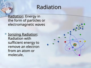
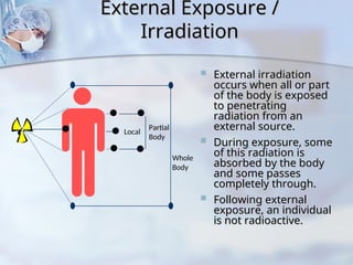
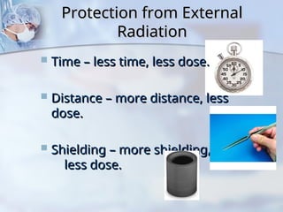
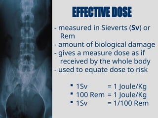
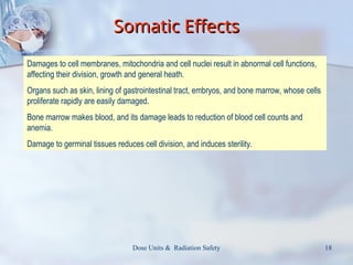
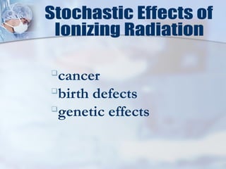
Radiation
Radiation
 Radiation
Radiation: Energy in
: Energy in
the form of particles or
electromagnetic waves
 Ionizing Radiation
Ionizing Radiation:
:
Radiation with
Radiation with
sufficient energy to
remove an electron
remove an electron
from an atom or
from an atom or
molecule
molecule.
.
Radioactivity
Radioactivity
 The process by which
The process by which
unstable atoms
unstable atoms
spontaneously
spontaneously
transform to new
transform to new
atoms* and in the
atoms* and in the
process emit
process emit
radiation
radiation.
.
$-
* The “new atom” may be the
same atom in a lower energy
state.
External Exposure /
External Exposure /
Irradiation
Irradiation
 External irradiation
External irradiation
occurs when all or part
of the body is exposed
to penetrating
to penetrating
radiation from an
radiation from an
external source.
external source.
 During exposure, some
During exposure, some
of this radiation is
absorbed by the body
and some passes
and some passes
completely through.
completely through.
 Following external
Following external
exposure, an individual
is not radioactive.
is not radioactive.
s





Local
Partial
Body
Whole
Body

Protection from External
Radiation
Radiation
 Time – less time, less dose.
Time – less time, less dose.
 Distance – more distance, less
Distance – more distance, less
dose.
dose.
 Shielding – more shielding,
Shielding – more shielding,
less dose.
less dose.
Internal Exposure
Internal Exposure
 Internal exposure is from
Internal exposure is from
radioactive materials that
have been taken into the
body.
body.
 Radioactive material can
Radioactive material can
enter the body through:
 Injection
Injection
 Inhalation
Inhalation
 Ingestion
Ingestion
 Absorption
Absorption
 Once radioactive materials
Once radioactive materials
are in the body, they
irradiate body tissues as long
as they remain in the body
Thyroid
Lung
Liver
Bone
Internal Exposure
Internal Exposure
• There are two ways for radioactive materials to be
There are two ways for radioactive materials to be
removed from the body:
 Biological clearance
Biological clearance
 Radiological decay
Radiological decay
• The term “committed dose” refers to the fact that the
The term “committed dose” refers to the fact that the
radioactive dose will continue as long as the
radioactive material is in the body
• The amount of radioactivity in the body can be
The amount of radioactivity in the body can be
assessed by Bioassay;(measurement of the
concentration or potency of a substance by its effect
on living cells or tissues)
- measured in C/kg or
Roentgen(R)
- amount of charge (electrons)
liberated per kilogram of Air
(Ionization)
 1R = 2.58x10⁻⁴
C/Kg
C- curie
- measured in Gray (Gy) or
Rad
- amount of energy
deposited/ absorbed per
kilogram of tissue
 1Gy = 1 Joule/Kg
 100 Rad = 1 Joule/Kg
 1Rad = 1/100 Gy
- measured in Sieverts (Sv) or
Rem
- amount of biological damage
- gives a measure dose as if
received by the whole body
- used to equate dose to risk
 1Sv = 1 Joule/Kg
 100 Rem = 1 Joule/Kg
 1Sv = 1/100 Rem
 More radiosensitive than adults due to sensitive cells
More radiosensitive than adults due to sensitive cells
and developing organs
 Gonad shielding important
Gonad shielding important
 Radiographic examinations difficult:
Radiographic examinations difficult:
 Patient movement
Patient movement
 Exposure technique more critical
Exposure technique more critical
 Embryo/fetus is rapidly developing so is more sensitive to a
Embryo/fetus is rapidly developing so is more sensitive to a
possible radiation effect than an adult.
 Effects vary with amount of radiation and stage of development
Effects vary with amount of radiation and stage of development
of the embryo/fetus.
 Principal effects are loss of pregnancy, malformations, and
Principal effects are loss of pregnancy, malformations, and
mental retardation.
mental retardation.
Dose Units & Radiation Safety 18
Somatic Effects
Somatic Effects
Damages to cell membranes, mitochondria and cell nuclei result in abnormal cell functions,
affecting their division, growth and general heath.
Organs such as skin, lining of gastrointestinal tract, embryos, and bone marrow, whose cells
proliferate rapidly are easily damaged.
Bone marrow makes blood, and its damage leads to reduction of blood cell counts and
anemia.
Damage to germinal tissues reduces cell division, and induces sterility.
 Genetic mutations occur from incorrect
Genetic mutations occur from incorrect
repair of damaged chromosomes in egg or
sperm cells.
sperm cells.
 Ovaries can repair mild radiation damage.
Ovaries can repair mild radiation damage.
 Genetic mutations may show up in future
Genetic mutations may show up in future
generations.
generations.
 Radiation-caused genetic mutations have
Radiation-caused genetic mutations have
been shown in animal studies at very high
radiation doses (>25 rem).
Genetic Effects
Genetic Effects

cancer

birth defects

genetic effects
Photograph of the patient’s
back 6-8 weeks after multiple coronary
angiography and angioplasty procedures.
Photograph of the injury 16-21 weeks after
the procedures. A small ulcerated area is
present.
Close-up of the lesion shown in C
Photograph of the patient’s back 18-21 months
after the procedures. Tissue necrosis is evident
Photograph of the patient’s
back after Grafting.
Summary of Biological Effects of
Radiation
Radiation
 Radiation may…
Radiation may…
 Deposit Energy in Body
Deposit Energy in Body
 Cause DNA Damage
Cause DNA Damage
 Create Ionizations in Body, Leading to Free
Create Ionizations in Body, Leading to Free
Radicals
Radicals
 Which may lead to biological damage
Which may lead to biological damage
Response to radiation depends
on
on:
:
 Total dose
Total dose
 Dose rate
Dose rate
 Radiation quality
Radiation quality
 Stage of development at the time of
Stage of development at the time of
exposure
exposure
Radiation exposure of personnel
and the general public should be kept
As Low As Reasonably Achievable.
A.L.A.R.A. policy
A.L.A.R.A. policy
RADIATION
RADIATION
PROTECTION
PROTECTION
correct exposure factors
correct radiographic technique
appropriate radiation protection
appropriate development/viewing techniques
appropriate radiographic positions for
examination
minimize repeat examinations
continuing education
Only a physician or a registered
x-ray technologist under the
direct
direct
supervision of a physician may
perform fluoroscopy.
 Time
Time
 Distance
Distance
 Shielding
Shielding
 Contamination
Contamination
Control
Control
4 Basic Categories
 Reduce of your exposure
 Increase from the source
 Make use of available
TIME
TIME
DISTANCE
DISTANCE
SHIELDING
SHIELDING
 minimize time in radiography or fluoroscopy rooms
minimize time in radiography or fluoroscopy rooms
 minimize time spent with patients who are undergoing
minimize time spent with patients who are undergoing
therapy treatment eg. nuclear medicine procedures,
radioactive implants
 Know Your Protocol
Know Your Protocol
 Read the procedure through carefully
Read the procedure through carefully
 Understand the steps clearly or
Understand the steps clearly or
 Have the protocol displayed where you can
Have the protocol displayed where you can
see it
see it
 Practice the technique beforehand
Practice the technique beforehand
Time
Time
 Radiation is only produced
Radiation is only produced
when the beam is on!
 Irradiate only when it is
Irradiate only when it is
necessary to observe motion.
 Last-image-hold and instant
Last-image-hold and instant
replay can usually save dose.
The distance between you and the isotope is of
paramount importance for high energy emitters and
penetrating radiations.
The intensity of radiation at different distances is
represented by the formula: law stating that a specified
physical quantity or intensity is inversely proportional to the
square of the distance from the source of that physical quantity.
This is the Inverse Square Law.
I2 d1
d2
I1
( )
=
2
 Inverse Square Law - double the
Inverse Square Law - double the distance
distance
from the source of
from the source of radiation -
radiation - reduce dose by a
reduce dose by a
factor of 4
factor of 4
 General rule - 3 meters
General rule - 3 meters (approximately
(approximately
10 ft) from the source
10 ft) from the source of radiation - dose is
of radiation - dose is
insignificant
insignificant
 Keep the x-ray source (the tube) as far away from
Keep the x-ray source (the tube) as far away from
you (the operator) as possible consistent with
optimal imaging
optimal imaging
Radiation Safety Principle
 Use the least amount of magnification
Use the least amount of magnification
consistent with seeing the object
adequately.
adequately.
 BIGGER IS NOT ALWAYS BETTER!!
BIGGER IS NOT ALWAYS BETTER!!
 A larger image means more radiation
A larger image means more radiation
 If it is necessary for adequate visualization,
If it is necessary for adequate visualization,
fine
fine
 If it does not improve procedure safety or
If it does not improve procedure safety or
performance, reduce the magnification
Fluoro only when viewing monitor
Use pulsed fluoroscopy when possible
Use last image hold
Personal shielding
Personal shielding
 lead aprons - at least 3 - 5mm Pb
lead aprons - at least 3 - 5mm Pb
equivalent provide up to 90%
shielding.
shielding.
 thyroid / eye shielding during
thyroid / eye shielding during
fluoroscopy
fluoroscopy
 lead glove (5mm + Pb eq.) if hands
lead glove (5mm + Pb eq.) if hands
are
are likely to be in the beam
likely to be in the beam
 Lead drape on fluoro tower provides
Lead drape on fluoro tower provides
an additional 90% protection of the
remaining
remaining 10% from lead aprons
10% from lead aprons
above.
above.
Protective barriers
Protective barriers
 lead glass / acrylic for windows, lead sheets
lead glass / acrylic for windows, lead sheets
in doors or plaster board walls, brick walls
 minimize directing primary beam at
minimize directing primary beam at
widows / doors
widows / doors
 best position to be located during x-ray
best position to be located during x-ray
exposure
exposure
 State law requires that, during
fluoroscopy, one badge must be
worn outside the apron at the
collar level.
Some institutions provide
additional badges, usually upon
request during pregnancy
Badge readings are
reviewed regularly by the RSO
Institutional investigation
levels are set below regulatory
limits
Personnel are notified
regularly and badge readings are
posted
Questions
Questions
 How much radiation does it take to
How much radiation does it take to
cause a person’s
cause a person’s risk for a
for a
health effect to increase?
to increase?
 What are the health effects that can
What are the health effects that can
occur if there is enough exposure?
 Is there a level of radiation that is
Is there a level of radiation that is
safe?
?
Risk
Risk
 Risk from a radiation dose is typically based on
Risk from a radiation dose is typically based on
calculations of the “real” effect of the radiation
dose that is absorbed.
 These calculations are based on
These calculations are based on:
:
 The type of radiation.
The type of radiation.
 Each type of radiation is different and affects tissues differently.
Each type of radiation is different and affects tissues differently.
 The energy that it leaves in the body.
The energy that it leaves in the body.
 More energy means a higher probability of an effect.
More energy means a higher probability of an effect.
 Where in the body the energy remains.
Where in the body the energy remains.
 Radiation exposure to a nonsensitive area of the body (i.e., wrist)
Radiation exposure to a nonsensitive area of the body (i.e., wrist)
really has no actual effect. Radiation exposure to a sensitive area
of the body (i.e., blood-forming organs) can have an effect if the
amount of energy left is high enough.

radiation-protec tion.ppt

  • 2.
    Radiation Radiation  Radiation Radiation: Energyin : Energy in the form of particles or the form of particles or electromagnetic waves electromagnetic waves  Ionizing Radiation Ionizing Radiation: : Radiation with Radiation with sufficient energy to sufficient energy to remove an electron remove an electron from an atom or from an atom or molecule molecule. .
  • 3.
    Radioactivity Radioactivity  The processby which The process by which unstable atoms unstable atoms spontaneously spontaneously transform to new transform to new atoms* and in the atoms* and in the process emit process emit radiation radiation. . $- * The “new atom” may be the same atom in a lower energy state.
  • 4.
    External Exposure / ExternalExposure / Irradiation Irradiation  External irradiation External irradiation occurs when all or part occurs when all or part of the body is exposed of the body is exposed to penetrating to penetrating radiation from an radiation from an external source. external source.  During exposure, some During exposure, some of this radiation is of this radiation is absorbed by the body absorbed by the body and some passes and some passes completely through. completely through.  Following external Following external exposure, an individual exposure, an individual is not radioactive. is not radioactive. s      Local Partial Body Whole Body 
  • 5.
    Protection from External Protectionfrom External Radiation Radiation  Time – less time, less dose. Time – less time, less dose.  Distance – more distance, less Distance – more distance, less dose. dose.  Shielding – more shielding, Shielding – more shielding, less dose. less dose.
  • 6.
    Internal Exposure Internal Exposure Internal exposure is from Internal exposure is from radioactive materials that radioactive materials that have been taken into the have been taken into the body. body.  Radioactive material can Radioactive material can enter the body through: enter the body through:  Injection Injection  Inhalation Inhalation  Ingestion Ingestion  Absorption Absorption  Once radioactive materials Once radioactive materials are in the body, they are in the body, they irradiate body tissues as long irradiate body tissues as long as they remain in the body as they remain in the body Thyroid Lung Liver Bone
  • 7.
    Internal Exposure Internal Exposure •There are two ways for radioactive materials to be There are two ways for radioactive materials to be removed from the body: removed from the body:  Biological clearance Biological clearance  Radiological decay Radiological decay • The term “committed dose” refers to the fact that the The term “committed dose” refers to the fact that the radioactive dose will continue as long as the radioactive dose will continue as long as the radioactive material is in the body radioactive material is in the body • The amount of radioactivity in the body can be The amount of radioactivity in the body can be assessed by Bioassay;(measurement of the assessed by Bioassay;(measurement of the concentration or potency of a substance by its effect concentration or potency of a substance by its effect on living cells or tissues) on living cells or tissues)
  • 10.
    - measured inC/kg or Roentgen(R) - amount of charge (electrons) liberated per kilogram of Air (Ionization)  1R = 2.58x10⁻⁴ C/Kg C- curie
  • 11.
    - measured inGray (Gy) or Rad - amount of energy deposited/ absorbed per kilogram of tissue  1Gy = 1 Joule/Kg  100 Rad = 1 Joule/Kg  1Rad = 1/100 Gy
  • 12.
    - measured inSieverts (Sv) or Rem - amount of biological damage - gives a measure dose as if received by the whole body - used to equate dose to risk  1Sv = 1 Joule/Kg  100 Rem = 1 Joule/Kg  1Sv = 1/100 Rem
  • 16.
     More radiosensitivethan adults due to sensitive cells More radiosensitive than adults due to sensitive cells and developing organs and developing organs  Gonad shielding important Gonad shielding important  Radiographic examinations difficult: Radiographic examinations difficult:  Patient movement Patient movement  Exposure technique more critical Exposure technique more critical  Embryo/fetus is rapidly developing so is more sensitive to a Embryo/fetus is rapidly developing so is more sensitive to a possible radiation effect than an adult. possible radiation effect than an adult.  Effects vary with amount of radiation and stage of development Effects vary with amount of radiation and stage of development of the embryo/fetus. of the embryo/fetus.  Principal effects are loss of pregnancy, malformations, and Principal effects are loss of pregnancy, malformations, and mental retardation. mental retardation.
  • 18.
    Dose Units &Radiation Safety 18 Somatic Effects Somatic Effects Damages to cell membranes, mitochondria and cell nuclei result in abnormal cell functions, affecting their division, growth and general heath. Organs such as skin, lining of gastrointestinal tract, embryos, and bone marrow, whose cells proliferate rapidly are easily damaged. Bone marrow makes blood, and its damage leads to reduction of blood cell counts and anemia. Damage to germinal tissues reduces cell division, and induces sterility.
  • 19.
     Genetic mutationsoccur from incorrect Genetic mutations occur from incorrect repair of damaged chromosomes in egg or repair of damaged chromosomes in egg or sperm cells. sperm cells.  Ovaries can repair mild radiation damage. Ovaries can repair mild radiation damage.  Genetic mutations may show up in future Genetic mutations may show up in future generations. generations.  Radiation-caused genetic mutations have Radiation-caused genetic mutations have been shown in animal studies at very high been shown in animal studies at very high radiation doses (>25 rem). radiation doses (>25 rem). Genetic Effects Genetic Effects
  • 20.
  • 21.
    Photograph of thepatient’s back 6-8 weeks after multiple coronary angiography and angioplasty procedures. Photograph of the injury 16-21 weeks after the procedures. A small ulcerated area is present.
  • 22.
    Close-up of thelesion shown in C Photograph of the patient’s back 18-21 months after the procedures. Tissue necrosis is evident
  • 23.
    Photograph of thepatient’s back after Grafting.
  • 25.
    Summary of BiologicalEffects of Summary of Biological Effects of Radiation Radiation  Radiation may… Radiation may…  Deposit Energy in Body Deposit Energy in Body  Cause DNA Damage Cause DNA Damage  Create Ionizations in Body, Leading to Free Create Ionizations in Body, Leading to Free Radicals Radicals  Which may lead to biological damage Which may lead to biological damage
  • 26.
    Response to radiationdepends Response to radiation depends on on: :  Total dose Total dose  Dose rate Dose rate  Radiation quality Radiation quality  Stage of development at the time of Stage of development at the time of exposure exposure
  • 27.
    Radiation exposure ofpersonnel Radiation exposure of personnel and the general public should be kept and the general public should be kept As Low As Reasonably Achievable. A.L.A.R.A. policy A.L.A.R.A. policy RADIATION RADIATION PROTECTION PROTECTION
  • 28.
    correct exposure factors correctradiographic technique appropriate radiation protection appropriate development/viewing techniques appropriate radiographic positions for examination minimize repeat examinations continuing education
  • 29.
    Only a physicianor a registered Only a physician or a registered x-ray technologist under the x-ray technologist under the direct direct supervision of a physician may supervision of a physician may perform fluoroscopy. perform fluoroscopy.
  • 30.
     Time Time  Distance Distance Shielding Shielding  Contamination Contamination Control Control 4 Basic Categories
  • 31.
     Reduce ofyour exposure  Increase from the source  Make use of available TIME TIME DISTANCE DISTANCE SHIELDING SHIELDING
  • 32.
     minimize timein radiography or fluoroscopy rooms minimize time in radiography or fluoroscopy rooms  minimize time spent with patients who are undergoing minimize time spent with patients who are undergoing therapy treatment eg. nuclear medicine procedures, therapy treatment eg. nuclear medicine procedures, radioactive implants radioactive implants  Know Your Protocol Know Your Protocol  Read the procedure through carefully Read the procedure through carefully  Understand the steps clearly or Understand the steps clearly or  Have the protocol displayed where you can Have the protocol displayed where you can see it see it  Practice the technique beforehand Practice the technique beforehand
  • 33.
    Time Time  Radiation isonly produced Radiation is only produced when the beam is on! when the beam is on!  Irradiate only when it is Irradiate only when it is necessary to observe motion. necessary to observe motion.  Last-image-hold and instant Last-image-hold and instant replay can usually save dose. replay can usually save dose.
  • 34.
    The distance betweenyou and the isotope is of paramount importance for high energy emitters and penetrating radiations. The intensity of radiation at different distances is represented by the formula: law stating that a specified physical quantity or intensity is inversely proportional to the square of the distance from the source of that physical quantity. This is the Inverse Square Law. I2 d1 d2 I1 ( ) = 2
  • 36.
     Inverse SquareLaw - double the Inverse Square Law - double the distance distance from the source of from the source of radiation - radiation - reduce dose by a reduce dose by a factor of 4 factor of 4  General rule - 3 meters General rule - 3 meters (approximately (approximately 10 ft) from the source 10 ft) from the source of radiation - dose is of radiation - dose is insignificant insignificant  Keep the x-ray source (the tube) as far away from Keep the x-ray source (the tube) as far away from you (the operator) as possible consistent with you (the operator) as possible consistent with optimal imaging optimal imaging
  • 37.
    Radiation Safety Principle RadiationSafety Principle  Use the least amount of magnification Use the least amount of magnification consistent with seeing the object consistent with seeing the object adequately. adequately.  BIGGER IS NOT ALWAYS BETTER!! BIGGER IS NOT ALWAYS BETTER!!  A larger image means more radiation A larger image means more radiation  If it is necessary for adequate visualization, If it is necessary for adequate visualization, fine fine  If it does not improve procedure safety or If it does not improve procedure safety or performance, reduce the magnification performance, reduce the magnification
  • 38.
    Fluoro only whenviewing monitor Use pulsed fluoroscopy when possible Use last image hold
  • 39.
    Personal shielding Personal shielding lead aprons - at least 3 - 5mm Pb lead aprons - at least 3 - 5mm Pb equivalent provide up to 90% equivalent provide up to 90% shielding. shielding.  thyroid / eye shielding during thyroid / eye shielding during fluoroscopy fluoroscopy  lead glove (5mm + Pb eq.) if hands lead glove (5mm + Pb eq.) if hands are are likely to be in the beam likely to be in the beam  Lead drape on fluoro tower provides Lead drape on fluoro tower provides an additional 90% protection of the an additional 90% protection of the remaining remaining 10% from lead aprons 10% from lead aprons above. above.
  • 40.
    Protective barriers Protective barriers lead glass / acrylic for windows, lead sheets lead glass / acrylic for windows, lead sheets in doors or plaster board walls, brick walls in doors or plaster board walls, brick walls  minimize directing primary beam at minimize directing primary beam at widows / doors widows / doors  best position to be located during x-ray best position to be located during x-ray exposure exposure
  • 41.
     State lawrequires that, during fluoroscopy, one badge must be worn outside the apron at the collar level. Some institutions provide additional badges, usually upon request during pregnancy
  • 43.
    Badge readings are reviewedregularly by the RSO Institutional investigation levels are set below regulatory limits Personnel are notified regularly and badge readings are posted
  • 45.
    Questions Questions  How muchradiation does it take to How much radiation does it take to cause a person’s cause a person’s risk for a for a health effect to increase? to increase?  What are the health effects that can What are the health effects that can occur if there is enough exposure? occur if there is enough exposure?  Is there a level of radiation that is Is there a level of radiation that is safe? ?
  • 46.
    Risk Risk  Risk froma radiation dose is typically based on Risk from a radiation dose is typically based on calculations of the “real” effect of the radiation calculations of the “real” effect of the radiation dose that is absorbed. dose that is absorbed.  These calculations are based on These calculations are based on: :  The type of radiation. The type of radiation.  Each type of radiation is different and affects tissues differently. Each type of radiation is different and affects tissues differently.  The energy that it leaves in the body. The energy that it leaves in the body.  More energy means a higher probability of an effect. More energy means a higher probability of an effect.  Where in the body the energy remains. Where in the body the energy remains.  Radiation exposure to a nonsensitive area of the body (i.e., wrist) Radiation exposure to a nonsensitive area of the body (i.e., wrist) really has no actual effect. Radiation exposure to a sensitive area really has no actual effect. Radiation exposure to a sensitive area of the body (i.e., blood-forming organs) can have an effect if the of the body (i.e., blood-forming organs) can have an effect if the amount of energy left is high enough. amount of energy left is high enough.

Editor's Notes

  • #4 External Irradiation. External irradiation occurs when all or part of the body is exposed to penetrating radiation from an external source. During exposure, some of this radiation is absorbed by the body and some passes completely through. The proportion absorbed depends on the type and energy of the radiation involved. A similar thing occurs during an ordinary chest x-ray. Following external exposure, an individual is not radioactive and can be treated like any other patient.
  • #6 Incorporation. Incorporation refers to the uptake of radioactive materials by body cells, tissues, and target organs such as bone, liver, thyroid, or kidney. In general, radioactive materials are distributed throughout the body based upon their chemical properties. Incorporation cannot occur unless internal contamination has occurred. These three types of events (i.e. external irradiation, contamination, and incorporation) can happen in combination and can be complicated by physical injury or illness. IRRADIATION OF THE WHOLE BODY OR SOME SPECIFIC BODY PART DOES NOT CONSTITUTE A MEDICAL EMERGENCY EVEN IF THE AMOUNT OF RADIATION RECEIVED IS HIGH. The effects of irradiation usually are not evident for days to weeks and while medical treatment is needed, it is not needed on an emergency basis. On the other hand, contamination accidents must be considered medical emergencies since they might lead to internal contamination and subsequent incorporation. Incorporation can result in adverse health effects several years later if the amount of incorporated radioactive material is sufficient.
  • #7 Incorporation. Incorporation refers to the uptake of radioactive materials by body cells, tissues, and target organs such as bone, liver, thyroid, or kidney. In general, radioactive materials are distributed throughout the body based upon their chemical properties. Incorporation cannot occur unless internal contamination has occurred. These three types of events (i.e. external irradiation, contamination, and incorporation) can happen in combination and can be complicated by physical injury or illness. IRRADIATION OF THE WHOLE BODY OR SOME SPECIFIC BODY PART DOES NOT CONSTITUTE A MEDICAL EMERGENCY EVEN IF THE AMOUNT OF RADIATION RECEIVED IS HIGH. The effects of irradiation usually are not evident for days to weeks and while medical treatment is needed, it is not needed on an emergency basis. On the other hand, contamination accidents must be considered medical emergencies since they might lead to internal contamination and subsequent incorporation. Incorporation can result in adverse health effects several years later if the amount of incorporated radioactive material is sufficient.