Rajiv Lal
s110677
Hirschsprung Disease
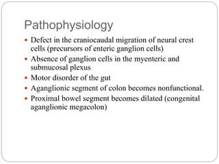
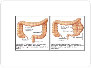
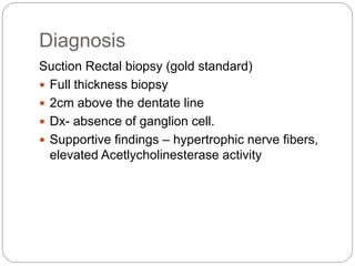
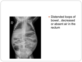
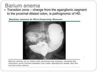
 Hirschsprung disease is a developmental disorder
characterized by absence of ganglia in the distal
intestine, resulting in a functional obstruction
 Most common cause of lower intestinal obstruction in
neonates.
Pathophysiology
 Defect in the craniocaudal migration of neural crest
cells (precursors of enteric ganglion cells)
 Absence of ganglion cells in the myenteric and
submucosal plexus
 Motor disorder of the gut
 Aganglionic segment of colon becomes nonfunctional.
 Proximal bowel segment becomes dilated (congenital
aganglionic megacolon)
The aganglionosis usually starts at the internal anal
sphincter and then extends proximally to
 Rectosigmoid area in about 80% (short segment
disease)
 Beyond sigmoid colon in 15-20%( long segment
disease)
 Whole colon 5% (total colonic aganglionosis)
Epidemiology
 Approximately 1/5000 live births
 Male to female ratio of 4:1 (SSD)
1:1(TCA)
 3% recurrence risk for siblings (SSD)
 Inc length of aganglionic segment….. RR to
siblings also inc and is almost 20% for TCA
 Association with genetic syndromes: downs
syndrome (10%), multiple endocrine neoplasia
type 2A, shah-waardenburg, santos, haddad.
Clinical features
Majority are diagnosed in the neonatal period
 Delayed passage of meconium greater than 48hrs of life
 Abdominal distension
 Bilious vomiting
 Constipation
 Poor feeding
 Squirt sign or blast sign- explosive expulsion of gas and stool after DRE
In older Infants or childhood
 Chronic constipation
 Abdominal distension
 Wasting
 Failure to thrive
If complicated by Enterocolitis
 Fever; vomiting; abdominal distension;
dehydration; peritonitis; explosive, foul-smelling
and sometimes bloody stool.
Diagnosis
Suction Rectal biopsy (gold standard)
 Full thickness biopsy
 2cm above the dentate line
 Dx- absence of ganglion cell.
 Supportive findings – hypertrophic nerve fibers,
elevated Acetlycholinesterase activity
 Distended loops of
bowel , decreased
or absent air in the
rectum
Barium enema
 Transition zone – change from the aganglionic segment
to the proximal dilated colon, is pathognomic of HD.
Anorectal manometry
 Lack of relaxation of the internal anal sphincter
with balloon rectal distension is suggestive of HD.
 PPV of 75-95%
 Less accurate in infants younger than 1/12.
Treatment
 Resuscitation
 NBM
 IV LINE
 NG TUBE
 HYDRATION, ELECTROLYTE IMBALANCE
 URETHRAL CATHETER
 Antibiotics – ampicillin, gentamycin, flagly
Surgery- mainstay of treatment. Goal are to resect
the aganglionic segment, bring the normal bowel
close to the anus and anastomose, and preserve
internal anal sphincter function.
complications
 Intra-op
 Anaesthetic complications
 Hemmorage
 Post-op
 Fecal incontinence
 Constipation (stricture, inc internal anal sphincter
tone)
 Enterocolitis
 Anastomotic leak,
Differentials
 Meconium ileus
 Meconium plug syndrome
 Atresias of the distal ileum and proximal colon
 Necrotising enterocolitis
 Hypothyroidism

Hirschsprung

  • 1.
  • 2.
     Hirschsprung diseaseis a developmental disorder characterized by absence of ganglia in the distal intestine, resulting in a functional obstruction  Most common cause of lower intestinal obstruction in neonates.
  • 3.
    Pathophysiology  Defect inthe craniocaudal migration of neural crest cells (precursors of enteric ganglion cells)  Absence of ganglion cells in the myenteric and submucosal plexus  Motor disorder of the gut  Aganglionic segment of colon becomes nonfunctional.  Proximal bowel segment becomes dilated (congenital aganglionic megacolon)
  • 5.
    The aganglionosis usuallystarts at the internal anal sphincter and then extends proximally to  Rectosigmoid area in about 80% (short segment disease)  Beyond sigmoid colon in 15-20%( long segment disease)  Whole colon 5% (total colonic aganglionosis)
  • 6.
    Epidemiology  Approximately 1/5000live births  Male to female ratio of 4:1 (SSD) 1:1(TCA)  3% recurrence risk for siblings (SSD)  Inc length of aganglionic segment….. RR to siblings also inc and is almost 20% for TCA  Association with genetic syndromes: downs syndrome (10%), multiple endocrine neoplasia type 2A, shah-waardenburg, santos, haddad.
  • 7.
    Clinical features Majority arediagnosed in the neonatal period  Delayed passage of meconium greater than 48hrs of life  Abdominal distension  Bilious vomiting  Constipation  Poor feeding  Squirt sign or blast sign- explosive expulsion of gas and stool after DRE In older Infants or childhood  Chronic constipation  Abdominal distension  Wasting  Failure to thrive
  • 8.
    If complicated byEnterocolitis  Fever; vomiting; abdominal distension; dehydration; peritonitis; explosive, foul-smelling and sometimes bloody stool.
  • 9.
    Diagnosis Suction Rectal biopsy(gold standard)  Full thickness biopsy  2cm above the dentate line  Dx- absence of ganglion cell.  Supportive findings – hypertrophic nerve fibers, elevated Acetlycholinesterase activity
  • 10.
     Distended loopsof bowel , decreased or absent air in the rectum
  • 11.
    Barium enema  Transitionzone – change from the aganglionic segment to the proximal dilated colon, is pathognomic of HD.
  • 12.
    Anorectal manometry  Lackof relaxation of the internal anal sphincter with balloon rectal distension is suggestive of HD.  PPV of 75-95%  Less accurate in infants younger than 1/12.
  • 13.
    Treatment  Resuscitation  NBM IV LINE  NG TUBE  HYDRATION, ELECTROLYTE IMBALANCE  URETHRAL CATHETER  Antibiotics – ampicillin, gentamycin, flagly Surgery- mainstay of treatment. Goal are to resect the aganglionic segment, bring the normal bowel close to the anus and anastomose, and preserve internal anal sphincter function.
  • 14.
    complications  Intra-op  Anaestheticcomplications  Hemmorage  Post-op  Fecal incontinence  Constipation (stricture, inc internal anal sphincter tone)  Enterocolitis  Anastomotic leak,
  • 15.
    Differentials  Meconium ileus Meconium plug syndrome  Atresias of the distal ileum and proximal colon  Necrotising enterocolitis  Hypothyroidism