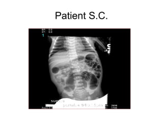
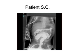
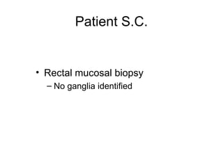
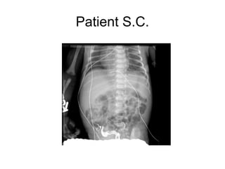
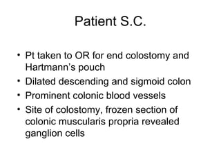
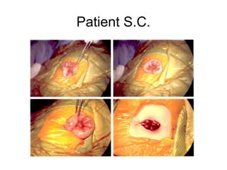
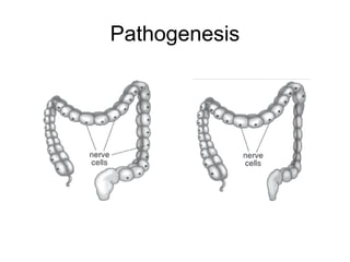
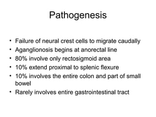
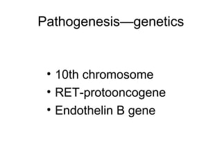
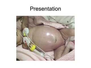
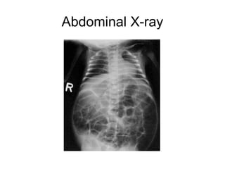
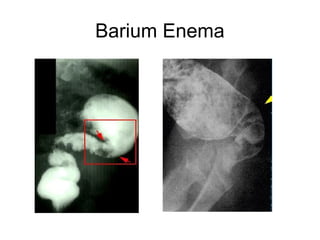
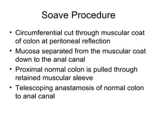
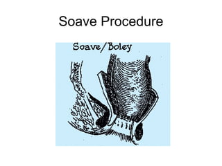
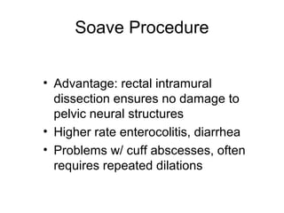
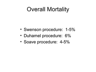
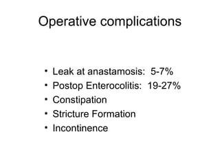
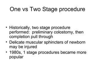
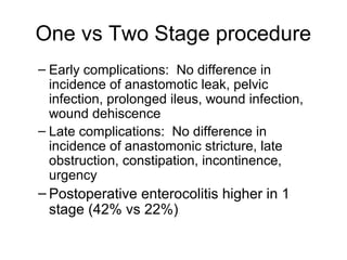
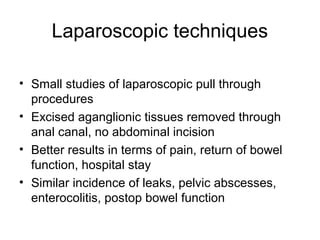
This document discusses the case of a newborn male patient presenting with symptoms of Hirschsprung's disease including abdominal distention and failure to pass meconium. Diagnostic testing revealed the absence of ganglion cells in the colon consistent with Hirschsprung's disease. The patient underwent an end colostomy and biopsy which confirmed the diagnosis. He recovered well and was discharged with plans for a future pull through surgery.
Hirschsprung Disease - Approach & ManagementVikas V
Hirschsprung Disease. - A developmental Disorder of Intrinsic Component of Enteric Nervous System.
Also known Congenital Megacolon.
This Presentation deals with The eitology, presentation, diagnosis, medical and surgical management & complications of the same.
Hirchsprung’s disease by Dr Afuye Olubunmi OlusolaAlade Olubunmi
In Hirchsprung's disease, Absence of ganglion cells in the myenteric and submucosal plexus
Upstream bowel becomes dilated secondary to functional obstruction.
NEONATAL BILIOUS VOMITING- PART 1 & 2
Dear Viewers,
Greetings from “Surgical Educator”
Today I have uploaded two videos on “Neonatal bilious Vomiting- Part 1 & 2. In this episode, I talked about various congenital causes for bowel obstruction in neonatal babies that also cause bilious vomiting. Since there are many causes, I have created two videos to cover everything. In Part1, I talked about duodenal atresia, annular pancreas, malrotation, jejunal & ileal atresia and necrotising enterocolitis. In Part2, I talked about Hirschsprung’s disease, meconium ileus, meconium plug, small left colon syndrome and meconium peritonitis. I request you to watch both videos together and I hope you will enjoy them. You can watch all my surgical teaching video casts in the following links:
Surgicaleducator.blogspot.com
Youtube.com/c/surgicaleducator
Thank you for your support.
HIRSCHSPRUNG DISEASE of neonate wrr.pptxShambelNegese
disease is a condition that affects the large intestine (colon) and causes problems with passing stool. The condition is present at birth (congenital) as a result of missing nerve cells in the muscles of the baby's colon.
Inguinal hernia presentation
by Shariatyfar MD
based on schwartz principles of surgery 11th edition
Qom university of medical sciences
winter 2017
email me at Mohammadali.shariatyfar@hotmail.com for Download
Good luck
Small intestine/Intestinal obstruction/crohns disease/ileostomy/viscous organ...RajeevPandit10
all about small intestine, anatomy, physiology, intestinal obstruction, crohns disease/ileostomy/viscous organ perforation, meckels diverticulum, mysenteric ischemia, short bowel syndrome, celiac disease
Information about Abdominal sepsis and peritonitis final by Dr Dhaval Mangukiya.
Details of Anatomy, intra abdominal infections, physiology, peritonitis, risks for failure of source control, management of critical issues.
https://drdhavalmangukiya.com/
http://www.youtube.com/c/DrDhavalMangukiyaGastrosurgeonSurat
https://gastrosurgerysurat.blogspot.com/
Approximately 75% of abdominal wall hernias occur in the groin.
The lifetime risk of inguinal hernia is 27% in men and 3% in women.
And hence Of inguinal hernia repairs, 90% are performed in men, and 10% are performed in women.
The incidence of inguinal hernia in men has a distribution, with peaks before the first year of life and after age 40.
Indirect inguinal and femoral hernias occur more commonly on the right side.
This is attributed to a delay in atrophy of the processus vaginalis after the normal slower descent of the right testis to the scrotum during fetal development.
The predominance of right-sided femoral hernias is thought to be caused by the tamponading effect of the sigmoid colon on the left femoral canal
The prevalence of hernias increases and the likelihood of strangulation and need for hospitalization increase with aging.
2. Patient S.C.
• Newborn male
• Full-term, uncomplicated vaginal
delivery
• Normal birth weight: 3115 g
• Apgars 91, 95
• Mother: 36 yo, G1P0, healthy
3. Patient S.C.
• Started breast feeding DOL 1
• DOL 2-3 noted to have increasing
abdominal distention
• No meconium passed in first 24 hrs of
life
• 1 episode Non-bilious emesis
5. Patient S.C.
• Pediatric Surgical Consult
• Rectal Exam
– Empty rectal ampulla
– Tight anal sphincter
– Large amount of stool and air upon
withdrawal of finger
9. Patient S.C.
• Pt taken to OR for end colostomy and
Hartmann’s pouch
• Dilated descending and sigmoid colon
• Prominent colonic blood vessels
• Site of colostomy, frozen section of
colonic muscularis propria revealed
ganglion cells
11. Patient S.C.
• Postoperative course uneventful
• Stool from colostomy POD 1
• Tolerated breast feeding
• Discharged POD 6
• 2nd stage pull through procedure
planned in several weeks
13. Hirschsprung’s Disease
• Neurogenic form of intestinal obstruction
• Absence of ganglion cells in the myenteric
and submucosal plexus
• Failure in relaxation of the internal anal
sphincter and affected bowel
• Upstream bowel becomes dilated
secondary to functional obstruction
14. History
• 1691 Ruysch latin texts
• 1886 Harald Hirschsprung – autopsy
• 1901 Tittel – histologic findings
• 1949 Swenson – pathophysiology and
definitive operative treatment
15. Epidemiology
• Prevalence: 1/5000 births
• 3-5% of pts have Down’s syndrome
• Definite family history
• 80% affected are boys
• Total colonic aganglionosis, 35% girls
• >95% cases are full term babies
17. Pathogenesis
• Failure of neural crest cells to migrate caudally
• Aganglionosis begins at anorectal line
• 80% involve only rectosigmoid area
• 10% extend proximal to splenic flexure
• 10% involves the entire colon and part of small
bowel
• Rarely involves entire gastrointestinal tract
20. Presentation
• Severe abdominal distention
• 95% - failure to pass meconium in first 24
hours life
• Bilious vomiting
• Older children - constipation, failure to thrive
• 10-15% - severe diarrhea alternating w/
constipation—enterocolitis of Hirschsprung’s
disease
24. Barium Enema
• Less sensitive for detecting short lesions,
total colon aganglionosis, and disease of
the newborn
• Many newborns do NOT show definitive
transition zone
• Delayed evacuation of contrast
30. Duhamel Procedure
• Posterior portion of defective colon
segment resected
• Side to side anastamosis to left over
portion of rectum
• Constipation a major problem d/t
remaining aganglionic tissue
• Simpler operation, less dissection
32. Soave Procedure
• Circumferential cut through muscular coat
of colon at peritoneal reflection
• Mucosa separated from the muscular coat
down to the anal canal
• Proximal normal colon is pulled through
retained muscular sleeve
• Telescoping anastamosis of normal colon
to anal canal
37. One vs Two Stage procedure
• Historically, two stage procedure
performed: preliminary colostomy, then
completion pull through
• Delicate muscular sphincters of newborn
may be injured
• 1980s, 1 stage procedures became more
popular
38. One vs Two Stage procedure
– Early complications: No difference in
incidence of anastomotic leak, pelvic
infection, prolonged ileus, wound infection,
wound dehiscence
– Late complications: No difference in
incidence of anastomonic stricture, late
obstruction, constipation, incontinence,
urgency
– Postoperative enterocolitis higher in 1
stage (42% vs 22%)
39. Laparoscopic techniques
• Small studies of laparoscopic pull through
procedures
• Excised aganglionic tissues removed through
anal canal, no abdominal incision
• Better results in terms of pain, return of bowel
function, hospital stay
• Similar incidence of leaks, pelvic abscesses,
enterocolitis, postop bowel function
Editor's Notes
Dilated small and large bowel loops, prominent transverse, descending, sigmoid
Narrow rectum, dilated sigmoid colon, normal appearing remaining large bowel.