Downloaded 141 times


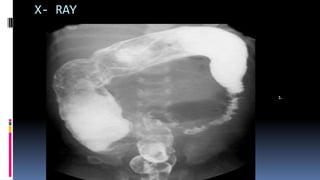

Hirschsprung's disease is a congenital condition in newborns characterized by the absence of intramural ganglion cells in the anorectum, which can affect varying lengths of the colon. It presents with symptoms such as delayed meconium passage and abdominal distention, primarily in males, and can be associated with genetic disorders. Diagnosis is confirmed through imaging, biopsies, and anorectal manometry, with treatment often requiring colostomy and surgical excision of the aganglionic segment.


![ONFH[AVN HIP] -TRIPLE REGIME -A NOVAL SURGICAL CONCEPT .pptx](https://cdn.slidesharecdn.com/ss_thumbnails/onfhavnhip2026koaconcalicutdrgokuldevdrmashraf-260210064517-213ec005-thumbnail.jpg?width=640&height=640&fit=bounds)


