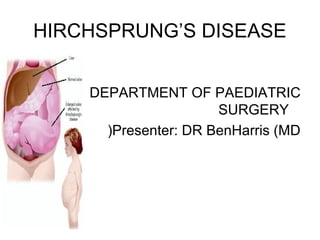
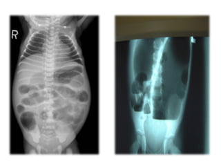
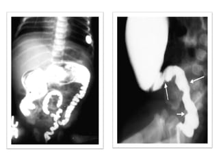
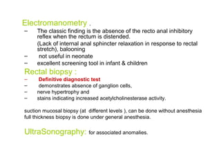
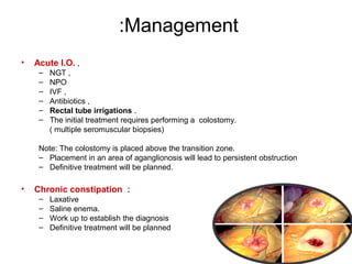
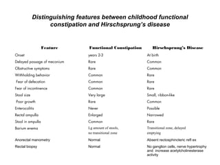
2. Introduction
• A.k,a Congenital aganglionic megacolon
• Discovered by Harald Hirchsprang (1886(
• Is a condition that affects GIT and usually causes problems with passing
stool.
• Present when a baby is born (congenital( and results from missing nerve
cells in the muscles of part or all of the baby’s colon.
• most common cause of lower intestinal obstruction in neonates.
• Familial tendency
• Risk factors;
- +ve family hx,
- being male,
- having other inherited conditions (heart problems, Down sydrome(
3. Incidence
• 15000 live birth newborn
• 70-80% is boys.
• (M / F. 4: 1 (
• Total colonic aganglionosis, 35% girls
• 3-5% have Down’s sydrome
• Less common in blacks.
• >95% cases are full term babies
• Prematurity is reported in as many as 10% of those children with HD
• Familial tendency (Dominant pattern of inheritance(
4. EMBRIYOLOGY
During normal fetal development cells from
neural crest migrate into the large intestine to
form the network of nerves called
Auerbach’s plexus (Muscularis externa( and
(Meissner’s plexus ( submucosa
Occurs in the end of first trimester-
Lack of these nerves causes failure of -
. relaxation of the involved part of the colon
Also supplied by sympathetic nerves, and -
(intrinsic component (enteric nervous system
5. HD, due to failure of neural crest cells to migrate
caudally
Aganglionosis begins at anorectal line
(internal anal sphincter (
extend only up to rectosigmoid junction 80%
(Short segment(
extend proximal to splenic flexure 10%
involves the entire colon and part of small bowel 10%
Rarely involves entire gastrointestinal tract
6. Types
1. Congenital :
Commonest
2. Acquired :
Degeneration of the ganglions may occur due to:
-Vascular causes like after pullthrough procedure due to
ischemia & tension.
- Non vascular causes like
Trypanosoma (chaga's disease(.
Vit B1 def.
Chronic infection ( TB.(.
7. Causes
No clear cause identified-
Due to mutations in one of several genes-
(RET proto-oncogene locus10q11.2 (most common
EDNRB, locus 13q22 >endothelin receptor type B
EDN3, locus 20q13.2-q13.3 >endothelin 3
RET gene
Provides instructions for producing a protein that is involved in -
signalling within cells,essential for the normal development of
(several kinds of nerve cells (cells of the neural crest
Ass with thyroid cancer and neuroblastoma -
EDNRB, EDN3 > coded proteins helps to connect the nerve
cells to to the digestive tract
Other: Nueregulin 3(NRG3( > formation of enteric nervous sytem
.
8. PATHOPHYSIOLOGY
• The gross pathologic feature of HD is a dilated proximal intestine with gradual or abrupt transition
to normal calibrated distal intestine . .
• The colon proximal to the aganglionic segment, in an effort to overcome the partial obstruction,
becomes distended and its wall markedly thickened because of muscle hypertrophy
• The degree of hypertrophy and dilatation depends upon the duration and degree of obstruction
and thus, indirectly to the age of the patient.
• TZ is typically funnel like or cone shaped
9. ASSOCIATED ANOMALIES
HD is usually a solitary anomaly in a full term, otherwise healthy infant
Associated anomalies do occur in nearly 20% of cases
• urogenital system (11%)
• cardiovascular system (6%)
• gastrointestinal system (6%),
• with 8% having various other malformations
Ass sydromes:
Waardenburg-Shah sydrome
Trisomy 21 occurs in approximately 5% of cases
Mowat-Wilson sydrome,
Goldberg-Shpritez megacolon sydrome, and
Congenital central hypoventilation sydrome.
MEN2 (Multiple endocrine neoplasia)
10. Waardeberg syndrome
An inherited auotosomal dominant disorder
hearing loss-
Pigmented anomalies affecting the eyes, -
hair, skin and various defects of neural
crest derived tissues
11. :Four variants
Types 1-4
Type 1: common cause of genetic deafness-
)Type 4:( ass with Hirchsprung’s disease-
12. Signs n symptoms
In newborns
-Failure to pass muconium
-Abd distension
-Vomiting (bilious)
-Constipation or gas
-Diarrhoea
In older children
-Chronic constipation
-abd distension
-failure to gain weight (growth retardation)
13. COMPLICATIONS
Neonatal Intestinal obstruction
-bilious vomiting,
-abdominal distension
-failure to pass muconium
Recurrent Enterocolitis
mainly in the 1st three months of life.
-fever,
-lethargy,
-anorexia,
-vomiting,
-abd distenon and
-diarrhoea
Tx: antibiotics, antpyretics, fluids
Spontanous perforation occurs in 3%, specially if long segment aganglionosis.
Chronic constipation
Growth retardation
Volvulus.
14. Diagnosis
History
Failure to pass meconium,
painless abdominal distension &
constipation
Physical examinations
Distended abdomen
Multiple fecal masses on abdominal examination
On DRE:
• Anal sphincter is hypertonic
• Rectal ampulla is typically empty.
• Hard fecal mass.
• Gash of air apon withdrawal of finger
15. Investigations
Radiology
1. Plain x-rays of the abdomen :Erect & supine
2. Contrast enema
– contrast enema should be done without preparation
of bowel
– Shows narrow distal segment,
– funnel-shaped dilatation at level of transition zone
with marked dilatation of the proximal colon.
24-hrs delayed films
(child with psychogenic stool holding)
16.
17.
18. Electromanometry .
– The classic finding is the absence of the recto anal inhibitory
reflex when the rectum is distended.
(Lack of internal anal sphincter relaxation in response to rectal
stretch), balooning
– not useful in neonate
– excellent screening tool in infant & children
Rectal biopsy :
– Definitive diagnostic test
– demonstrates absence of ganglion cells,
– nerve hypertrophy and
– stains indicating increased acetylcholinesterase activity.
suction mucosal biopsy (at different levels ), can be done without anesthesia
full thickness biopsy is done under general anesthesia.
UltraSonography: for associated anomalies.
19. :Management
• Acute I.O. ,
– NGT ,
– NPO
– IVF ,
– Antibiotics ,
– Rectal tube irrigations .
– The initial treatment requires performing a colostomy.
( multiple seromuscular biopsies)
Note: The colostomy is placed above the transition zone.
– Placement in an area of aganglionosis will lead to persistent obstruction
– Definitive treatment will be planned.
• Chronic constipation :
– Laxative
– Saline enema.
– Work up to establish the diagnosis
– Definitive treatment will be planned
20. :Definitive procedures
By the age of 6-12 months; 9kg or more), a formal pull-through procedure is done
1. Open surgery :
There are many surgical options for Pull-through operation.
All aiming at resection of aganglionic segment
They give excellent result in 90%.
a.swenson.
b.soave.
c.Rehbein.
d.Duhamel.
e. Boley's.
21. Swenson Procedure
Sharp extrarectal dissection down
to 2 cm above the anal canal
Aganglionic colonic segment
resected
End-to-end anastamosis of normal
proximal colon to anal canal
Completely removes defective
aganglionic colon
22. Duhamel Procedure
Posterior portion of defective colon
segment resected
Side to side anastamosis to left
over portion of rectum
Constipation a major problem d/t
remaining aganglionic tissue
Simpler operation, less dissection
23. Soave Procedure
Circumferential cut through
muscular coat of colon at peritoneal
reflection
Mucosa separated from the
muscular coat down to the anal
canal
Proximal normal colon is pulled
through retained muscular sleeve
Telescoping anastamosis of normal
colon to anal canal
Advantage: rectal intramural dissection
ensures no damage to pelvic neural
structures
Higher rate enterocolitis, diarrhoea, often
requires repeated dilations
24. 2. LAPAROSCOPY .
Transanal endorectal pullthrough
Excised aganglionic tissues removed through anal -
canal
no abdominal incision-
Better results in terms of pain, return of bowel -
function, shortens hospital stay
Similar incidence of leaks, pelvic abscesses, -
.enterocolitis
25. One vs Two Stage procedure
,Historically
Two stage procedure performed:
preliminary colostomy, then completion
pull through
Delicate muscular sphincters of newborn -
may be injured
1980s, 1 stage procedures became more
popular
26. Comparison by complications
Early complications: No difference in incidence
of anastomotic leak, pelvic infection,
prolonged ileus, wound infection, wound
dehiscence
Late complications: No difference in incidence
of anastomonic stricture, late obstruction,
constipation, incontinence, urgency
Postoperative enterocolitis higher in 1
(stage (42% vs 22%
27. COMPLICATIONS
1. anastomotic leak.
2. stricture .
3. retraction of the colon.
4. fecal incontinence (soiling/encopresis/paradoxical
diarrhoea (.
5. persistant constipation.
NOTE:
-Afebrile Dirrhoea soon after pullthrough is
expected
-Fluid and observation, Avoid antemetics
28. Distinguishing features between childhood functional
constipation and Hirschsprung’s disease
Feature Functional Constipation Hirschsprung’s Disease
Onset years 2-3 At birth
Delayed passage of meconium Rare Common
Obstructive symptoms Rare Common
Withholding behavior Common Rare
Fear of defecation Common Rare
Fear of incontinence Common Rare
Stool size Very large Small, ribbon-like
Poor growth Rare Common
Enterocolitis Never Possible
Rectal ampulla Enlarged Narrowed
Stool in ampulla Common Rare
Barium enema Lg amount of stools,
no transitional zone
Transitional zone, delayed
emptying
Anorectal manometry Normal Absent rectosphincteric refl ex
Rectal biopsy Normal No ganglion cells, nerve hypertrophy
and increase acetylcholinesterase
activity