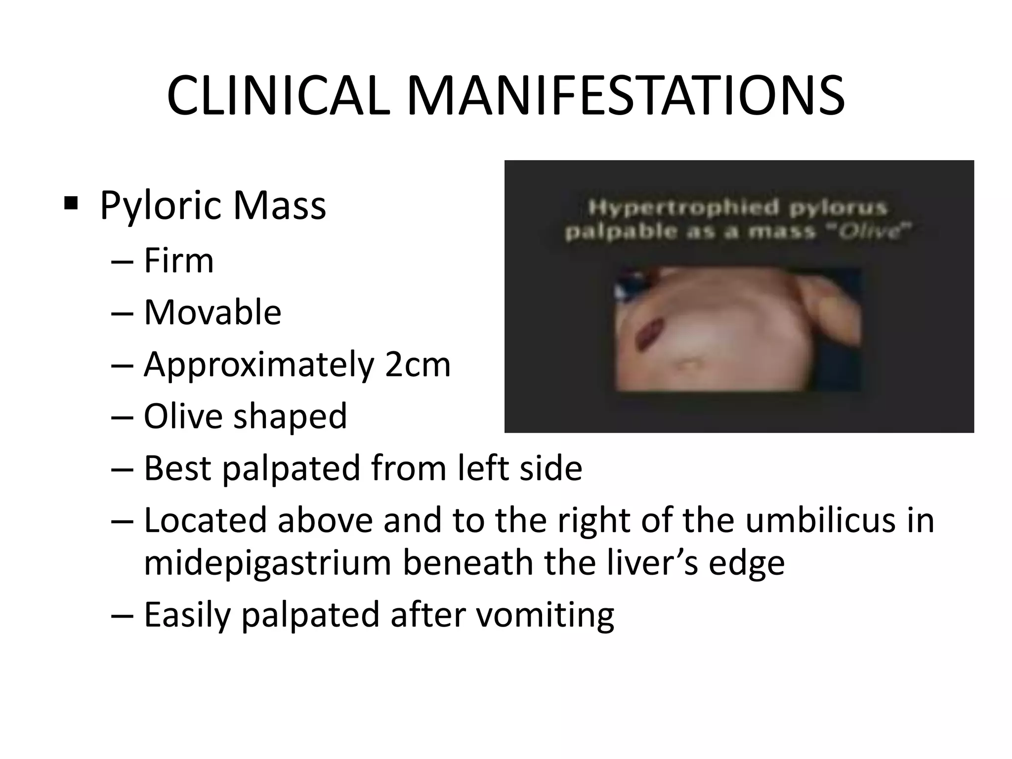
Kelly, a 6-month old infant, has been vomiting
whenever she is fed. Her parents thought that she is
just overfed, but this has continued for 2 weeks
already. They also noticed an olive-shaped mass
whenever her abdomen is gently palpated. They
consulted at their local pediatrician.
What may be the problem?
Hypertrophic Pyloric Stenosis
Presented by-
N/Cdt P. Soorya Suresh
Guided by-
Maj Kavita Rajwar
INTRODUCTION
Pyloric stenosis, also known as infantile
hypertrophic pyloric stenosis (IHPS), is the
most common cause of intestinal
obstruction in infancy.
Anatomy
DEFINITION
Hypertrophic pyloric stenosis is the marked
or progressive overgrowth or enlargement of
circular muscle fibers of pylorus causing
partial or total obstruction of the stomach
outlet due to narrowing of the lumen.
EPIDEMIOLOGY
o Occurs in 1-3 per 1,000 infants.
o More common in whites of Northern Europe, less in
blacks and rare in Asians.
o Males (especially first borns) are affected
approximately 4-6 times as compared to females.
o 20% of male child and 10% of female child of affected
mother develops pyloric stenosis.
o Increased incidence is seen in blood groups B and O
o Monozygotic twins are more affected than the
dizygotic twins.
ETIOLOGY AND PATHOPHYSIOLOGY
• Exact cause is unknown.
• Initially thought to be a congenital condition,
but it is an acquired condition.
• Risk factors for development of Hypertrophic
Pyloric Stenosis-
o Erythromycin exposure in neonates with highest
risk if medication is given within the first 2wks of
life.
o Female infants of mothers treated with
macrolide antibiotics (e.g,. Azithromycin)
during pregnancy and breastfeeding.
o Abnormal muscle innervation
o Increased serum prostglandin level
ETIOLOGY AND PATHOPHYSIOLOGY
(Contd..)
o Decreased
level of
neuronal
nitric oxide
synthase
Decreased
expression of
neuronal nitric
oxide synthase
exon 1c
regulatory
region
Affects
expression of
neuronal nitric
oxide synthase
gene
Decreased nitric
oxide levels
Development of
Hypertrophic Pyloric
Stenosis.
ETIOLOGY AND PATHOPHYSIOLOGY
(Contd..)
VOMITING HYPERBILIRUBINEMIA
PYLORIC MASS
VISIBLE GASTRIC PERISTALSIS
AFTER FEEDING
CLINICAL
MANIFESTATIONS
CLINICAL MANIFESTATIONS
 Vomiting
– Non bilious
– May or may not be projectile
– Immediately after feeding
– May occur after each feeds or may be intermittent
– Starts after 3 wks of age but can develop as soon as 1 wk
or as late as 5 months
– After vomiting infant is hungry and wants to feed again.
– Leads to progressive loss of
fluids, H+ and Cl- hypochloremic metabolic alkalosis.
CLINICAL MANIFESTATIONS
 Hyperbilirubinemia
– Most common clinical association
– also known as icteropyloric syndrome
– Unconjugated > conjugated hyperbilirubinemia;
corrects by surgery
– Associated with decreased levels of glucuronyl
transferase (5% of patients) due to mutation in
bilirubin uridine diphosphate glucuronosyl-
transferase gene (UGT1A1)
CLINICAL MANIFESTATIONS
 Pyloric Mass
– Firm
– Movable
– Approximately 2cm
– Olive shaped
– Best palpated from left side
– Located above and to the right of the umbilicus in
midepigastrium beneath the liver’s edge
– Easily palpated after vomiting
CLINICAL MANIFESTATIONS
 Visible gastric peristalsis after feeding
DIAGNOSIS
• History & physical
examination
• Clinical findings
• USG (criteria for
diagnosis) Pyloric-
o thickness 3-4cm
o length 15-17cm
o diameter- >13cm
• Contrast studies-
o String sign-
-elongated pyloric channel
o Shoulder sign-
-bulge of pyloric muscle
into antrum
o Double tract sign-
-parallel streaks of barium
seen in narrow channel
RADIOGRAPHIC FEATURES
1) Plain radiograph
• Abdominal x-ray findings are
non-specific but may show a
distended stomach with
minimal distal intestinal bowel
gas.
2) Fluoroscopy
An UGI (barium meal) excludes other, more serious causes
of pathology, but the findings of an upper gastrointestinal series
infer, rather than directly visualize, the hypertrophied muscle.
On upper gastrointestinal fluoroscopy:
• delayed gastric emptying
• peristaltic waves (caterpillar sign)
• elongated pylorus with a narrow lumen (string sign) which may
appear duplicated due to puckering of the mucosa (double-track
sign)
• the pylorus indents the contrast-filled antrum (shoulder sign)
and (tit sign) or base of the duodenal bulb (mushroom sign)
• the entrance to the pylorus may be beak-shaped (beak sign)
RADIOGRAPHIC FEATURES (contd…)
Fluoroscopy
Bulge in the distal antrum
with streak of barium
pointing towards pyloric
canal- BEAK SIGN
The barium may outline
the crowded mucosal
folds as parallel lines-
DOUBLE TRACT SIGN
RADIOGRAPHIC FEATURES (contd…)
3) Ultrasound
• Ultrasound is the modality of choice
• Advantages over a barium meal are-
o it directly visualizes the pyloric muscle,
o does not use ionizing radiation.
• Disadvantages- it is incapable of
excluding other diagnoses such as midgut
volvulus.
• The hypertrophied muscle is hypoechoic,
and the central mucosa is hyperechoic.
• Diagnostic measurements include:
– pyloric muscle thickness (diameter of a
single muscular wall on a transverse
image): >3 mm
– length: >15-17 mm
– pyloric volume: >1.5 cm3
– pyloric transverse diameter: >13 mm
DIFFERENTIAL DIAGNOSIS
1) GERD with/ without hiatal hernia.
2) Adrenal insufficiency from adreno-genital syndrome
can stimulate pyloric stenosis.
– absence of metabolic acidosis
– increased serum K+
– urinary Na+ concentration of adrenal insufficiency
3) Inborn errors of metabolism can cause recurrent
episodes of emesis with alkalosis( urea cycle) or
acidosis( organic acidemia) & lethargy, coma,
seizures.
DIFFERENTIAL DIAGNOSIS (CONTD…)
4) Pyloric membrane/ pyloric duplication
-projectile vomiting
-visible peristalsis and
-palpable mass
5) Duodenal stenosis proximal to ampulla of Vater
- Clinical features of HPS
- Differentiate by physical examination or USG
(absence of mass).
In duplication
COMPLICATIONS
Post-op complications include-
 infections
 fluid-electrolyte imbalances
 persistent obstruction
 duodenal perforation
PROGNOSIS
o Prognosis is excellent, unless diagnosis is
delayed and prolonged severe dehydration
occurs.
o Mortality rare is after pyloromytomy.
MANAGEMENT
PRE-OPERATIVE
• Ramstedt’s Pyloromyotomy
• Laparoscopic Pyoloromyotomy
SURGICAL
CONSERVATIVE
• Pre-Operative
• Post-Operative
NURSING
PREOPERATIVE MANAGEMENT
• Correcting imbalances of-
─ Fluid
─ Acid-base
– Electrolyes
• Correction of alkalosis is essential to prevent
post operative apnea due to anesthesia
SURGICAL MANAGEMENT
• Ramstedt’s Pyloromytomy
─ Through a short transverse skin incision
─ Underlying pyloric mass is cut longitudinally to
the layer of submucosa
─ Incision is closed
• Laparoscopic pyloromyotomy
─ Equally successful
─ Shorter time to full feeding, discharge & great
parental satisfaction.
─ Success depends on surgeon’s skills
SURGICAL MANAGEMENT (contd..)
o Post- op vomiting occurs in half of the infants;
maybe because of edema at the incision in the
pylorus.
o Feedings can be started in 12-24 hrs after surgery
o Persistent vomiting indicates incomplete surgery,
gastritis, GERD or any other obstruction
o Endoscopic balloon dilation is done for
incomplete pyloromyotomy
SURGICAL MANAGEMENT (contd..)
CONSERVATIVE MANAGEMENT
• Nasoduodenal feedings in patients who aren’t good candidates for
surgery
• Oral & IV atropine sulphate (pyloric muscle relaxant); 80% success
rate.
• IV atropine
– Dose- 0.01 mg/kg
– 6 times a day 5min before feeding
– Monitor HR & ECG
• Oral feeding-
– Started at a volume of 10ml, 6 times/day
– Volume is increased until patient tolerates 150ml/kg/day unless
vomiting occurs > 2 times/day.
– When feeds are tolerated without vomiting not more than 2
times/day; 0.02mg/kg oral atropine is started before feeding.
NURSING MANAGEMENT
I. Pre-Op:
 Maintenance of fluid & electrolyte along with nutritional intake of
breastfeeding (if not contraindicated).
 Relief of parental anxiety
 Nasogastric aspiration of gastric distension for vomiting relief.
 During feeding, baby should be on upright position, slightly
towards right.
 Small; frequent feedings
 Prevention of HAI
 Continuous monitoring
 Vitals, hydration, body weight, vomiting, stool, urine, signs of
compications.
 Parental involvement in care of the baby.
NURSING MANAGEMENT (contd..)
II. Post-Op:
 Basic anesthesia care and post-op care to be provided
with warmth, feeding, wound care, medications, hygienic
care, emotional support to the parents and health
education with discharge advice and follow-up.
 Special attention on oral feeds, usually after 8-12 hrs of
surgery, small frequent feeds with EBM.
 Initially, feedings can be started in a stepwise manner
with 1-2 tsp of clear solution (5% glucose) q12hrly for 8
hrs, after anesthesia effect.
 After feeding, baby should be placed in an upright
position towards the right for 45-60 min.
 Usually discharged on 3rd-4th post-op day.
Hypertrophic Pyloric Stenosis

Hypertrophic Pyloric Stenosis

  • 1.
    Kelly, a 6-monthold infant, has been vomiting whenever she is fed. Her parents thought that she is just overfed, but this has continued for 2 weeks already. They also noticed an olive-shaped mass whenever her abdomen is gently palpated. They consulted at their local pediatrician. What may be the problem?
  • 2.
    Hypertrophic Pyloric Stenosis Presentedby- N/Cdt P. Soorya Suresh Guided by- Maj Kavita Rajwar
  • 3.
    INTRODUCTION Pyloric stenosis, alsoknown as infantile hypertrophic pyloric stenosis (IHPS), is the most common cause of intestinal obstruction in infancy.
  • 4.
  • 5.
    DEFINITION Hypertrophic pyloric stenosisis the marked or progressive overgrowth or enlargement of circular muscle fibers of pylorus causing partial or total obstruction of the stomach outlet due to narrowing of the lumen.
  • 6.
    EPIDEMIOLOGY o Occurs in1-3 per 1,000 infants. o More common in whites of Northern Europe, less in blacks and rare in Asians. o Males (especially first borns) are affected approximately 4-6 times as compared to females. o 20% of male child and 10% of female child of affected mother develops pyloric stenosis. o Increased incidence is seen in blood groups B and O o Monozygotic twins are more affected than the dizygotic twins.
  • 7.
    ETIOLOGY AND PATHOPHYSIOLOGY •Exact cause is unknown. • Initially thought to be a congenital condition, but it is an acquired condition. • Risk factors for development of Hypertrophic Pyloric Stenosis- o Erythromycin exposure in neonates with highest risk if medication is given within the first 2wks of life.
  • 8.
    o Female infantsof mothers treated with macrolide antibiotics (e.g,. Azithromycin) during pregnancy and breastfeeding. o Abnormal muscle innervation o Increased serum prostglandin level ETIOLOGY AND PATHOPHYSIOLOGY (Contd..)
  • 9.
    o Decreased level of neuronal nitricoxide synthase Decreased expression of neuronal nitric oxide synthase exon 1c regulatory region Affects expression of neuronal nitric oxide synthase gene Decreased nitric oxide levels Development of Hypertrophic Pyloric Stenosis. ETIOLOGY AND PATHOPHYSIOLOGY (Contd..)
  • 10.
    VOMITING HYPERBILIRUBINEMIA PYLORIC MASS VISIBLEGASTRIC PERISTALSIS AFTER FEEDING CLINICAL MANIFESTATIONS
  • 11.
    CLINICAL MANIFESTATIONS  Vomiting –Non bilious – May or may not be projectile – Immediately after feeding – May occur after each feeds or may be intermittent – Starts after 3 wks of age but can develop as soon as 1 wk or as late as 5 months – After vomiting infant is hungry and wants to feed again. – Leads to progressive loss of fluids, H+ and Cl- hypochloremic metabolic alkalosis.
  • 12.
    CLINICAL MANIFESTATIONS  Hyperbilirubinemia –Most common clinical association – also known as icteropyloric syndrome – Unconjugated > conjugated hyperbilirubinemia; corrects by surgery – Associated with decreased levels of glucuronyl transferase (5% of patients) due to mutation in bilirubin uridine diphosphate glucuronosyl- transferase gene (UGT1A1)
  • 13.
    CLINICAL MANIFESTATIONS  PyloricMass – Firm – Movable – Approximately 2cm – Olive shaped – Best palpated from left side – Located above and to the right of the umbilicus in midepigastrium beneath the liver’s edge – Easily palpated after vomiting
  • 14.
    CLINICAL MANIFESTATIONS  Visiblegastric peristalsis after feeding
  • 15.
    DIAGNOSIS • History &physical examination • Clinical findings • USG (criteria for diagnosis) Pyloric- o thickness 3-4cm o length 15-17cm o diameter- >13cm • Contrast studies- o String sign- -elongated pyloric channel o Shoulder sign- -bulge of pyloric muscle into antrum o Double tract sign- -parallel streaks of barium seen in narrow channel
  • 16.
    RADIOGRAPHIC FEATURES 1) Plainradiograph • Abdominal x-ray findings are non-specific but may show a distended stomach with minimal distal intestinal bowel gas.
  • 17.
    2) Fluoroscopy An UGI(barium meal) excludes other, more serious causes of pathology, but the findings of an upper gastrointestinal series infer, rather than directly visualize, the hypertrophied muscle. On upper gastrointestinal fluoroscopy: • delayed gastric emptying • peristaltic waves (caterpillar sign) • elongated pylorus with a narrow lumen (string sign) which may appear duplicated due to puckering of the mucosa (double-track sign) • the pylorus indents the contrast-filled antrum (shoulder sign) and (tit sign) or base of the duodenal bulb (mushroom sign) • the entrance to the pylorus may be beak-shaped (beak sign) RADIOGRAPHIC FEATURES (contd…)
  • 18.
    Fluoroscopy Bulge in thedistal antrum with streak of barium pointing towards pyloric canal- BEAK SIGN The barium may outline the crowded mucosal folds as parallel lines- DOUBLE TRACT SIGN
  • 20.
    RADIOGRAPHIC FEATURES (contd…) 3)Ultrasound • Ultrasound is the modality of choice • Advantages over a barium meal are- o it directly visualizes the pyloric muscle, o does not use ionizing radiation. • Disadvantages- it is incapable of excluding other diagnoses such as midgut volvulus. • The hypertrophied muscle is hypoechoic, and the central mucosa is hyperechoic. • Diagnostic measurements include: – pyloric muscle thickness (diameter of a single muscular wall on a transverse image): >3 mm – length: >15-17 mm – pyloric volume: >1.5 cm3 – pyloric transverse diameter: >13 mm
  • 21.
    DIFFERENTIAL DIAGNOSIS 1) GERDwith/ without hiatal hernia. 2) Adrenal insufficiency from adreno-genital syndrome can stimulate pyloric stenosis. – absence of metabolic acidosis – increased serum K+ – urinary Na+ concentration of adrenal insufficiency 3) Inborn errors of metabolism can cause recurrent episodes of emesis with alkalosis( urea cycle) or acidosis( organic acidemia) & lethargy, coma, seizures.
  • 22.
    DIFFERENTIAL DIAGNOSIS (CONTD…) 4)Pyloric membrane/ pyloric duplication -projectile vomiting -visible peristalsis and -palpable mass 5) Duodenal stenosis proximal to ampulla of Vater - Clinical features of HPS - Differentiate by physical examination or USG (absence of mass). In duplication
  • 23.
    COMPLICATIONS Post-op complications include- infections  fluid-electrolyte imbalances  persistent obstruction  duodenal perforation
  • 24.
    PROGNOSIS o Prognosis isexcellent, unless diagnosis is delayed and prolonged severe dehydration occurs. o Mortality rare is after pyloromytomy.
  • 25.
    MANAGEMENT PRE-OPERATIVE • Ramstedt’s Pyloromyotomy •Laparoscopic Pyoloromyotomy SURGICAL CONSERVATIVE • Pre-Operative • Post-Operative NURSING
  • 26.
    PREOPERATIVE MANAGEMENT • Correctingimbalances of- ─ Fluid ─ Acid-base – Electrolyes • Correction of alkalosis is essential to prevent post operative apnea due to anesthesia
  • 27.
    SURGICAL MANAGEMENT • Ramstedt’sPyloromytomy ─ Through a short transverse skin incision ─ Underlying pyloric mass is cut longitudinally to the layer of submucosa ─ Incision is closed
  • 28.
    • Laparoscopic pyloromyotomy ─Equally successful ─ Shorter time to full feeding, discharge & great parental satisfaction. ─ Success depends on surgeon’s skills SURGICAL MANAGEMENT (contd..)
  • 29.
    o Post- opvomiting occurs in half of the infants; maybe because of edema at the incision in the pylorus. o Feedings can be started in 12-24 hrs after surgery o Persistent vomiting indicates incomplete surgery, gastritis, GERD or any other obstruction o Endoscopic balloon dilation is done for incomplete pyloromyotomy SURGICAL MANAGEMENT (contd..)
  • 30.
    CONSERVATIVE MANAGEMENT • Nasoduodenalfeedings in patients who aren’t good candidates for surgery • Oral & IV atropine sulphate (pyloric muscle relaxant); 80% success rate. • IV atropine – Dose- 0.01 mg/kg – 6 times a day 5min before feeding – Monitor HR & ECG • Oral feeding- – Started at a volume of 10ml, 6 times/day – Volume is increased until patient tolerates 150ml/kg/day unless vomiting occurs > 2 times/day. – When feeds are tolerated without vomiting not more than 2 times/day; 0.02mg/kg oral atropine is started before feeding.
  • 31.
    NURSING MANAGEMENT I. Pre-Op: Maintenance of fluid & electrolyte along with nutritional intake of breastfeeding (if not contraindicated).  Relief of parental anxiety  Nasogastric aspiration of gastric distension for vomiting relief.  During feeding, baby should be on upright position, slightly towards right.  Small; frequent feedings  Prevention of HAI  Continuous monitoring  Vitals, hydration, body weight, vomiting, stool, urine, signs of compications.  Parental involvement in care of the baby.
  • 32.
    NURSING MANAGEMENT (contd..) II.Post-Op:  Basic anesthesia care and post-op care to be provided with warmth, feeding, wound care, medications, hygienic care, emotional support to the parents and health education with discharge advice and follow-up.  Special attention on oral feeds, usually after 8-12 hrs of surgery, small frequent feeds with EBM.  Initially, feedings can be started in a stepwise manner with 1-2 tsp of clear solution (5% glucose) q12hrly for 8 hrs, after anesthesia effect.  After feeding, baby should be placed in an upright position towards the right for 45-60 min.  Usually discharged on 3rd-4th post-op day.