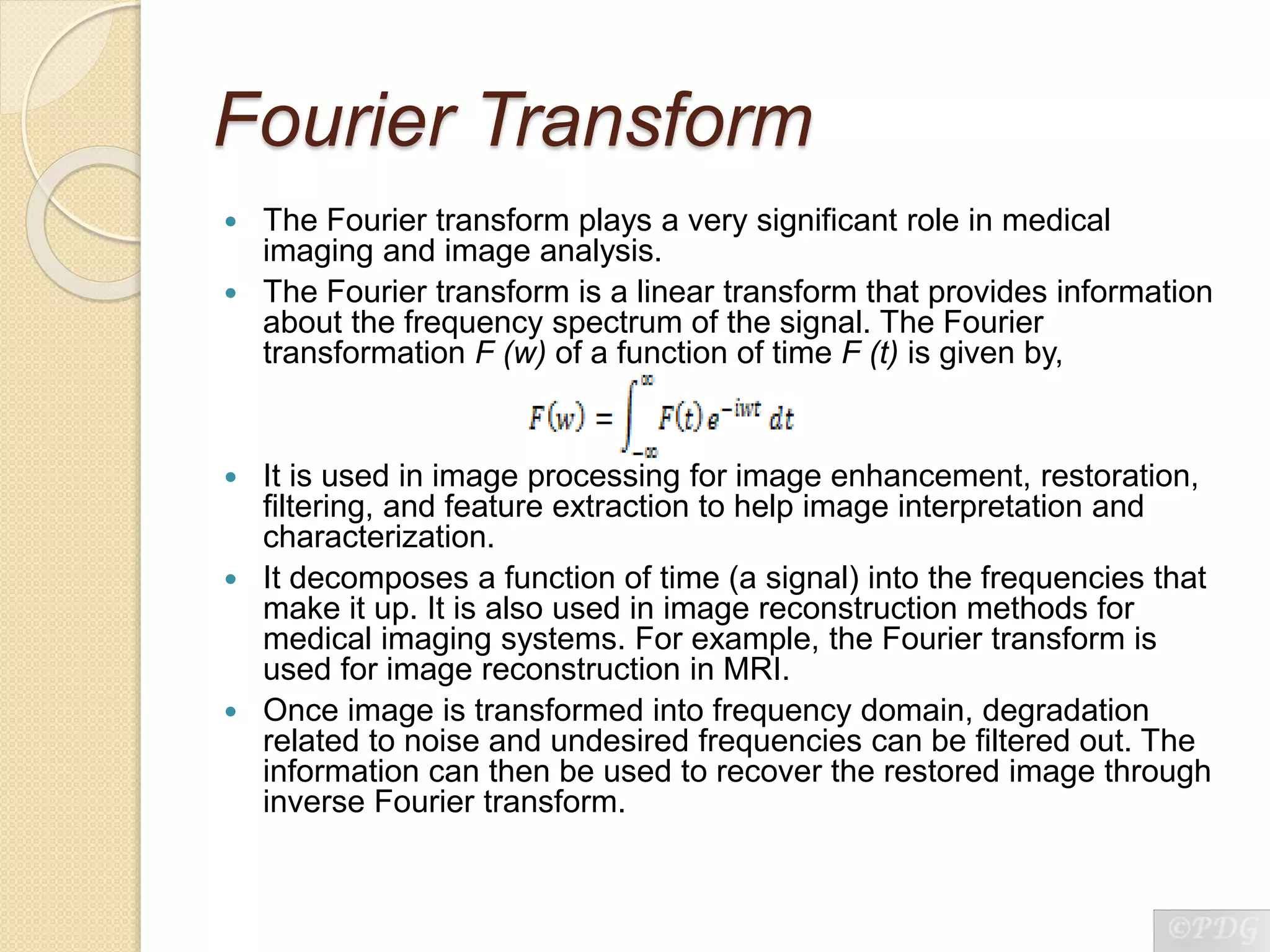
BIOMEDICAL IMAGE
PROCESSING
INTRODUCTION
 Image (from Latin word ‘imago’), is an artifact like
a two dimensional picture, that has a similar
appearance to some subject like a physical
object or a person.
 Image processing is any form of signal
processing for which the input is an image and
the output may either be an image or a set of
characteristics or parameters related to the
image.
 Image processing is used in areas such as
multimedia, computing, secured image
communication, biomedical imaging, remote
sensing, pattern recognition, image compression
and retrieval, etc.
BIOMEDICAL IMAGING
 It is the technique and process used to create
images of the human body or parts of it for
clinical purposes or for studying anatomy and
physiology.
 A multitude of diagnostic medical imaging
systems are used to probe the human body.
They comprise both microscopic (viz. cellular
level) and macroscopic (viz. organ and systems
level) modalities.
 Biomedical image processing includes the
analysis, enhancement and display of images
captured via instruments such as X-Ray,
Ultrasound, MRI (Magnetic Resonance Imaging),
CT scanners, nuclear medicine and optical
imaging technologies.
NEED OF IMAGE
PROCESSING IN MEDICINE
 The images produced by equipments (like CT scanner,
MRI, etc.) are composed of pixels, to which discrete
brightness and color values are assigned.
 Through image processing they can be efficiently
processed, evaluated and analyzed, and through
compression stored and made available to many places
at the same time through appropriate communication
networks and protocols.
 It is possible for doctors to see the interior portions of
the human body, with extreme clarity, ease and detail,
thus facilitating easy detection and diagnosis of various
diseases.
 It has also helped doctors to make keyhole surgeries
without opening too much of the body.
 Image processing techniques that were originally
developed for analyzing remote sensing data can be
modified to analyze the outputs of medical imaging
systems to get the best advantage to analyze
symptoms of patients with ease.
NEED OF IMAGE
PROCESSING IN MEDICINE
Main tasks performed by the image processing unit in medicine are:
 Interfacing analog outputs of sensors such as microscopes,
endoscopes, ultrasound etc., to digitizers and in turn to Image
Processing systems.
 Image enhancements.
 Changing density dynamic range of B/W images.
 Color correction and manipulating of colors within a color image.
 Contour detection and area calculations of the cells of a biomedical
image.
 Restoration and smoothing of images.
 Registration of multiple images and creating mosaic of multiple
images.
 Construction of 3-D images from 2-D images.
 Generation of negative images.
 Zooming of images.
 Removal of artifacts from the image.
PRINCIPLES OF IMAGE
PROCESSING
 An image is usually a function of two spatial
variables, e.g. f[x, y], which represents the
brightness f at the Cartesian location [x, y].
 It can also be defined as an array, or a matrix,
of square pixels (picture elements) arranged
in columns and rows.
 After converting image information into an
array of integers, the image can be
manipulated, processed, and displayed by
computer.
 Computer processing is used for image
enhancement, restoration, segmentation,
description, recognition, coding,
reconstruction, transformation.
Types Of Images
1. Analog image
 An analog image is described by the spatial
distribution of brightness or gray levels that
reflect a distribution of detected energy.
 The image can be displayed using a
medium such as paper or film.
 Black and white images require only one
gray level or intensity variable while color
images require multiple variable like the
three basic colors red, blue, green(RGB).
 When combined together, the RGB
intensities can produce a selected color at a
spatial location of the image.
Types Of Images
2. Digital image
 A digital image is discrete in both spatial
and intensity (gray level) domains.
 A discrete spatial location of finite size with
a discrete gray-level value is called a pixel.
 For example, an image of 1024 x 1024
pixels may be displayed in 8-bit gray-level
resolution. This means that each pixel in the
image may have any value from 0 to 255
(i.e. total of 256 gray levels).
 The pixel dimensions would depend on the
spatial sampling.
Color Formats Used In Image
Processing
1. The RGB color model
 It relates very closely to the way we perceive
color with the r, g and b receptors in our retinas.
 RGB uses additive color mixing and is the basic
color model used in television or any other
medium that projects color with light as in
computers and for web graphics, but it cannot be
used for print production.
Color Formats Used In Image
Processing
2. The CMYK color model
 The 4-colour CMYK model used in printing lays
down overlapping layers of varying percentages
of transparent cyan (C), magenta (M) and yellow
(Y) inks. In addition a layer of black (K) ink can be
added.
 The CMYK model uses the subtractive color
model. Cya
n
YellowMagent
a
Fourier Transform
 The Fourier transform plays a very significant role in medical
imaging and image analysis.
 The Fourier transform is a linear transform that provides information
about the frequency spectrum of the signal. The Fourier
transformation F (w) of a function of time F (t) is given by,
 It is used in image processing for image enhancement, restoration,
filtering, and feature extraction to help image interpretation and
characterization.
 It decomposes a function of time (a signal) into the frequencies that
make it up. It is also used in image reconstruction methods for
medical imaging systems. For example, the Fourier transform is
used for image reconstruction in MRI.
 Once image is transformed into frequency domain, degradation
related to noise and undesired frequencies can be filtered out. The
information can then be used to recover the restored image through
inverse Fourier transform.
COMPONENTS OF IMAGE
PROCESSING
 Biomedical image processing covers
biomedical signal gathering, image forming,
picture processing, and image display to
medical diagnosis based on features
extracted from images. Some basic image
processing techniques include outlining, de-
blurring, noise cleaning, filtering, search and
texture analysis. Image processing covers
four main areas:
Image formation.
Visualization.
Analysis of image.
Management of the acquired information.
COMPONENTS OF IMAGE
PROCESSING
COMPONENTS OF IMAGE
PROCESSING
A. Image Formation
Image formation includes all the steps from capturing the image to forming a
digital image matrix. The main steps are:
 Acquisition: It is defined as the action of retrieving an image from some
source, usually a hardware-based source (For example: a CT scanner).
Performing image acquisition is the first step in the workflow sequence
because, without an image, no processing is possible. The image that is
acquired is completely unprocessed and is the result of whatever hardware
was used to generate it. Acquisition methods vary for different medical
instruments.
 Digitization: It is the process of converting information into a digital format .
In this format, information is organized into discrete units of data (called bits)
that can be separately addressed (usually in multiple-bit groups called bytes).
This is the binary data that computers and many devices with computing
capacity can process. Different analog to digital convertors are used for
converting the acquired data from instruments to digital format. The type of
convertor used depends upon the required resolution, speed, application and
cost. Some commonly available convertors are:
◦ Dual Slope ADC
◦ Successive Approximation ADC
◦ Flash ADC
◦ Serial or ripple ADC
◦ Sigma Delta Convertor Type ADC
◦ Combination of flash and successive approximation type ADC
There is a need to digitize the acquired analog data so that it can be processed
with the help of different software such as MATLAB, etc.
COMPONENTS OF IMAGE
PROCESSINGB. Image Enhancement And Visualization
 It refers to all types of manipulation that is done on the data acquired
in digital format, finally resulting in an optimized output of the image.
 The purpose of image enhancement methods is to process an
acquired image for better contrast and visibility of features of interest
for visual examination as well as subsequent computer-aided
analysis and diagnosis.
 There is no unique general theory or method for processing all kinds
of medical images for feature enhancement. Specific medical
imaging applications (such as cardiac, neurological, muscular,
mammography, etc.) present different challenges in image
processing for feature enhancement and analysis. Medical images
show characteristic information about the physiological properties of
the structures and tissues. However, the quality and visibility of
information depends on the imaging modality and the response
functions of the imaging scanner. Hence the goal of this step is to:
◦ Eliminate the extraneous components such as noise from the signal. Often this
is done using linear filters. Types of filters used are: High pass Filters, Low
pass Filters and Notch pass filters.
◦ Adjust the different parameters of the image such as brightness, contrast,
visibility, color saturation, etc.
 Image enhancement can be accomplished using Adobe Photoshop,
Corel PHOTO-PAINT and Origin software in order to achieve good
COMPONENTS OF IMAGE
PROCESSING
C. Image Analysis
Image analysis includes all the steps of
processing, which are used for
quantitative measurement as well as
interpretation of biomedical images.
These steps require a prior knowledge of
the nature and content of the images,
which is integrated into the algorithm on
a high level of abstraction.
Thus the process of image analysis is
very specific, and developed algorithms
can be transferred directly to application
domains.
COMPONENTS OF IMAGE
PROCESSING
D. Image Management
 Image management sums up all the techniques that provide
the efficient storage, communication, transmission, archiving,
and access (retrieval) of image data.
 The methods of telemedicine are also a part of image
management. There are different formats in which the digital
image can be stored in memory. Some of the most common
file formats used for saving images in the digital form (in hard
drive or memory) is:
◦ GIF: An 8-bit (256 color), non-destructively compressed bitmap
format. Mostly used for web. It has several sub-standards one of
which is the animated GIF.
◦ JPEG: It is a very efficient (i.e. much information per byte)
destructively compressed 24 bit (16 million colors) bitmap format.
◦ TIFF: It is the standard 24 bit publication bitmap format.
 Image compression is also a part of image management.
Through image compression large no. of images can be
stored and made available to many places at the same time
through appropriate communication networks and protocols
such as the Digital Imaging and Communications in Medicine
(DICOM) protocol.
X-Ray Machine and Digital
Radiography
 Two-dimensional projection radiography is the oldest medical imaging
modality and is still one of the most widely used imaging methods in
diagnostics.
 Conventional film radiography uses an X-Ray tube to focus a beam on the
imaging area of a patient's body to record an image on a film. The image
recorded on the film is a 2-D projection of the three-dimensional (3-D)
anatomical structure of the human body.
 Scattering can create a major problem in projection radiography. The
scattered photons can create artifacts and artificial structures in the image
that can lead to an incorrect interpretation or at least create a difficult
situation for diagnosis.
 In case of digital radiography, the combination of intensifying screen and film
is replaced by a phosphor layered screen coupled with a charge-coupled
device (CCD)-based panel. A solid-state detector system in digital
radiography uses a structured thallium-doped cesium iodide (CsI) scintillation
material to convert X-Ray photons into light photon, which are then converted
into electrical signal by CCDs through a fiber optics coupling interface.
 Electrical output signal sensitivity can be controlled much more efficiently
than in a film-based system. The digital detector system also provides
excellent linearity and gain control, which directly affects the SNR of the
acquired data. For this reason, a digital detection system provides a superior
dynamic range compared with the film-based systems. However, the
Magnetic Resonance Imaging
System Magnetic resonance imaging (MRI) is a noninvasive medical test
that helps physicians diagnose and treat medical conditions.
 MRI uses a powerful magnetic field, radio frequency pulses and a
computer to produce detailed pictures of organs, soft tissues, bone
and virtually all other internal body structures.
 The hydrogen proton is the most common form of nuclei used in
MRI. The three properties of hydrogen nuclei (protons) mapping are
the spin-lattice relaxation time Ti, Spin-spin relaxation time T 2, and
the spin density p.
 Magnetic resonance imaging is a complex multidimensional imaging
modality that produces extensive amounts of data. Imaging methods
and techniques applied in signal acquisition allow reconstruction of
images with multiple parameters that represent various physical and
chemical properties of the matter of the object.
 The imager system includes the computer for image processing,
display system and the control console. The computer system
collects the signal after analog to digital conversion, corrects,
recomposes and stores the image.
 Analog to digital convertors of 16 bits or higher are used and during
data acquisition, information is acquired at the rate of about 800
kbps.
 Algorithms like the fast Fourier transformation is used to convert the
time domain data to image data. Data is stored on high speed disks.
Ultrasonic Imaging Systems
 In this method a piezoelectric crystal-based transducer can be used
as a source to form an ultrasound beam as well as a detector to
receive the returned signal from the tissue. In a plastic casing, a
piezoelectric crystal is used along with a damping material layer and
acoustic insulation layer inside the plastic casing. An
electromagnetic tuning coil is used to apply a controlled
voltage pulse to produce ultrasound waves. In the receiver mode,
the pressure wave of the returning ultrasound signal is used to
create an electric signal through the tuned electromagnetic coil.
 The total travel distance traveled by the ultrasound pulse at the time
of return to the transducer is twice the depth of the tissue boundary
from the transducer. Thus, the maximum range of the echo
formation can be determined by the speed of sound in the tissue
multiplied by half of the pulse-repetition period.
 When the echoes are received by the transducer crystal, their
intensity is converted into a voltage signal that generates the raw
data for imaging.
 The voltage signal then can be digitized and processed according to
the need to display on a computer monitor as an image.
 Ultrasound images appear noisy with speckles, lacking a continuous
boundary definition of the object structure. The interpretation and
quantification of the object structure in ultrasound images is more
challenging than in X-ray computed tomography (X-ray CT) or
magnetic resonance (MR) images.
 The operator in ultrasound imaging has a great ability to control the
X-Ray Computed
Tomography It uses computer-processed X-rays to produce images of specific areas of a
scanned object, allowing the user to see inside the object without cutting.
 Digital geometry processing is used to generate a 3-D image of the inside of
the object from a large series of two-dimensional radiographic images taken
around a single axis of rotation. The cross-sectional images are used
for diagnostic and therapeutic purposes in various medical disciplines. CT
produces a volume of data that can be manipulated in order to demonstrate
various bodily structures based on their ability to block the X-ray beam.
 In conventional CT machines, an X-ray tube and detector are physically
rotated behind a circular shroud. Sometimes contrast materials such as
intravenous iodinated contrast are used. This is useful to highlight structures
such as blood vessels that otherwise would be difficult to delineate from their
surroundings. Using contrast material can also help to obtain functional
information about tissues.
 A visual representation of the raw data obtained is called a sinogram, yet it is
not sufficient for interpretation. Once the scan data has been acquired, the
data must be processed using a form of tomographic reconstruction, which
produces a series of cross-sectional images.
 In terms of mathematics, the raw data acquired by the scanner consists of
multiple "projections" of the object being scanned. The technique of filter
backed projection is one of the most established algorithmic techniques for
this problem. It is conceptually simple, tunable and deterministic. It is also
X-Ray Computed
Tomography
 Earlier methods, such as filtered back projection, assume a perfect
scanner and highly simplified physics, which leads to a number of
artifacts, high noise and impaired image resolution.
 Iterative techniques provide images with improved resolution,
reduced noise and fewer artifacts, as well as the ability to greatly
reduce the radiation dose in certain circumstances.
 The disadvantage is a very high computational requirement, but
advances in computer technology and computing techniques, such
as use of highly parallel GPU algorithms or use of specialized
hardware such as FPGAs or ASICs, now allow practical use.
 For three dimensional reconstruction of the image obtained Multi
Planar Reconstruction(MPR) is the simplest method of
reconstruction. A volume is built by stacking the axial slices. The
software then cuts slices through the volume in a different plane
(usually orthogonal). MPR is frequently used for examining the
spine. Axial images through the spine will only show one vertebral
body at a time and cannot reliably show the inter vertebral discs. By
reformatting the volume, it becomes much easier to visualize the
position of one vertebral body in relation to the others.
CONCLUSION
 In medical sciences, image processing has enabled for
accurate and fast quantitative analysis and visualization
of medical images of numerous modalities such as MRI,
CT, X-Ray, etc.
 It has also enabled doctors and researchers at remote
sites to easily share data and analyze, thereby
enhancing their ability to diagnose, monitor and treat
various medical disorders.
 Due to advancement in image processing tools, it has
become possible to acquire high quality images of
different parts of the human body and analyze the
images using various softwares, thereby facilitating the
early detection of many diseases such as cancer,
abnormalities in organs, etc. thus enabling accurate
diagnosis which has helped in saving human life.
REFERENCES
 Biomedical Image Processing, Thomas
Martin Deserno, Springer
 Medical Image Processing, K.M.M Rao and
V.D.P Rao
 Medical Image Analysis, Second edition,
Atam P. Dhawan, IEEE Press Series in
Biomedical Engineering
 Image Processing And Data Analysis In
Computed Tomography, E. D. Seleþchi1, O.
G. Duliu, University Of Bucharest, Romania
 Handbook of Biomedical Instrumentation,
Second Edition, R S Khandpur, Tata McGraw-
Hill Education

Biomedical image processing ppt

  • 1.
  • 2.
    INTRODUCTION  Image (fromLatin word ‘imago’), is an artifact like a two dimensional picture, that has a similar appearance to some subject like a physical object or a person.  Image processing is any form of signal processing for which the input is an image and the output may either be an image or a set of characteristics or parameters related to the image.  Image processing is used in areas such as multimedia, computing, secured image communication, biomedical imaging, remote sensing, pattern recognition, image compression and retrieval, etc.
  • 3.
    BIOMEDICAL IMAGING  Itis the technique and process used to create images of the human body or parts of it for clinical purposes or for studying anatomy and physiology.  A multitude of diagnostic medical imaging systems are used to probe the human body. They comprise both microscopic (viz. cellular level) and macroscopic (viz. organ and systems level) modalities.  Biomedical image processing includes the analysis, enhancement and display of images captured via instruments such as X-Ray, Ultrasound, MRI (Magnetic Resonance Imaging), CT scanners, nuclear medicine and optical imaging technologies.
  • 4.
    NEED OF IMAGE PROCESSINGIN MEDICINE  The images produced by equipments (like CT scanner, MRI, etc.) are composed of pixels, to which discrete brightness and color values are assigned.  Through image processing they can be efficiently processed, evaluated and analyzed, and through compression stored and made available to many places at the same time through appropriate communication networks and protocols.  It is possible for doctors to see the interior portions of the human body, with extreme clarity, ease and detail, thus facilitating easy detection and diagnosis of various diseases.  It has also helped doctors to make keyhole surgeries without opening too much of the body.  Image processing techniques that were originally developed for analyzing remote sensing data can be modified to analyze the outputs of medical imaging systems to get the best advantage to analyze symptoms of patients with ease.
  • 5.
    NEED OF IMAGE PROCESSINGIN MEDICINE Main tasks performed by the image processing unit in medicine are:  Interfacing analog outputs of sensors such as microscopes, endoscopes, ultrasound etc., to digitizers and in turn to Image Processing systems.  Image enhancements.  Changing density dynamic range of B/W images.  Color correction and manipulating of colors within a color image.  Contour detection and area calculations of the cells of a biomedical image.  Restoration and smoothing of images.  Registration of multiple images and creating mosaic of multiple images.  Construction of 3-D images from 2-D images.  Generation of negative images.  Zooming of images.  Removal of artifacts from the image.
  • 6.
    PRINCIPLES OF IMAGE PROCESSING An image is usually a function of two spatial variables, e.g. f[x, y], which represents the brightness f at the Cartesian location [x, y].  It can also be defined as an array, or a matrix, of square pixels (picture elements) arranged in columns and rows.  After converting image information into an array of integers, the image can be manipulated, processed, and displayed by computer.  Computer processing is used for image enhancement, restoration, segmentation, description, recognition, coding, reconstruction, transformation.
  • 7.
    Types Of Images 1.Analog image  An analog image is described by the spatial distribution of brightness or gray levels that reflect a distribution of detected energy.  The image can be displayed using a medium such as paper or film.  Black and white images require only one gray level or intensity variable while color images require multiple variable like the three basic colors red, blue, green(RGB).  When combined together, the RGB intensities can produce a selected color at a spatial location of the image.
  • 8.
    Types Of Images 2.Digital image  A digital image is discrete in both spatial and intensity (gray level) domains.  A discrete spatial location of finite size with a discrete gray-level value is called a pixel.  For example, an image of 1024 x 1024 pixels may be displayed in 8-bit gray-level resolution. This means that each pixel in the image may have any value from 0 to 255 (i.e. total of 256 gray levels).  The pixel dimensions would depend on the spatial sampling.
  • 9.
    Color Formats UsedIn Image Processing 1. The RGB color model  It relates very closely to the way we perceive color with the r, g and b receptors in our retinas.  RGB uses additive color mixing and is the basic color model used in television or any other medium that projects color with light as in computers and for web graphics, but it cannot be used for print production.
  • 10.
    Color Formats UsedIn Image Processing 2. The CMYK color model  The 4-colour CMYK model used in printing lays down overlapping layers of varying percentages of transparent cyan (C), magenta (M) and yellow (Y) inks. In addition a layer of black (K) ink can be added.  The CMYK model uses the subtractive color model. Cya n YellowMagent a
  • 11.
    Fourier Transform  TheFourier transform plays a very significant role in medical imaging and image analysis.  The Fourier transform is a linear transform that provides information about the frequency spectrum of the signal. The Fourier transformation F (w) of a function of time F (t) is given by,  It is used in image processing for image enhancement, restoration, filtering, and feature extraction to help image interpretation and characterization.  It decomposes a function of time (a signal) into the frequencies that make it up. It is also used in image reconstruction methods for medical imaging systems. For example, the Fourier transform is used for image reconstruction in MRI.  Once image is transformed into frequency domain, degradation related to noise and undesired frequencies can be filtered out. The information can then be used to recover the restored image through inverse Fourier transform.
  • 12.
    COMPONENTS OF IMAGE PROCESSING Biomedical image processing covers biomedical signal gathering, image forming, picture processing, and image display to medical diagnosis based on features extracted from images. Some basic image processing techniques include outlining, de- blurring, noise cleaning, filtering, search and texture analysis. Image processing covers four main areas: Image formation. Visualization. Analysis of image. Management of the acquired information.
  • 13.
  • 14.
    COMPONENTS OF IMAGE PROCESSING A.Image Formation Image formation includes all the steps from capturing the image to forming a digital image matrix. The main steps are:  Acquisition: It is defined as the action of retrieving an image from some source, usually a hardware-based source (For example: a CT scanner). Performing image acquisition is the first step in the workflow sequence because, without an image, no processing is possible. The image that is acquired is completely unprocessed and is the result of whatever hardware was used to generate it. Acquisition methods vary for different medical instruments.  Digitization: It is the process of converting information into a digital format . In this format, information is organized into discrete units of data (called bits) that can be separately addressed (usually in multiple-bit groups called bytes). This is the binary data that computers and many devices with computing capacity can process. Different analog to digital convertors are used for converting the acquired data from instruments to digital format. The type of convertor used depends upon the required resolution, speed, application and cost. Some commonly available convertors are: ◦ Dual Slope ADC ◦ Successive Approximation ADC ◦ Flash ADC ◦ Serial or ripple ADC ◦ Sigma Delta Convertor Type ADC ◦ Combination of flash and successive approximation type ADC There is a need to digitize the acquired analog data so that it can be processed with the help of different software such as MATLAB, etc.
  • 15.
    COMPONENTS OF IMAGE PROCESSINGB.Image Enhancement And Visualization  It refers to all types of manipulation that is done on the data acquired in digital format, finally resulting in an optimized output of the image.  The purpose of image enhancement methods is to process an acquired image for better contrast and visibility of features of interest for visual examination as well as subsequent computer-aided analysis and diagnosis.  There is no unique general theory or method for processing all kinds of medical images for feature enhancement. Specific medical imaging applications (such as cardiac, neurological, muscular, mammography, etc.) present different challenges in image processing for feature enhancement and analysis. Medical images show characteristic information about the physiological properties of the structures and tissues. However, the quality and visibility of information depends on the imaging modality and the response functions of the imaging scanner. Hence the goal of this step is to: ◦ Eliminate the extraneous components such as noise from the signal. Often this is done using linear filters. Types of filters used are: High pass Filters, Low pass Filters and Notch pass filters. ◦ Adjust the different parameters of the image such as brightness, contrast, visibility, color saturation, etc.  Image enhancement can be accomplished using Adobe Photoshop, Corel PHOTO-PAINT and Origin software in order to achieve good
  • 16.
    COMPONENTS OF IMAGE PROCESSING C.Image Analysis Image analysis includes all the steps of processing, which are used for quantitative measurement as well as interpretation of biomedical images. These steps require a prior knowledge of the nature and content of the images, which is integrated into the algorithm on a high level of abstraction. Thus the process of image analysis is very specific, and developed algorithms can be transferred directly to application domains.
  • 17.
    COMPONENTS OF IMAGE PROCESSING D.Image Management  Image management sums up all the techniques that provide the efficient storage, communication, transmission, archiving, and access (retrieval) of image data.  The methods of telemedicine are also a part of image management. There are different formats in which the digital image can be stored in memory. Some of the most common file formats used for saving images in the digital form (in hard drive or memory) is: ◦ GIF: An 8-bit (256 color), non-destructively compressed bitmap format. Mostly used for web. It has several sub-standards one of which is the animated GIF. ◦ JPEG: It is a very efficient (i.e. much information per byte) destructively compressed 24 bit (16 million colors) bitmap format. ◦ TIFF: It is the standard 24 bit publication bitmap format.  Image compression is also a part of image management. Through image compression large no. of images can be stored and made available to many places at the same time through appropriate communication networks and protocols such as the Digital Imaging and Communications in Medicine (DICOM) protocol.
  • 18.
    X-Ray Machine andDigital Radiography  Two-dimensional projection radiography is the oldest medical imaging modality and is still one of the most widely used imaging methods in diagnostics.  Conventional film radiography uses an X-Ray tube to focus a beam on the imaging area of a patient's body to record an image on a film. The image recorded on the film is a 2-D projection of the three-dimensional (3-D) anatomical structure of the human body.  Scattering can create a major problem in projection radiography. The scattered photons can create artifacts and artificial structures in the image that can lead to an incorrect interpretation or at least create a difficult situation for diagnosis.  In case of digital radiography, the combination of intensifying screen and film is replaced by a phosphor layered screen coupled with a charge-coupled device (CCD)-based panel. A solid-state detector system in digital radiography uses a structured thallium-doped cesium iodide (CsI) scintillation material to convert X-Ray photons into light photon, which are then converted into electrical signal by CCDs through a fiber optics coupling interface.  Electrical output signal sensitivity can be controlled much more efficiently than in a film-based system. The digital detector system also provides excellent linearity and gain control, which directly affects the SNR of the acquired data. For this reason, a digital detection system provides a superior dynamic range compared with the film-based systems. However, the
  • 19.
    Magnetic Resonance Imaging SystemMagnetic resonance imaging (MRI) is a noninvasive medical test that helps physicians diagnose and treat medical conditions.  MRI uses a powerful magnetic field, radio frequency pulses and a computer to produce detailed pictures of organs, soft tissues, bone and virtually all other internal body structures.  The hydrogen proton is the most common form of nuclei used in MRI. The three properties of hydrogen nuclei (protons) mapping are the spin-lattice relaxation time Ti, Spin-spin relaxation time T 2, and the spin density p.  Magnetic resonance imaging is a complex multidimensional imaging modality that produces extensive amounts of data. Imaging methods and techniques applied in signal acquisition allow reconstruction of images with multiple parameters that represent various physical and chemical properties of the matter of the object.  The imager system includes the computer for image processing, display system and the control console. The computer system collects the signal after analog to digital conversion, corrects, recomposes and stores the image.  Analog to digital convertors of 16 bits or higher are used and during data acquisition, information is acquired at the rate of about 800 kbps.  Algorithms like the fast Fourier transformation is used to convert the time domain data to image data. Data is stored on high speed disks.
  • 20.
    Ultrasonic Imaging Systems In this method a piezoelectric crystal-based transducer can be used as a source to form an ultrasound beam as well as a detector to receive the returned signal from the tissue. In a plastic casing, a piezoelectric crystal is used along with a damping material layer and acoustic insulation layer inside the plastic casing. An electromagnetic tuning coil is used to apply a controlled voltage pulse to produce ultrasound waves. In the receiver mode, the pressure wave of the returning ultrasound signal is used to create an electric signal through the tuned electromagnetic coil.  The total travel distance traveled by the ultrasound pulse at the time of return to the transducer is twice the depth of the tissue boundary from the transducer. Thus, the maximum range of the echo formation can be determined by the speed of sound in the tissue multiplied by half of the pulse-repetition period.  When the echoes are received by the transducer crystal, their intensity is converted into a voltage signal that generates the raw data for imaging.  The voltage signal then can be digitized and processed according to the need to display on a computer monitor as an image.  Ultrasound images appear noisy with speckles, lacking a continuous boundary definition of the object structure. The interpretation and quantification of the object structure in ultrasound images is more challenging than in X-ray computed tomography (X-ray CT) or magnetic resonance (MR) images.  The operator in ultrasound imaging has a great ability to control the
  • 21.
    X-Ray Computed Tomography Ituses computer-processed X-rays to produce images of specific areas of a scanned object, allowing the user to see inside the object without cutting.  Digital geometry processing is used to generate a 3-D image of the inside of the object from a large series of two-dimensional radiographic images taken around a single axis of rotation. The cross-sectional images are used for diagnostic and therapeutic purposes in various medical disciplines. CT produces a volume of data that can be manipulated in order to demonstrate various bodily structures based on their ability to block the X-ray beam.  In conventional CT machines, an X-ray tube and detector are physically rotated behind a circular shroud. Sometimes contrast materials such as intravenous iodinated contrast are used. This is useful to highlight structures such as blood vessels that otherwise would be difficult to delineate from their surroundings. Using contrast material can also help to obtain functional information about tissues.  A visual representation of the raw data obtained is called a sinogram, yet it is not sufficient for interpretation. Once the scan data has been acquired, the data must be processed using a form of tomographic reconstruction, which produces a series of cross-sectional images.  In terms of mathematics, the raw data acquired by the scanner consists of multiple "projections" of the object being scanned. The technique of filter backed projection is one of the most established algorithmic techniques for this problem. It is conceptually simple, tunable and deterministic. It is also
  • 22.
    X-Ray Computed Tomography  Earliermethods, such as filtered back projection, assume a perfect scanner and highly simplified physics, which leads to a number of artifacts, high noise and impaired image resolution.  Iterative techniques provide images with improved resolution, reduced noise and fewer artifacts, as well as the ability to greatly reduce the radiation dose in certain circumstances.  The disadvantage is a very high computational requirement, but advances in computer technology and computing techniques, such as use of highly parallel GPU algorithms or use of specialized hardware such as FPGAs or ASICs, now allow practical use.  For three dimensional reconstruction of the image obtained Multi Planar Reconstruction(MPR) is the simplest method of reconstruction. A volume is built by stacking the axial slices. The software then cuts slices through the volume in a different plane (usually orthogonal). MPR is frequently used for examining the spine. Axial images through the spine will only show one vertebral body at a time and cannot reliably show the inter vertebral discs. By reformatting the volume, it becomes much easier to visualize the position of one vertebral body in relation to the others.
  • 23.
    CONCLUSION  In medicalsciences, image processing has enabled for accurate and fast quantitative analysis and visualization of medical images of numerous modalities such as MRI, CT, X-Ray, etc.  It has also enabled doctors and researchers at remote sites to easily share data and analyze, thereby enhancing their ability to diagnose, monitor and treat various medical disorders.  Due to advancement in image processing tools, it has become possible to acquire high quality images of different parts of the human body and analyze the images using various softwares, thereby facilitating the early detection of many diseases such as cancer, abnormalities in organs, etc. thus enabling accurate diagnosis which has helped in saving human life.
  • 24.
    REFERENCES  Biomedical ImageProcessing, Thomas Martin Deserno, Springer  Medical Image Processing, K.M.M Rao and V.D.P Rao  Medical Image Analysis, Second edition, Atam P. Dhawan, IEEE Press Series in Biomedical Engineering  Image Processing And Data Analysis In Computed Tomography, E. D. Seleþchi1, O. G. Duliu, University Of Bucharest, Romania  Handbook of Biomedical Instrumentation, Second Edition, R S Khandpur, Tata McGraw- Hill Education