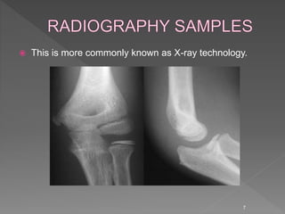
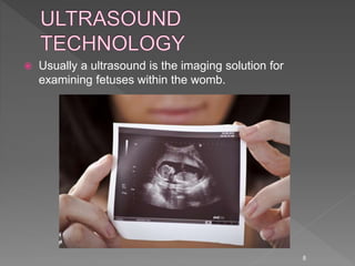
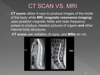
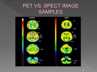
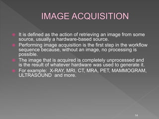
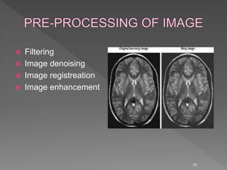
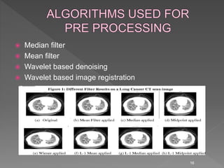
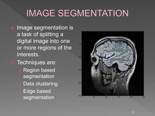
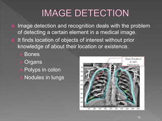
2. • INTRODUCTION
• WHAT IS MEDICAL IMAGE
PROCESSING(MIP)?
• IMAGING MODALITIES AND THEIR SHORT
DESCRIPTION
• MAIN COMPONENTS OF MIP
• APPLICATIONS OF MEDICAL
INDUSTRIES
• CONCLUSION
• REFERENCES
• ACKNOWLEDGEMENT
• QUESTIONS
2
3. Based on digital imaging techniques, the entire
spectrum of digital image processing is now
applicable to the study of medicine. The commonly
used term “medical image processing" means the
provision of digital image processing for medicine.
Medical image processing deals with the development
of problem-specific approaches to the enhancement of
raw medical image data for the purposes of selective
visualization as well as further analysis.
3
4. The study of medical imaging is concerned with
interaction of all forms of radiation with
tissue.
The development of appropriate technology to
extract clinically useful information, usually in
the form of an image from the observed
technology.
Non-invasive visualization of internal organs, tissues
etc.
4
5. Many different imaging techniques have been
developed and are in clinical use. In practice they are
complementary in that they offer different insights into
the same underlying reality.
In medical imaging, these different imaging techniques
are called modalities.
Anatomical modalities provide insight into the
anatomical morphology.
They include:
Projection x-ray or radiography
Ultrasound (US)
Computed tomography (CT)
Magnetic resonance imagery (MRI)
Magnetic resonance angiography (MRA)
5
6. Functional modalities, on the other hand, depict the
metabolism of the underlying tissues or organs.
They include:
Nuclear magnetic resonance
Single photon emission computed tomography
(SPECT)
Positron emission tomography (PET)
Functional magnetic resonance imagery (FMRI)
Fundus photography
6
7. This is more commonly known as X-ray technology.
7
8. Usually a ultrasound is the imaging solution for
examining fetuses within the womb.
8
9. CT scans utilize X-rays to produce images of the inside
of the body while MRI (magnetic resonance imaging)
uses powerful magnetic fields and radio frequency
pulses to produce detailed pictures of organs and other
internal body structures.
CT scans use radiation (X-rays), and MRIs do not.
9
10. Nuclear medicine is a branch of medical imaging that
uses small amounts of radioactive material to diagnose
and determine the severity of or treat a variety of
diseases, including many types of cancers, heart
disease, gastrointestinal, endocrine, neurological
disorders and other abnormalities within the body.
Nuclear Imaging is two types :
Positron emission tomography (PET)
Single photon emission computed
tomography(SPECT)
10
11. 11
PET SPECT
It emits positrons It emits gamma radiations.
Higher resolution Lower resolution
High sensitivity Low sensitivity
Limited half life of radio isotopes Longer lived radio isotopes
High cost Less cost
Cardiology, neurology and oncology
use PET scanning
It provide information about blood
flow to tissue
14. It is defined as the action of retrieving an image from some
source, usually a hardware-based source.
Performing image acquisition is the first step in the workflow
sequence because, without an image, no processing is
possible.
The image that is acquired is completely unprocessed and
is the result of whatever hardware was used to generate it.
For example: X-RAY, MRI, CT, MRA, PET, MAMMOGRAM,
ULTRASOUND and more.
14
16. Median filter
Mean filter
Wavelet based denoising
Wavelet based image registration
16
17. Image segmentation is
a task of splitting a
digital image into one
or more regions of the
interests.
Techniques are:
Region based
segmentation
Data clustering
Edge based
segmentation
17
19. Image detection and recognition deals with the problem
of detecting a certain element in a medical image.
It finds location of objects of interest without prior
knowledge of about their location or existence.
Bones
Organs
Polyps in colon
Nodules in lungs
19
20. Image analysis includes all the steps of processing,
which are used for quantitative measurements as well
as abstract interpretations of medical images.
The process of image analysis is very specific, precise,
fast and developed algorithms can be transferred
directly to application domains.
20
21. Medical image classification is one of the most
important problems in the image recognition area, and
its aim is to classify medical images into different
categories to help doctors in disease diagnosis or
further research.
Comparison of developed atlases
Use of knowledge databases
Classify the normal/abnormal objects
Determine the disease
21
22. Medical image compression
Medical image denoising
Image filtering
Medical image retrieval
Mammograms
Tumor detection
Medical image fusion
Monitoring bone strength
Tortuosity or integrated curvature
22
23. 23
In medical sciences, image processing has enabled for
accurate and fast quantitative analysis and visualization
of medical images of numerous modalities.
It has also enabled doctors and researchers at remote
sites to easily share data and analyze, thereby
enhancing their ability to diagnose, monitor and treat
various medical disorders.
It also helps in the field of image-based surgeries.