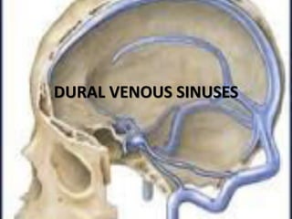
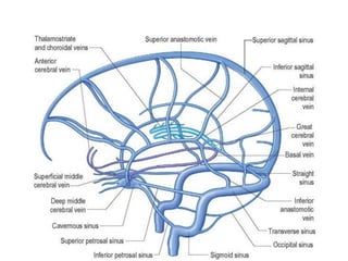
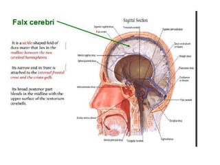
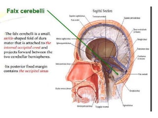
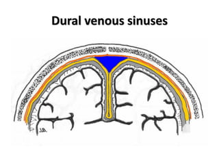
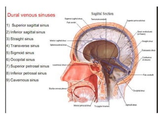
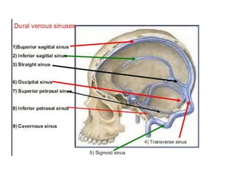
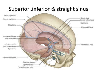
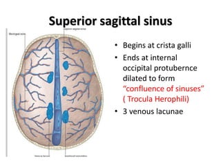
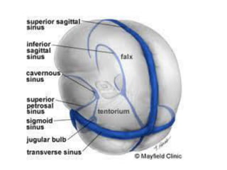
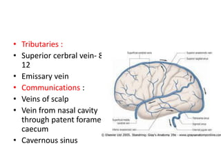
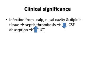
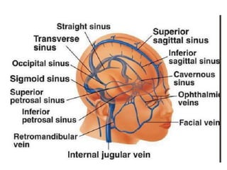
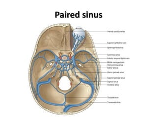
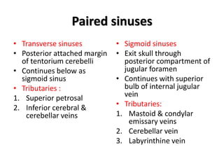
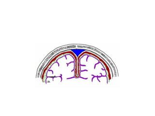
DURAL VENOUS SINUSES
Dural venous sinuses
Characteristic feature of dural venous
sinuses
• Lined by endothelium, no muscular coat &
valveless.
• Collect blood from brain,meninges, orbit,internal
ear & diploe.
• Connected to valveless emissary veins to
maintain the internal & external venous pressure.
• Projection of arachnoid granulation into it for CSF
absorption.
Superior ,inferior & straight sinus
Superior sagittal sinus
• Begins at crista galli
• Ends at internal
occipital protubernce
dilated to form
confluence of sinuses
( Trocula Herophili)
• 3 venous lacunae
• Tributaries :
• Superior cerbral vein- 8-
12
• Emissary vein
• Communications :
• Veins of scalp
• Vein from nasal cavity
through patent foramen
caecum
• Cavernous sinus
Clinical significance
• Infection from scalp, nasal cavity & diploic
tissue  septic thrombosis  CSF
absorption  ICT
Straight sinus
Straight sinus
• Location : junction of falx cerebri & tentorium
cerebelli
• Terminate into left transvese sinus &
connected to confluence
• Tributaries :
1. Inferior sagittal sinus
2. Few superior cerebellar veins
3. Great cerebral vein ( vein of Galen)
Paired sinus
Paired sinuses
• Transverse sinuses
• Posterior attached margin
of tentorium cerebelli
• Continues below as
sigmoid sinus
• Tributaries :
1. Superior petrosal
2. Inferior cerebral &
cerebellar veins
• Sigmoid sinuses
• Exit skull through
posterior compartment of
jugular foramen
• Continues with superior
bulb of internal jugular
vein
• Tributaries:
1. Mastoid & condylar
emissary veins
2. Cerebellar vein
3. Labyrinthine vein
Cavernous sinus
• Formation : cephalic
part of primary head
vein
• 2x 1 cm
• Roof & lateral wall :
meningeal layer
• Floor & medial wall :
endosteal layer
• RELATIONS
Carotid siphon
CLINICAL SIGNIFICANCE
• Septic thrombosis
• Pulsating exophthalmous
Cavernous sinus
Dural venous sinuses

Dural venous sinuses

  • 1.
  • 13.
  • 14.
    Characteristic feature ofdural venous sinuses • Lined by endothelium, no muscular coat & valveless. • Collect blood from brain,meninges, orbit,internal ear & diploe. • Connected to valveless emissary veins to maintain the internal & external venous pressure. • Projection of arachnoid granulation into it for CSF absorption.
  • 19.
    Superior ,inferior &straight sinus
  • 20.
    Superior sagittal sinus •Begins at crista galli • Ends at internal occipital protubernce dilated to form confluence of sinuses ( Trocula Herophili) • 3 venous lacunae
  • 22.
    • Tributaries : •Superior cerbral vein- 8- 12 • Emissary vein • Communications : • Veins of scalp • Vein from nasal cavity through patent foramen caecum • Cavernous sinus
  • 23.
    Clinical significance • Infectionfrom scalp, nasal cavity & diploic tissue  septic thrombosis  CSF absorption  ICT
  • 24.
  • 25.
    Straight sinus • Location: junction of falx cerebri & tentorium cerebelli • Terminate into left transvese sinus & connected to confluence • Tributaries : 1. Inferior sagittal sinus 2. Few superior cerebellar veins 3. Great cerebral vein ( vein of Galen)
  • 27.
  • 28.
    Paired sinuses • Transversesinuses • Posterior attached margin of tentorium cerebelli • Continues below as sigmoid sinus • Tributaries : 1. Superior petrosal 2. Inferior cerebral & cerebellar veins • Sigmoid sinuses • Exit skull through posterior compartment of jugular foramen • Continues with superior bulb of internal jugular vein • Tributaries: 1. Mastoid & condylar emissary veins 2. Cerebellar vein 3. Labyrinthine vein
  • 29.
    Cavernous sinus • Formation: cephalic part of primary head vein • 2x 1 cm • Roof & lateral wall : meningeal layer • Floor & medial wall : endosteal layer • RELATIONS
  • 31.
  • 34.
    CLINICAL SIGNIFICANCE • Septicthrombosis • Pulsating exophthalmous
  • 37.