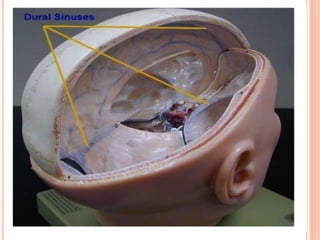
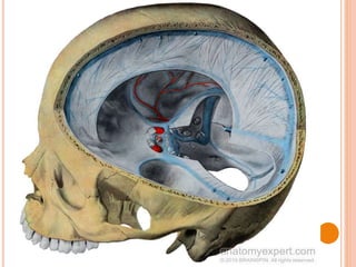
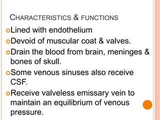
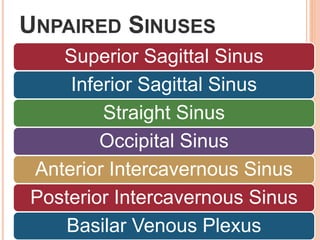
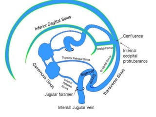
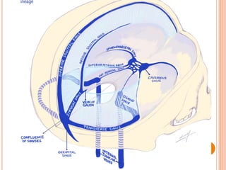
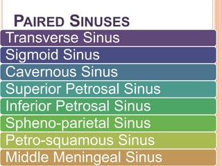
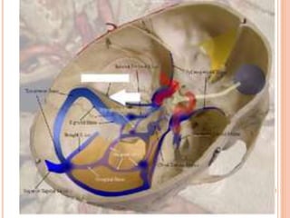
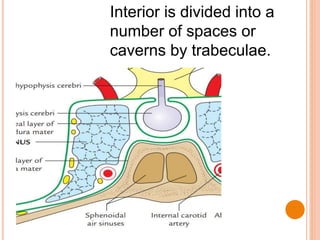
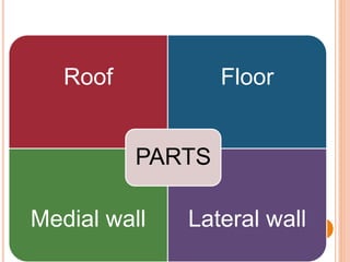
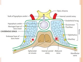
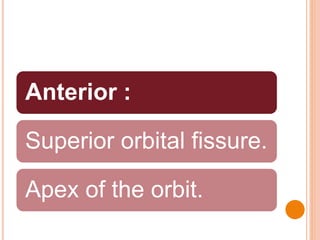
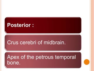
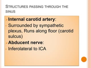
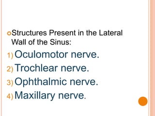
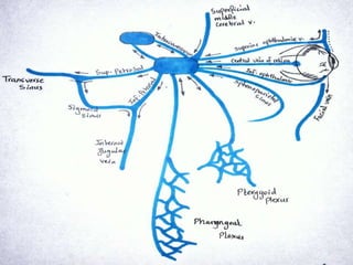
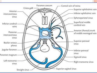
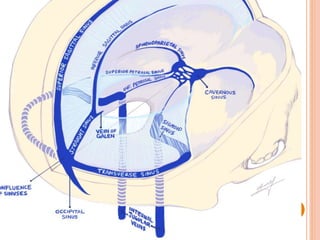
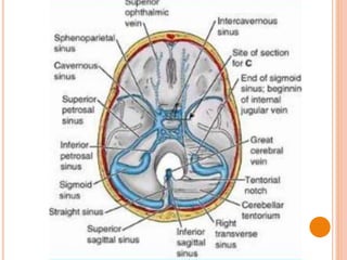
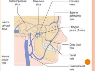
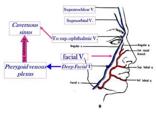
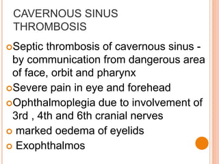
DURAL VENOUS SINUSES &
CAVERNOUS SINUS
OBJECTIVES
Dural venous sinuses –
characteristics & functions
Classification
Cavernous sinus in detail
Applied anatomy
DURAL VENOUS SINUSES
These are the various venous
channels present in the cranial
dura.
They are formed in the following
two ways:
1. By the separation of two layers of
cranial dura.
2. By the reduplication of the
meningeal layer.
CHARACTERISTICS & FUNCTIONS
Lined with endothelium
Devoid of muscular coat & valves.
Drain the blood from brain, meninges &
bones of skull.
Some venous sinuses also receive
CSF.
Receive valveless emissary vein to
maintain an equilibrium of venous
pressure.
CLASSIFICATION
Unpaired
sinuses
Paired
sinuses
UNPAIRED SINUSES
Superior Sagittal Sinus
Inferior Sagittal Sinus
Straight Sinus
Occipital Sinus
Anterior Intercavernous Sinus
Posterior Intercavernous Sinus
Basilar Venous Plexus
PAIRED SINUSES
Transverse Sinus
Sigmoid Sinus
Cavernous Sinus
Superior Petrosal Sinus
Inferior Petrosal Sinus
Spheno-parietal Sinus
Petro-squamous Sinus
Middle Meningeal Sinus
CAVERNOUS SINUS
Paired dural venous sinus
Situated in the middle cranial fossa,
on either side of body of the sphenoid
bone and sella turcica in the middle
cranial fossa.
Interior is divided into a
number of spaces or
caverns by trabeculae.
Formation: separation between
meningeal & endosteal layer of
duramater.
Size:
Length : 2 cm
Breadth : 1cm
Extent:
Front – superior orbital fissure
Behind - apex of petrous part of
temporal bone
Roof Floor
Medial wall Lateral wall
PARTS
Roof & Lateral wall
– meningeal layer
Floor & Medial wall
– endosteal layer
RELATIONS
Superior
• Optic chiasma
• Optic tract
• Internal carotid artery
• Anterior perforated
substance
Inferiorly:
Foramen lacerum.
Junction of the body and the
greater wing of the sphenoid.
Medially:
Pituitary gland
Sphenoid air sinus.
Laterally:
Cavum trigeminale
Temporal lobe of the cerebral
hemisphere.
Anterior :
Superior orbital fissure.
Apex of the orbit.
Posterior :
Crus cerebri of midbrain.
Apex of the petrous temporal
bone.
STRUCTURES PASSING THROUGH THE
SINUS
Internal carotid artery:
• Surrounded by sympathetic
plexus, Runs along floor (carotid
sulcus)
Abducent nerve:
• Inferolateral to ICA
Structures Present in the Lateral
Wall of the Sinus:
1) Oculomotor nerve.
2) Trochlear nerve.
3) Ophthalmic nerve.
4) Maxillary nerve.
TRIBUTARIES
The cavernous sinus receives
blood from three sources: orbit,
meninges, and brain.
 Hence, its tributaries come from
these sources as follows:
FROM ORBIT
 Superior ophthalmic vein.
 Inferior ophthalmic vein.
 Central vein of retina.
FROM BRAIN
 Superficial middle cerebral vein
 Inferior cerebral veins
FROM MENINGES
Sphenoparietal sinus
Anterior (frontal) trunk of the
middle meningeal vein
COMMUNICATIONS
With transverse sinus – via
superior petrosal sinus
With IJV –via inferior petrosal
sinus
With pterygoid venous plexus –
via emissary vein pass thro’ ovale,
spinosum, vesalii & lacerum
4. Facial vein via two routes:
(a) Superior ophthalmic vein →
angular vein → facial vein
(b) Emissary veins → pterygoid
venous plexus → deep facial vein
→ facial vein.
 Opposite cavernous sinuses via
anterior and posterior
intercavernous sinuses.
 Superior sagittal sinus via
superficial middle cerebral vein and
superior anastomotic vein.
Internal vertebral venous plexus,
via basilar venous plexus.
APPLIED ANATOMY
CAVERNOUS SINUS
THROMBOSIS
Septic thrombosis of cavernous sinus -
by communication from dangerous area
of face, orbit and pharynx
Severe pain in eye and forehead
Ophthalmoplegia due to involvement of
3rd , 4th and 6th cranial nerves
 marked oedema of eyelids
 Exophthalmos
PULSATING EXOPHTHALMOS
Pulsating exophthalmos
 internal carotid artery is ruptured as
a result of fracture of base of skull –
arterio- venous communication is
established.
ligation of inernal carotid artery may
be helpful, but patient may develop
contralateral hemiplegia.

Dural venous sinuses & cavernous sinus