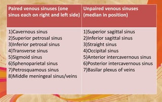
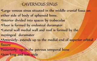
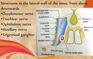
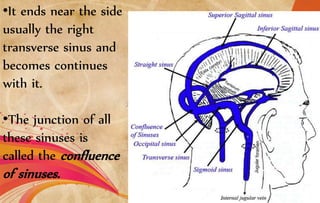
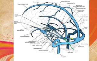
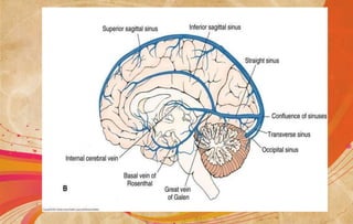
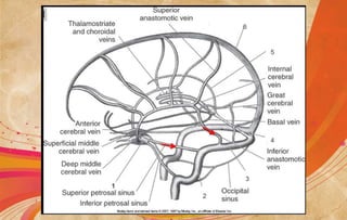
CEREBRAL VENOUS
SINUSES
•Venous spaces, walls are formed by duramater.
•Have an inner lining of endothelium, no muscles in
their wall and have no valves
•Receives venous blood from brain, meanings and bones
of skull.
VENOUS SINUSES OF DURAMATER
•Cranial venous sinuses communicates with veins
outside the skull by emissary veins – helps to
keep the pressure of blood in the sinuses
constant
•23 venous sinuses
8paired
7unpaired
Paired venous sinuses (one
sinus each on right and left side)
Unpaired venous sinuses
(median in position)
1)Cavernous sinus
2)Superior petrosal sinus
3)Inferior petrosal sinus
4)Transverse sinus
5)Sigmoid sinus
6)Sphenoparietal sinus
7)Petrosquamous sinus
8)Middle meningeal sinus/veins
1)Superior sagittal sinus
2)Inferior sagittal sinus
3)Straight sinus
4)Occipital sinus
5)Anterior intercavernous sinus
6)Posterior intercavernous sinus
7)Basilar plexus of veins
CAVERNOUS SINUS
•Large venous sinus situated in the middle cranial fossa on
either side of body of sphenoid bone.
•Interior divided into spaces by trabeculae
•Floor is formed by endosteal duramater
•Lateral wall medial wall and roof is formed by the
meningeal duramater
•Anteriorly- extends up to the medial end of superior orbital
fissure
Posteriorly- up to the petrous temporal bone
•2cm long and 1cm wide
RELATIONS
Structures outside the sinus
•Superiorly- optic tract, optic chiasma, olfactory tract, internal
carotid artery and anterior perforated substance.
•Inferiorly- foramen lacerum and the junction of the body and
greater wing of the sphenoid bone
•Medially- hypophysis cerebri and sphenoidal air sinus
•Laterally- temporal lobe with uncus
•Anteriorly- superior orbital fissure and the apex of the orbit
•Posteriorly- apex of the petrous temporal and the crus
cerebri of the midbrain
Structures in the lateral wall of the sinus, from above
downwards
•Occulomotor nerve
•Trochlear nerve
•Ophthalmic nerve
•Maxillary nerve
•Trigeminal ganglion
Structures passing through the center of the sinus
•Internal carotid artery with the venous and sympathetic
plexus around it
•Abducent nerve, inferolateral to the internal carotid artery
The structures in the lateral wall and in the center of the
sinus are separated from blood by the endothelial lining
TRIBUTARIES OR INCOMING CHANNELS
•From the orbit
1)Superior ophthalmic vein
2)A branch of inferior ophthalmic vein or sometimes the
vein itself
3)the central vein of the retina may drain either into the
superior ophthalmic vein or into the cavernous sinus.
•From the brain
1)superior middle cerebral vein
2)inferior cerebral veins from the temporal vein
•From the meninges
1)sphenoparietal sinus
2)the frontal trunk of middle meningeal vein may
drain either into the pterygoid plexus through the
foramen ovale or into the cavernous sinus.
Draining channels and communications
The cavernous sinus drains
•Into the transverse sinus through the superior petrosal sinus
•Into the internal jugular vein through the inferior petrosal sinus and
through the plexus around the internal carotid artery
•Into the pterygoid plexus of veins through the emissary veins passing
through the foramen ovale, the foramen lacerum and the emissary
sphenoidal foramen.
•Into the facial vein through the superior ophthalmic vein
•The right and left cavernous sinuses communicate with each other
through the anterior and posterior inter cavernous sinuses and
through the basilar plexus of veins.
•All these communications are valve less and blood can flow through
them in either direction
Superior sagittal sinus
•Occupies the upper convex attached margin of the falx cerebri
•Begins anteriorly at the crista gali by the union of tiny meningeal
veins
•Here it communicates with veins of frontal sinus and occasionally
with the veins of nose through foramen caecum.
•As it runs upwards and backwards it becomes progressively larger in
size
•It is triangular in cross section.
•It ends near the side
usually the right
transverse sinus and
becomes continues
with it.
•The junction of all
these sinuses is
called the confluence
of sinuses.
The interior of sinus shows
•Openings of the superior cerebral veins
•Openings of venous lacunae, usually three on each side
•Arachnoid villi and granulations projecting into the lacunae as well
as into the sinus
•Numerous fibrous bands crossing the inferior angle of the sinus
Tributaries
•Superior cerebral sinus which never open into the
venous lacunae
•Parietal emissary veins
•Venous lacunae, usually three on each side which first
receive the diploic and meningeal veins and then open
into the sinus
•Occasionally a vein from the nose opens into the sinus
when the foramen caecum is patent.
INFERIOR SAGITTAL SINUS
•The inferior saggital sinus a small channel lies
in the posterior two thirds of the lower, concave
free margin of the falx cerebri.
•It ends by joining the great cerebral vein to
form the straight sinus
STRAIGHT SINUS
•Lies in the median plane within the junction of falx
cerebri and the tentorium cerebelli
•It is formed anteriorly by the union of the inferior
sagittal sinus with the great cerebral vein and ends at
the internal occipital protuberance by continuing as the
transverse sinus(usually left).
•Receives a few of the superior cerebellar veins
TRANSVERSE SINUS
•Large sinuses –right sinus is larger than left
•Right sinus is situated in the posterior part of the
attached margin of the tentoriun cerebelli
•It is usually a continuation of superior saggital sinus
•Left sinus is a continuation of straight sinus
•Each sinus extends from the internal occipital
protuberance to the posteroinferior angle of the parietal
bone at the base of mastoid process where it bends
downwards and becomes the sigmoid sinus
Its tributaries are
•Superior petrosal sinus
•Inferior cerebral veins
•Inferior cerebellar veins
•Diploic (posterior temporal) vein
•Inferior anastomotic vein
Sigmoid sinus
•Each sinus right and left is the direct continuation of
the transverse sinus
•S-shaped
•Extends from the posterioinferior angle of the parietal
bone to the posterior part of jugular foramen where it
becomes the superior bulb of the internal jugular vein
•It grooves the mastoid part of the temporal bone
where it is separated anteriorly from the mastoid
antrum and mastoid air cells by only a thin plate of
bone
Tributaries are
•The mastoid and condylar emissary veins
•Cerebellar veins
•The internal auditory vein
Other sinuses
Occipital sinus
is small and lies in attached margin of the falx cerebri
It begins near the foramen magnum and ends in the confluence of
sinuses
Sphenoparietal sinus
Right and left lies along the free margin of the lesser wing of
sphenoid bone and drain into the anterior part of the cavernous sinus
Each sinus may receive the frontal trunk of the middle meningeal vein
Superior petrosal sinus
Lie in the anterior part of the attached margin of the tentorium
cerebelli along the upper border of the petrous temporal bone. It
drains the cavernous sinus into the transverse sinus
Inferior petrosal sinus
Right and left lie in the corresponding petro occipital fissure, and
drain the cavernous sinus into the superior bulb of the internal
jugular vein
Basilar plexus of veins
Lies over the clivus of the skull. It connects two inferior petrosal
sinuses and communicates with the internal vertebral venous plexus
Middle meningeal veins
Form two main trunks one frontal or anterior and one parietal or
posterior which accompany the two branches of the middle meningeal
artery.
Frontal trunk end either in the pterygoid plexus through th foramen
ovale or in sphenoparietal or cavernous sinus
Parietal trunk ends in the pterygoid plexus through the foramen
spinosum
The meningeal veins are nearer to the bone than artries and are
therfore more pliable to injury in fractures of skull
anterior and posterior intercavernous sinuses
They connect the cavernous sinuses

Cerebral venous sinuses

  • 1.
  • 2.
    •Venous spaces, wallsare formed by duramater. •Have an inner lining of endothelium, no muscles in their wall and have no valves •Receives venous blood from brain, meanings and bones of skull. VENOUS SINUSES OF DURAMATER
  • 3.
    •Cranial venous sinusescommunicates with veins outside the skull by emissary veins – helps to keep the pressure of blood in the sinuses constant •23 venous sinuses 8paired 7unpaired
  • 4.
    Paired venous sinuses(one sinus each on right and left side) Unpaired venous sinuses (median in position) 1)Cavernous sinus 2)Superior petrosal sinus 3)Inferior petrosal sinus 4)Transverse sinus 5)Sigmoid sinus 6)Sphenoparietal sinus 7)Petrosquamous sinus 8)Middle meningeal sinus/veins 1)Superior sagittal sinus 2)Inferior sagittal sinus 3)Straight sinus 4)Occipital sinus 5)Anterior intercavernous sinus 6)Posterior intercavernous sinus 7)Basilar plexus of veins
  • 5.
    CAVERNOUS SINUS •Large venoussinus situated in the middle cranial fossa on either side of body of sphenoid bone. •Interior divided into spaces by trabeculae •Floor is formed by endosteal duramater •Lateral wall medial wall and roof is formed by the meningeal duramater •Anteriorly- extends up to the medial end of superior orbital fissure Posteriorly- up to the petrous temporal bone •2cm long and 1cm wide
  • 6.
    RELATIONS Structures outside thesinus •Superiorly- optic tract, optic chiasma, olfactory tract, internal carotid artery and anterior perforated substance. •Inferiorly- foramen lacerum and the junction of the body and greater wing of the sphenoid bone
  • 7.
    •Medially- hypophysis cerebriand sphenoidal air sinus •Laterally- temporal lobe with uncus •Anteriorly- superior orbital fissure and the apex of the orbit •Posteriorly- apex of the petrous temporal and the crus cerebri of the midbrain
  • 8.
    Structures in thelateral wall of the sinus, from above downwards •Occulomotor nerve •Trochlear nerve •Ophthalmic nerve •Maxillary nerve •Trigeminal ganglion
  • 9.
    Structures passing throughthe center of the sinus •Internal carotid artery with the venous and sympathetic plexus around it •Abducent nerve, inferolateral to the internal carotid artery The structures in the lateral wall and in the center of the sinus are separated from blood by the endothelial lining
  • 10.
    TRIBUTARIES OR INCOMINGCHANNELS •From the orbit 1)Superior ophthalmic vein 2)A branch of inferior ophthalmic vein or sometimes the vein itself 3)the central vein of the retina may drain either into the superior ophthalmic vein or into the cavernous sinus. •From the brain 1)superior middle cerebral vein 2)inferior cerebral veins from the temporal vein
  • 12.
    •From the meninges 1)sphenoparietalsinus 2)the frontal trunk of middle meningeal vein may drain either into the pterygoid plexus through the foramen ovale or into the cavernous sinus.
  • 13.
    Draining channels andcommunications The cavernous sinus drains •Into the transverse sinus through the superior petrosal sinus •Into the internal jugular vein through the inferior petrosal sinus and through the plexus around the internal carotid artery •Into the pterygoid plexus of veins through the emissary veins passing through the foramen ovale, the foramen lacerum and the emissary sphenoidal foramen.
  • 16.
    •Into the facialvein through the superior ophthalmic vein •The right and left cavernous sinuses communicate with each other through the anterior and posterior inter cavernous sinuses and through the basilar plexus of veins. •All these communications are valve less and blood can flow through them in either direction
  • 17.
    Superior sagittal sinus •Occupiesthe upper convex attached margin of the falx cerebri •Begins anteriorly at the crista gali by the union of tiny meningeal veins •Here it communicates with veins of frontal sinus and occasionally with the veins of nose through foramen caecum. •As it runs upwards and backwards it becomes progressively larger in size •It is triangular in cross section.
  • 18.
    •It ends nearthe side usually the right transverse sinus and becomes continues with it. •The junction of all these sinuses is called the confluence of sinuses.
  • 19.
    The interior ofsinus shows •Openings of the superior cerebral veins •Openings of venous lacunae, usually three on each side •Arachnoid villi and granulations projecting into the lacunae as well as into the sinus •Numerous fibrous bands crossing the inferior angle of the sinus
  • 20.
    Tributaries •Superior cerebral sinuswhich never open into the venous lacunae •Parietal emissary veins •Venous lacunae, usually three on each side which first receive the diploic and meningeal veins and then open into the sinus •Occasionally a vein from the nose opens into the sinus when the foramen caecum is patent.
  • 21.
    INFERIOR SAGITTAL SINUS •Theinferior saggital sinus a small channel lies in the posterior two thirds of the lower, concave free margin of the falx cerebri. •It ends by joining the great cerebral vein to form the straight sinus
  • 23.
    STRAIGHT SINUS •Lies inthe median plane within the junction of falx cerebri and the tentorium cerebelli •It is formed anteriorly by the union of the inferior sagittal sinus with the great cerebral vein and ends at the internal occipital protuberance by continuing as the transverse sinus(usually left). •Receives a few of the superior cerebellar veins
  • 25.
    TRANSVERSE SINUS •Large sinuses–right sinus is larger than left •Right sinus is situated in the posterior part of the attached margin of the tentoriun cerebelli •It is usually a continuation of superior saggital sinus •Left sinus is a continuation of straight sinus •Each sinus extends from the internal occipital protuberance to the posteroinferior angle of the parietal bone at the base of mastoid process where it bends downwards and becomes the sigmoid sinus
  • 26.
    Its tributaries are •Superiorpetrosal sinus •Inferior cerebral veins •Inferior cerebellar veins •Diploic (posterior temporal) vein •Inferior anastomotic vein
  • 28.
    Sigmoid sinus •Each sinusright and left is the direct continuation of the transverse sinus •S-shaped •Extends from the posterioinferior angle of the parietal bone to the posterior part of jugular foramen where it becomes the superior bulb of the internal jugular vein •It grooves the mastoid part of the temporal bone where it is separated anteriorly from the mastoid antrum and mastoid air cells by only a thin plate of bone
  • 29.
    Tributaries are •The mastoidand condylar emissary veins •Cerebellar veins •The internal auditory vein
  • 30.
    Other sinuses Occipital sinus issmall and lies in attached margin of the falx cerebri It begins near the foramen magnum and ends in the confluence of sinuses Sphenoparietal sinus Right and left lies along the free margin of the lesser wing of sphenoid bone and drain into the anterior part of the cavernous sinus Each sinus may receive the frontal trunk of the middle meningeal vein Superior petrosal sinus Lie in the anterior part of the attached margin of the tentorium cerebelli along the upper border of the petrous temporal bone. It drains the cavernous sinus into the transverse sinus
  • 31.
    Inferior petrosal sinus Rightand left lie in the corresponding petro occipital fissure, and drain the cavernous sinus into the superior bulb of the internal jugular vein Basilar plexus of veins Lies over the clivus of the skull. It connects two inferior petrosal sinuses and communicates with the internal vertebral venous plexus Middle meningeal veins Form two main trunks one frontal or anterior and one parietal or posterior which accompany the two branches of the middle meningeal artery.
  • 32.
    Frontal trunk endeither in the pterygoid plexus through th foramen ovale or in sphenoparietal or cavernous sinus Parietal trunk ends in the pterygoid plexus through the foramen spinosum The meningeal veins are nearer to the bone than artries and are therfore more pliable to injury in fractures of skull anterior and posterior intercavernous sinuses They connect the cavernous sinuses