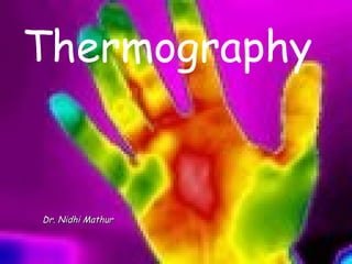
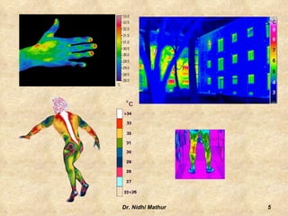
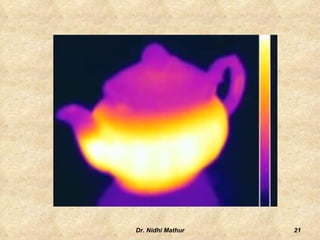
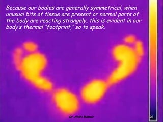
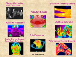
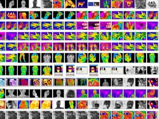
ThermographyDr. Nidhi Mathur
ThermogaphyA medical science that performs diagnostics using highly detailed and sensitive infrared images.2Dr. Nidhi Mathur
ThermogramA regional temperature map of the surface of a part of the body made by a thermograph.3Dr. Nidhi Mathur
 Thermal readings have been used in medicine for thousands of years.
 The Greek physician Hippocrates wrote, In whatever part of the body excess of heat or cold is felt, the disease is there to be discovered.4Dr. Nidhi Mathur
5Dr. Nidhi Mathur
Medical Thermography                                           Medical Thermography (digital infrared thermal imaging - DITI ) is used as a method of research for early pre-clinical diagnosis and control during treatment.
 The intrinsic safety of this method makes infrared Thermography free from any limitations or contra- indications.
Thermography is a non-invasive, non-contact tool that uses the heat from your body to aid in making diagnosis of a host of health care conditions.
 Thermography is completely safe and uses no radiation.6Dr. Nidhi Mathur
 It has two parts,
 the IR camera or detector and
 a standard PC or laptop computer. These systems have only a few controls and relatively easy to use. 7Dr. Nidhi Mathur
Two categories of detectors are commonly found.  based on thermal effects and include thermocouples, bolometers, thermopiles and pyroelectric detectors.
 relies on quantum effects and include photoconductors, and photovoltaic diodes.Uncooled detector which can be fabricated into high resolution FPA's. These are currently becoming available in a range of IR viewers. 8Dr. Nidhi Mathur
Basics9Dr. Nidhi Mathur
 any object whose temperature is above 0 °K radiates infrared energy.
The amount of radiated energy is a function of the object's temperature and its relative efficiency of thermal radiation, known as emissivity.
 Radiated energy (power) is proportional to the body's temperature, raised to the 4th power.
 This energy can be measured and an instrument calibrated to indicate the corresponding temperature of the surface it's "looking at."
 Instruments which "scan" an object and create an image or spatial map of surface temperatures are referred to as thermal imagers. 10Dr. Nidhi Mathur
Difference between infrared film and thermographyIR film is sensitive to infrared (IR) radiation in the 250°C to 500°C range, while the range of thermography is approximately -50°C to over 2,000°C.
So, for an IR film to show something, it must be over 250°C or be reflecting infrared radiation from something that is at least that hot.11Dr. Nidhi Mathur
12Dr. Nidhi Mathur
Thermography makes use of the infrared (IR) spectral band of the electromagnetic spectrum.
IR involves four bands:
near infrared (0.75-3 μm),
middle infrared (3-6 μm),
far infrared (6-15 μm) and
extreme infrared (15-100 μm).
Infrared video cameras are passive (emits no energy), but merely collects the thermal radiation emitted from the surface of the human body.In active Thermography, an energy source is required to produce a thermal contrast between the feature of interest and the background. The active approach is necessary in many cases given that the inspected parts are usually in equilibrium with the surroundings.13Dr. Nidhi Mathur
 IR cameras don’t see temperature.
 The IR camera captures the radiosity of the target it is viewing.
Radiosity is defined as the infrared energy coming from a target modulated by the intervening atmosphere, and consists of

Thermography

  • 1.
  • 2.
    ThermogaphyA medical sciencethat performs diagnostics using highly detailed and sensitive infrared images.2Dr. Nidhi Mathur
  • 3.
    ThermogramA regional temperaturemap of the surface of a part of the body made by a thermograph.3Dr. Nidhi Mathur
  • 4.
    Thermal readingshave been used in medicine for thousands of years.
  • 5.
    The Greekphysician Hippocrates wrote, In whatever part of the body excess of heat or cold is felt, the disease is there to be discovered.4Dr. Nidhi Mathur
  • 6.
  • 7.
    Medical Thermography                                         Medical Thermography (digital infrared thermal imaging - DITI ) is used as a method of research for early pre-clinical diagnosis and control during treatment.
  • 8.
    The intrinsicsafety of this method makes infrared Thermography free from any limitations or contra- indications.
  • 9.
    Thermography is anon-invasive, non-contact tool that uses the heat from your body to aid in making diagnosis of a host of health care conditions.
  • 10.
    Thermography iscompletely safe and uses no radiation.6Dr. Nidhi Mathur
  • 11.
    It hastwo parts,
  • 12.
    the IRcamera or detector and
  • 13.
    a standardPC or laptop computer. These systems have only a few controls and relatively easy to use. 7Dr. Nidhi Mathur
  • 14.
    Two categories ofdetectors are commonly found. based on thermal effects and include thermocouples, bolometers, thermopiles and pyroelectric detectors.
  • 15.
    relies onquantum effects and include photoconductors, and photovoltaic diodes.Uncooled detector which can be fabricated into high resolution FPA's. These are currently becoming available in a range of IR viewers. 8Dr. Nidhi Mathur
  • 16.
  • 17.
    any objectwhose temperature is above 0 °K radiates infrared energy.
  • 18.
    The amount ofradiated energy is a function of the object's temperature and its relative efficiency of thermal radiation, known as emissivity.
  • 19.
    Radiated energy(power) is proportional to the body's temperature, raised to the 4th power.
  • 20.
    This energycan be measured and an instrument calibrated to indicate the corresponding temperature of the surface it's "looking at."
  • 21.
    Instruments which"scan" an object and create an image or spatial map of surface temperatures are referred to as thermal imagers. 10Dr. Nidhi Mathur
  • 22.
    Difference between infraredfilm and thermographyIR film is sensitive to infrared (IR) radiation in the 250°C to 500°C range, while the range of thermography is approximately -50°C to over 2,000°C.
  • 23.
    So, for anIR film to show something, it must be over 250°C or be reflecting infrared radiation from something that is at least that hot.11Dr. Nidhi Mathur
  • 24.
  • 25.
    Thermography makes useof the infrared (IR) spectral band of the electromagnetic spectrum.
  • 26.
  • 27.
  • 28.
  • 29.
  • 30.
  • 31.
    Infrared video camerasare passive (emits no energy), but merely collects the thermal radiation emitted from the surface of the human body.In active Thermography, an energy source is required to produce a thermal contrast between the feature of interest and the background. The active approach is necessary in many cases given that the inspected parts are usually in equilibrium with the surroundings.13Dr. Nidhi Mathur
  • 32.
    IR camerasdon’t see temperature.
  • 33.
    The IRcamera captures the radiosity of the target it is viewing.
  • 34.
    Radiosity is definedas the infrared energy coming from a target modulated by the intervening atmosphere, and consists of