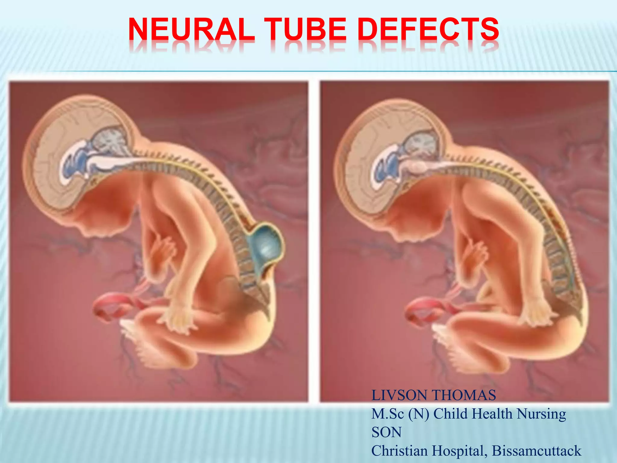
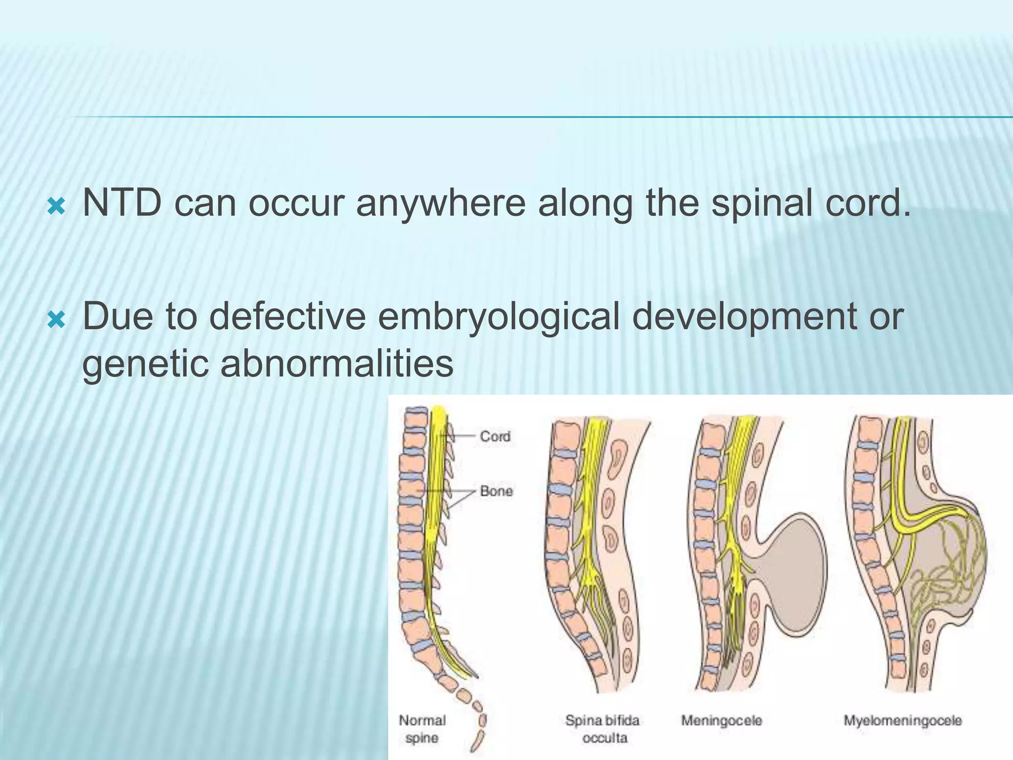
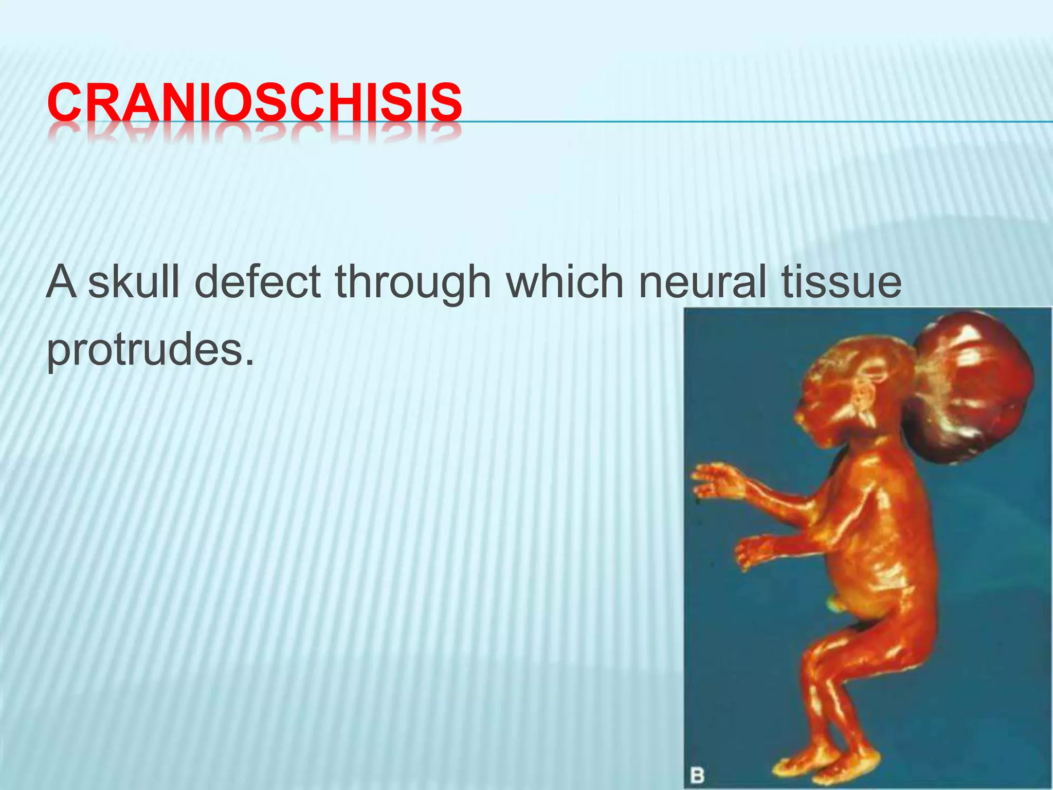
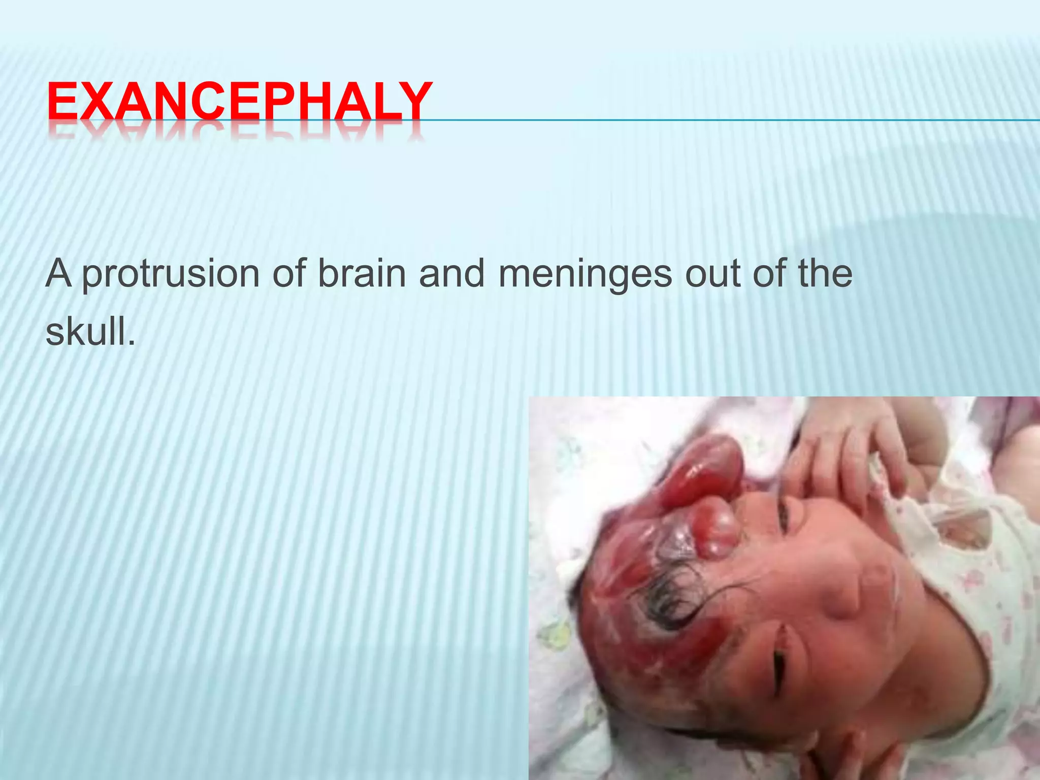
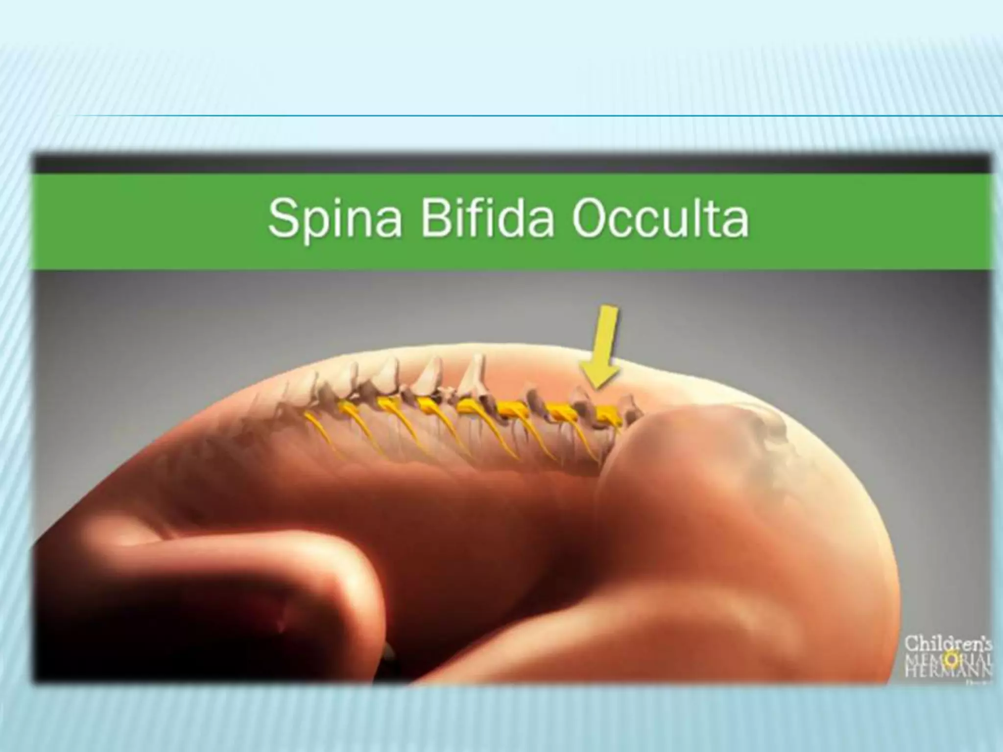
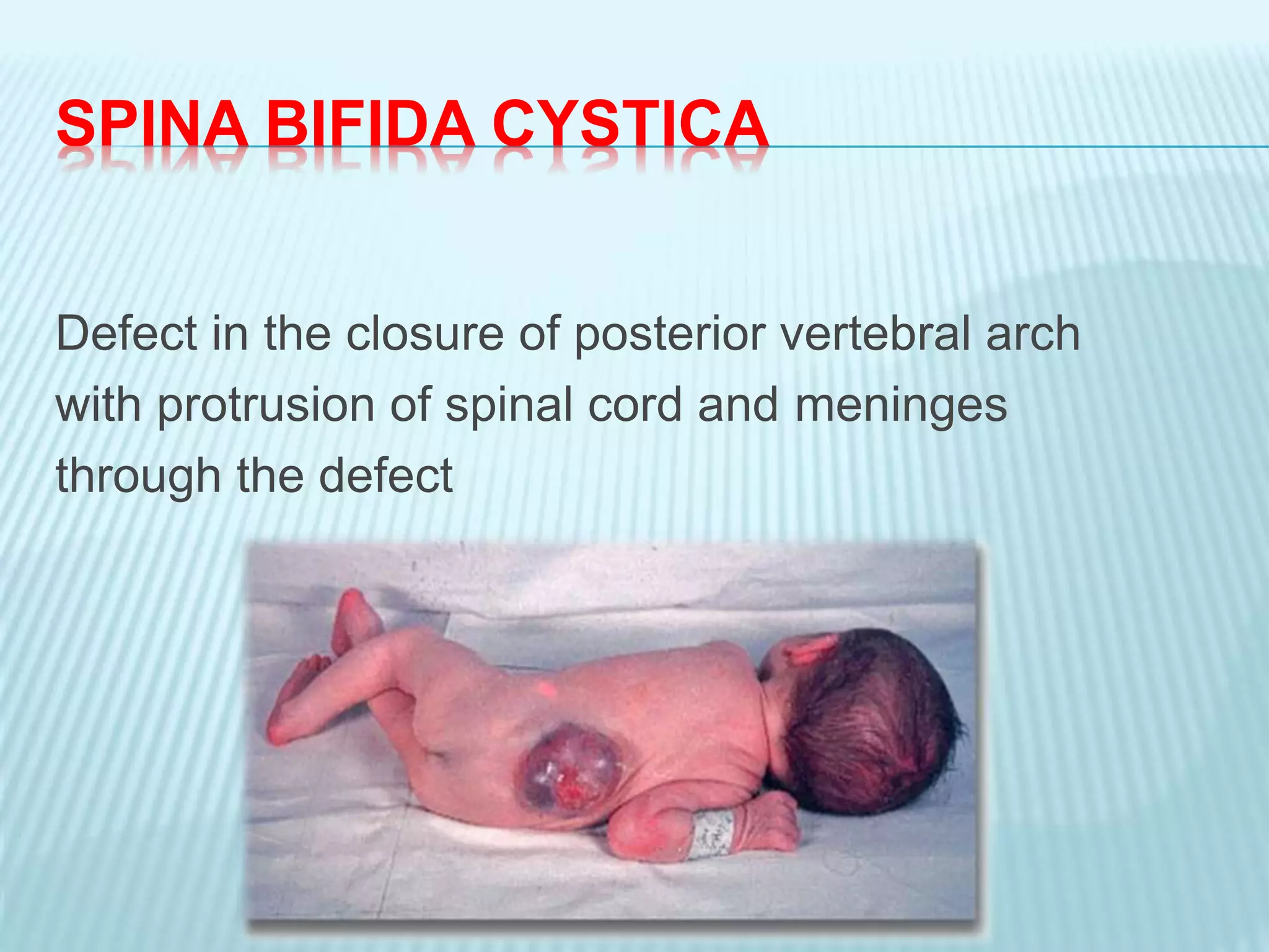
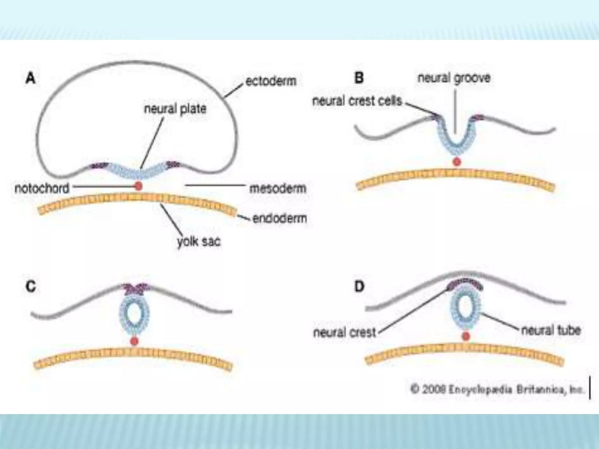
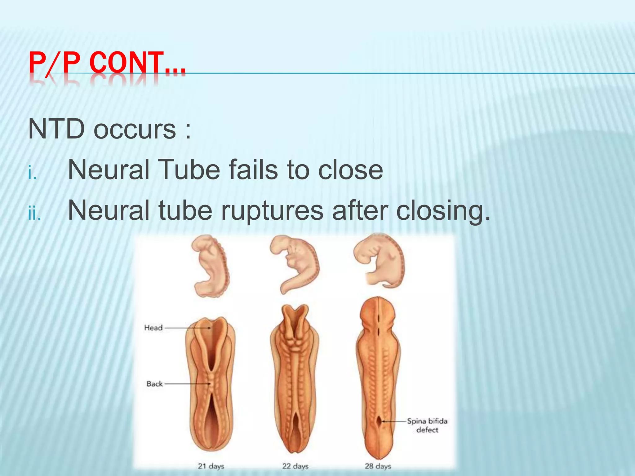
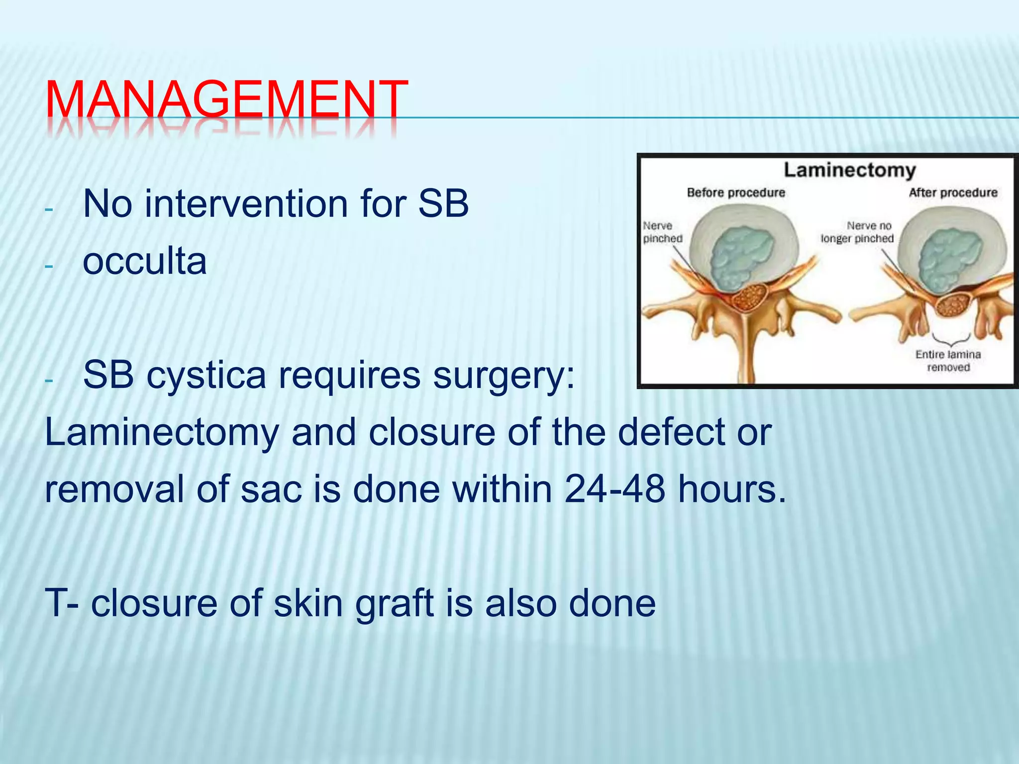
NEURAL TUBE DEFECTS
LIVSON THOMAS
M.Sc (N) Child Health Nursing
SON
Christian Hospital, Bissamcuttack
 NTD can occur anywhere along the spinal cord.
 Due to defective embryological development or
genetic abnormalities
ANENCEPHALY
There is absence of cranial vault with cerebral
hemispheres completely absent or greatly
reduced in size
CRANIOSCHISIS
A skull defect through which neural tissue
protrudes.
EXANCEPHALY
A protrusion of brain and meninges out of the
skull.
ENCEPHALOCELE
A protrusion of brain and meninges into a fluid-
filled sac through a skull defect.
RACHISCHISIS
Meninges and spinal cord are exposed through a
fissure in the vertebral coloumn.
SPINA BIFIDA
It is a NTD where there is an incomplete closure
of the vertebrae and neural tube.
SB is a malformation of spine, in which posterior
portion of lamina of vertebrae fail to close with or
defective development of spinal cord.
1 or 2 per 1000 live births
ETIOLOGY
 Genetic: 15 times more chance if parents had
SB
 Maternal Age: over 35 years or below 20 years
 Environmental: Radiations, more in Europe
 Diet: Folic acid deficiency in pregnant mothers
increases the risk
CLASSIFICATION
SPINA BIFIDA
MYELOMENINGOCELEMENINGOCELE
SB CYSTICASB OCCULTA
SPINA BIFIDA OCCULTA (5%)
Failure of formation of bony arch around
the spinal cord.
Spinal cord & meninges are normal
Not visible externally
Asymptomatic
SPINA BIFIDA CYSTICA
Defect in the closure of posterior vertebral arch
with protrusion of spinal cord and meninges
through the defect
MENINGOCELE
It is a sac like herniation through the bony
malformation, containing meninges and CSF
The covering of the sac maybe thin and
transluscent or membranous.
MYELOMENINGOCELE
It is a sac- like protrusion of spinal cord ,
CSF and meninges through spinal cleft.
Most cases found in lumbar or lumbosacral
region.
SB Cystica occurs in
- Lumbosacral region
- Lower lumbar region
- Upper lumbar region
- Sacral region
PATHOPHYSIOLOGY
- Neural tube development start at 3rd week of
gestation
1. Depression forms at dorsal of ectoderm
2. Neural tube closes by end of 4th week.
3. Walls of neural tube thicken and become spinal
cord and brain.
4. Neural canal  ventricles & central canal of SC
5. Vertebral coloumn is formed simultaneosly
P/P CONT…
NTD occurs :
i. Neural Tube fails to close
ii. Neural tube ruptures after closing.
CLINICAL FEATURES
1. Spina Bifida Occulta
- Most of the patients are asymptomatic
- A dimple in the skin
- Growth of hair over malformed area (Tuft of hairs)
- May experience foot weakness
- Disturbance in bladder and bowel sphincter.
2. Spina Bifida Cystica
a. MENINGOCELE
- Visible cystic defect at the back.
- Sac contains: meninges and CSF.
- Weakness of legs.
- Lack of sphincter control.
b. Myelomeningocele
- Depends on location of defect, higher the defect
more the neurological deficit.
- Herniated mass present over vertebral coloumn
- Associated hydrocephalus
- Loss of motor control
- Loss of sensation
- Thoracic lesion: Flaccid paralysis
- Sacral Lesion: Weakness of lower limbs
- Bowel and bladder may or may not be affected.
- UTI
- Skeletal anomalies
- Developmental delays
DIAGNOSTIC EVALUATION
Prenatal diagnosis
- USG
- Fetal MRI
- Amniocentesis
After birth
- Sac at the back
- CT and MRI scan
TRANSILLUMINATION TEST
To determine the content of the sac:
Translucent: Meningocele
Non- translucent: Myelomeningocele
MANAGEMENT
- No intervention for SB
- occulta
- SB cystica requires surgery:
Laminectomy and closure of the defect or
removal of sac is done within 24-48 hours.
T- closure of skin graft is also done
NURSING MANAGEMENT
1. Preventing injury to the sac.
2. Preventing skin break down
3. Prevention of Infection
4. Providing adequate nutrition
5. Preventing UTI
6. Parent education and support to reduce
parent’s anxiety
PROGNOSIS
 Myelomeningocele can be surgically corrected in
most of the cases.
 Length of the life is not affected
 Quality of life can be improved
 Can cause lower limb deformities in future
PREVENTION
 Genetic counselling
 Therapeutic abortion if detected during early
pregnancy
 Incidence can be reduced if folic acid
supplementation is taken. 1mg folic acid/ day.
Neural tube defects

Neural tube defects

  • 1.
    NEURAL TUBE DEFECTS LIVSONTHOMAS M.Sc (N) Child Health Nursing SON Christian Hospital, Bissamcuttack
  • 2.
     NTD canoccur anywhere along the spinal cord.  Due to defective embryological development or genetic abnormalities
  • 3.
    ANENCEPHALY There is absenceof cranial vault with cerebral hemispheres completely absent or greatly reduced in size
  • 4.
    CRANIOSCHISIS A skull defectthrough which neural tissue protrudes.
  • 5.
    EXANCEPHALY A protrusion ofbrain and meninges out of the skull.
  • 6.
    ENCEPHALOCELE A protrusion ofbrain and meninges into a fluid- filled sac through a skull defect.
  • 7.
    RACHISCHISIS Meninges and spinalcord are exposed through a fissure in the vertebral coloumn.
  • 8.
    SPINA BIFIDA It isa NTD where there is an incomplete closure of the vertebrae and neural tube. SB is a malformation of spine, in which posterior portion of lamina of vertebrae fail to close with or defective development of spinal cord. 1 or 2 per 1000 live births
  • 9.
    ETIOLOGY  Genetic: 15times more chance if parents had SB  Maternal Age: over 35 years or below 20 years  Environmental: Radiations, more in Europe  Diet: Folic acid deficiency in pregnant mothers increases the risk
  • 10.
  • 11.
    SPINA BIFIDA OCCULTA(5%) Failure of formation of bony arch around the spinal cord. Spinal cord & meninges are normal Not visible externally Asymptomatic
  • 14.
    SPINA BIFIDA CYSTICA Defectin the closure of posterior vertebral arch with protrusion of spinal cord and meninges through the defect
  • 15.
    MENINGOCELE It is asac like herniation through the bony malformation, containing meninges and CSF The covering of the sac maybe thin and transluscent or membranous.
  • 17.
    MYELOMENINGOCELE It is asac- like protrusion of spinal cord , CSF and meninges through spinal cleft. Most cases found in lumbar or lumbosacral region.
  • 19.
    SB Cystica occursin - Lumbosacral region - Lower lumbar region - Upper lumbar region - Sacral region
  • 20.
    PATHOPHYSIOLOGY - Neural tubedevelopment start at 3rd week of gestation 1. Depression forms at dorsal of ectoderm 2. Neural tube closes by end of 4th week. 3. Walls of neural tube thicken and become spinal cord and brain. 4. Neural canal  ventricles & central canal of SC 5. Vertebral coloumn is formed simultaneosly
  • 22.
    P/P CONT… NTD occurs: i. Neural Tube fails to close ii. Neural tube ruptures after closing.
  • 23.
    CLINICAL FEATURES 1. SpinaBifida Occulta - Most of the patients are asymptomatic - A dimple in the skin - Growth of hair over malformed area (Tuft of hairs) - May experience foot weakness - Disturbance in bladder and bowel sphincter.
  • 24.
    2. Spina BifidaCystica a. MENINGOCELE - Visible cystic defect at the back. - Sac contains: meninges and CSF. - Weakness of legs. - Lack of sphincter control.
  • 25.
    b. Myelomeningocele - Dependson location of defect, higher the defect more the neurological deficit. - Herniated mass present over vertebral coloumn - Associated hydrocephalus - Loss of motor control - Loss of sensation - Thoracic lesion: Flaccid paralysis - Sacral Lesion: Weakness of lower limbs
  • 26.
    - Bowel andbladder may or may not be affected. - UTI - Skeletal anomalies - Developmental delays
  • 27.
    DIAGNOSTIC EVALUATION Prenatal diagnosis -USG - Fetal MRI - Amniocentesis After birth - Sac at the back - CT and MRI scan
  • 28.
    TRANSILLUMINATION TEST To determinethe content of the sac: Translucent: Meningocele Non- translucent: Myelomeningocele
  • 29.
    MANAGEMENT - No interventionfor SB - occulta - SB cystica requires surgery: Laminectomy and closure of the defect or removal of sac is done within 24-48 hours. T- closure of skin graft is also done
  • 31.
    NURSING MANAGEMENT 1. Preventinginjury to the sac. 2. Preventing skin break down 3. Prevention of Infection 4. Providing adequate nutrition 5. Preventing UTI 6. Parent education and support to reduce parent’s anxiety
  • 32.
    PROGNOSIS  Myelomeningocele canbe surgically corrected in most of the cases.  Length of the life is not affected  Quality of life can be improved  Can cause lower limb deformities in future
  • 33.
    PREVENTION  Genetic counselling Therapeutic abortion if detected during early pregnancy  Incidence can be reduced if folic acid supplementation is taken. 1mg folic acid/ day.