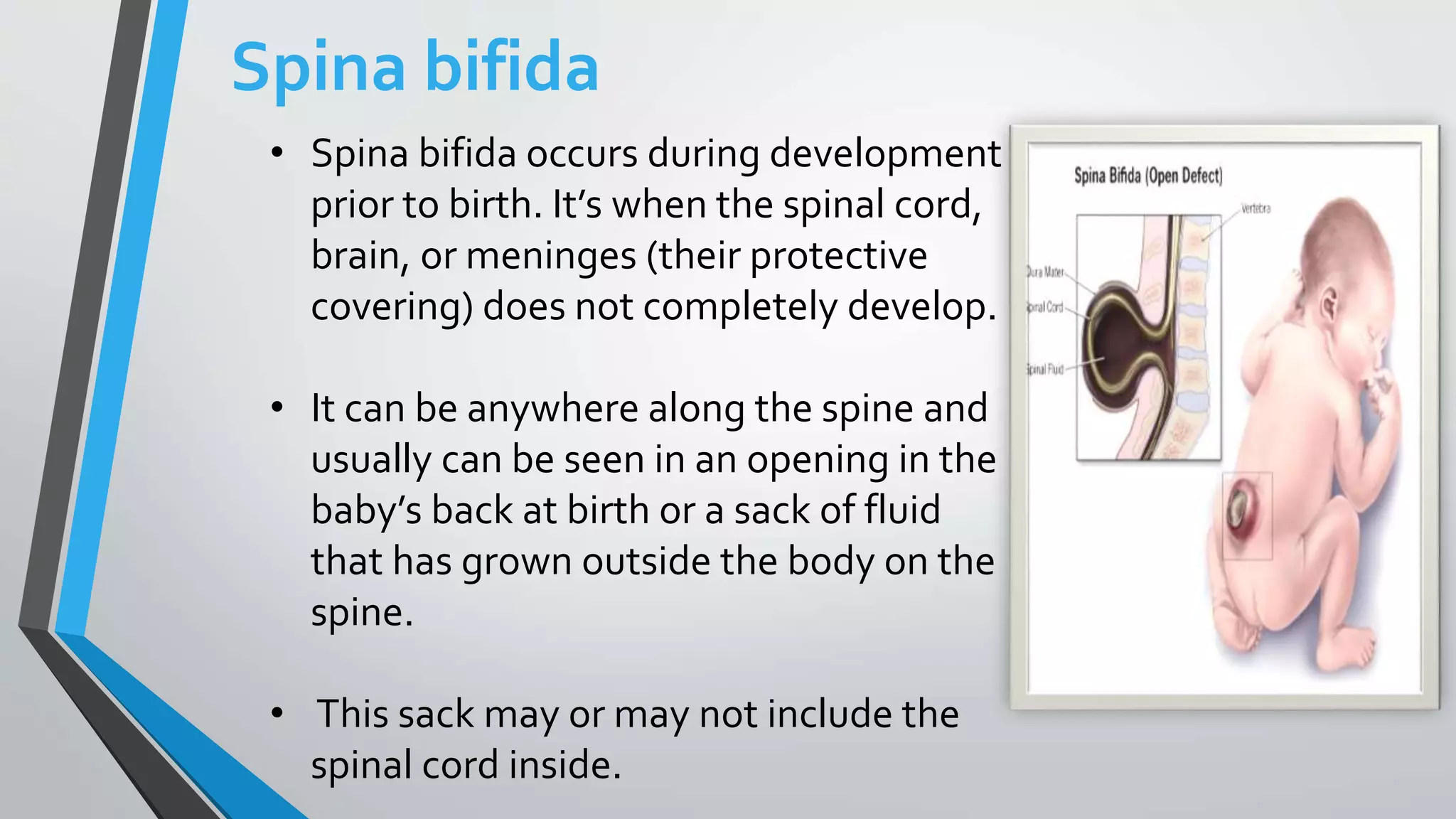
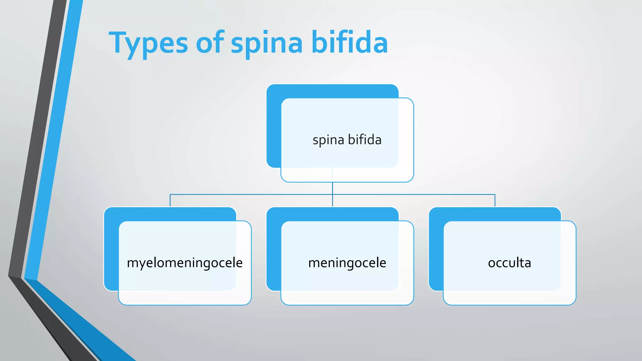
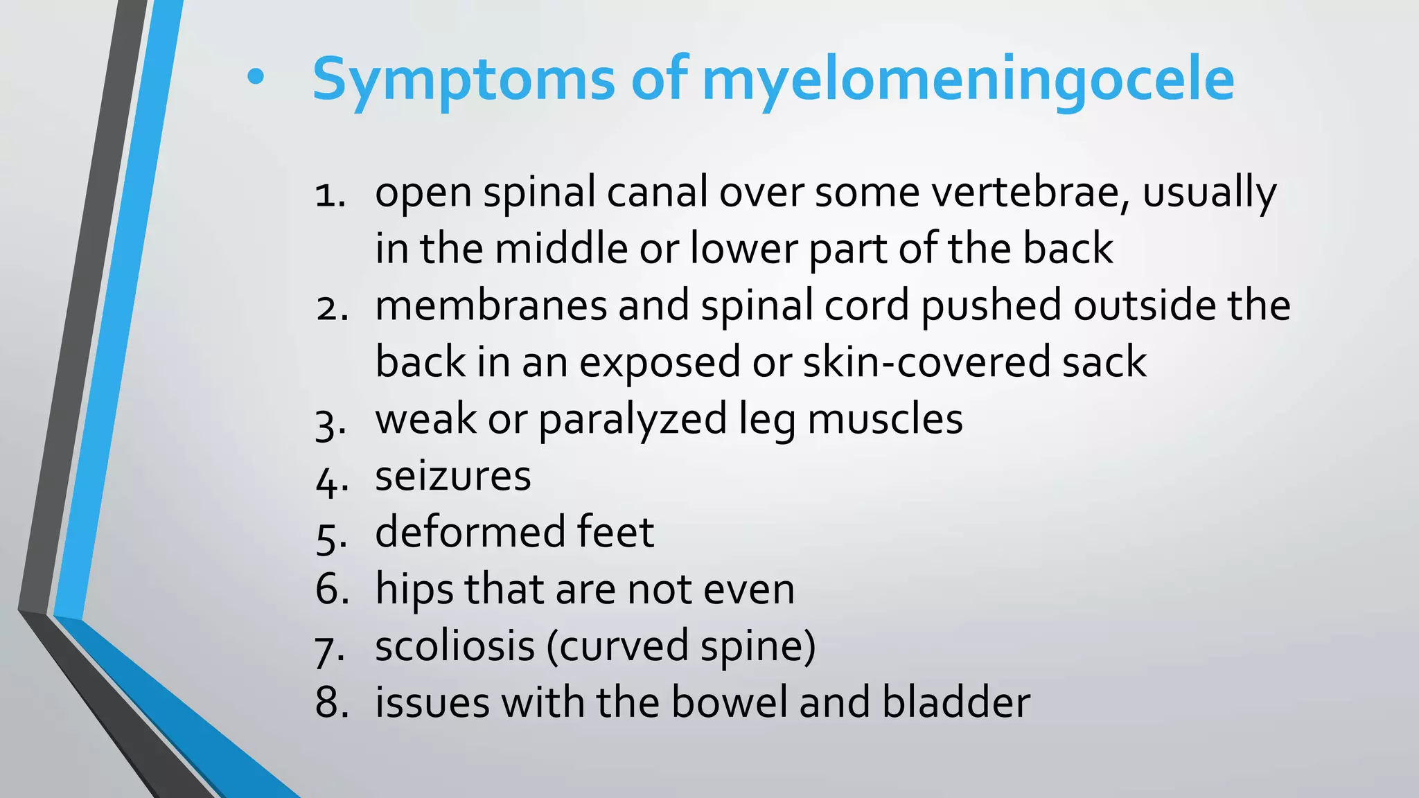
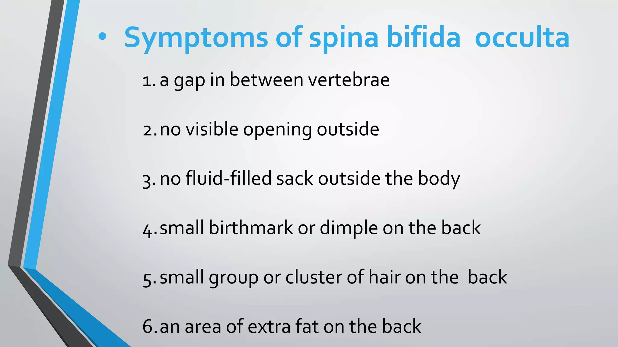
Neural Tube Defects
Group 6
Introduction
•the neural tube is the
embryonic precursor to
the central nervous
system, which is made
up of the brain and spinal
cord
Formation of the neural tube
• By the third week of development ,the
edges of the neural plate start to thicken
and lift upward , forming the neural folds.
• The center of the neural plate remains
grounded, allowing a U-shaped neural
groove .
• The neural folds pinch in towards the
midline of the embryo and fuse
together to form the neural tube.
• the neural tube is open both
cranially and caudally.
• These openings, called
neuropores, close during the
fourth week in humans.
• Improper closure of the neuropores
can result in neural tube defects
such as anencephaly or spina
bifida.
Causes
• A mother’s exposure to certain
environmental toxins, medications,
exposure to high temperatures
diabetes and obesity may be risk
factors for pregnancy complications,
inadequate intake of folic acid.
• A lack of this key nutrient may
raise your risk of having a baby with
other neural tube defects in addition
to anencephaly, such as spina
bifida. Pregnant women can
minimize this risk with folic acid
supplements or diet changes.
Anencephaly
•In anencephaly, most of the
brain and skull do not develop.
• This often results in a baby
being born without the front
part of the brain (forebrain)
and the thinking and
coordinating
•There are three types of anencephaly:
 Meroanencephaly : where there is rudimentary
brain tissue and partial formation of the cranium
 Holoanencephaly : the most common type,
in which the brain is completely absent
 Craniorachischisis : the most severe,
where area cerebrovasculosa and
area medullovasculosa fill both cranial defects and the
spinal column.
Diagnosis
• Anencephaly can be
diagnosed during pregnancy
or after the baby is born.
During Pregnancy
• During pregnancy, there are screening
tests (prenatal tests) to check for birth defects
and other conditions.
• Anencephaly would result in an abnormal result
on a blood or serum screening test or it might be
seen during an ultrasound (which creates
pictures of the body).
After the Baby is Born
• In some cases, anencephaly might
not be diagnosed until after the
baby is born. Anencephaly is
immediately seen at birth.
Treatments
• There is no known cure or
standard treatment
for anencephaly.
• Almost all babies born with
anencephaly will die shortly after
birth.
• Spina bifida occurs during development
prior to birth. It’s when the spinal cord,
brain, or meninges (their protective
covering) does not completely develop.
• It can be anywhere along the spine and
usually can be seen in an opening in the
baby’s back at birth or a sack of fluid
that has grown outside the body on the
spine.
• This sack may or may not include the
spinal cord inside.
Spina bifida
Types of spina bifida
spina bifida
myelomeningocele meningocele occulta
 Myelomeningocele
• This is the most common and serious type of
spina bifida.
• It involves a sack outside the opening in the
baby’s back somewhere on the spine.
• This sack contains parts of the spinal cord and
nerves.The spinal cord and nerves in the sack
will be damaged.
• People with myelomeningocele have physical
disabilities that range from moderate to severe.
These disabilities may include:
 incontinence
 difficulty going to
the bathroom
 inability to move or feel
their legs or feet
 Meningocele
• This type of spina bifida also involves a
sack of fluid outside an opening in the
baby’s back.
• The sack does not contain any part of the
spinal cord.
• Because there isn’t a lot of nerve
damage, meningocele causes only minor
disabilities.
 Spina bifida occulta
• This is a mild type of spina bifida. It may also
go by the term “hidden” spina bifida.
• It does not cause any disabilities and may go
unnoticed until later in life
• .There is usually no opening in the baby’s
back, but only a gap in the spine.
• In this type, there is no damage to the spinal
cord or the nerves.
Symptoms of spina bifida
• The symptoms of spina bifida
are different for each type.They
can also vary from person to
person within each type.
• Symptoms of myelomeningocele
1. open spinal canal over some vertebrae, usually
in the middle or lower part of the back
2. membranes and spinal cord pushed outside the
back in an exposed or skin-covered sack
3. weak or paralyzed leg muscles
4. seizures
5. deformed feet
6. hips that are not even
7. scoliosis (curved spine)
8. issues with the bowel and bladder
• Symptoms of meningocele
1.small opening in the back
2.sack that’s visible at birth
3.membranes pushing out through the
opening in the vertebrae into sack
4.normal development of the spinal cord
• Symptoms of spina bifida occulta
1.a gap in between vertebrae
2.no visible opening outside
3.no fluid-filled sack outside the body
4.small birthmark or dimple on the back
5.small group or cluster of hair on the back
6.an area of extra fat on the back
Treatment
• The treatment for spina bifida will be
different for each person because
symptoms and severity can vary. In
some cases, especially in spina bifida
occulta, there may not be any
treatment needed.
• However, myelomeningocele and
meningocele require surgery to put the
exposed sack and nerves back in place.
• The surgeon will then close the
opening over the vertebrae.
• This surgery may be performed shortly after
the child’s birth.
• In some cases, prenatal surgery may be
done while the baby is still in the womb.
• Even after surgery is performed, some symptoms
and disability can remain.They will need to be
managed based on the severity of each symptom.
• Paralysis and bowel and bladder issues typically
remain throughout life.Treatment for remaining
symptoms can include:
1. additional surgeries
2. medications
3. physical therapy
4. rehabilitation services
5. walking aids
Thank yo
u

The neural tube defects

  • 1.
  • 2.
    Introduction •the neural tubeis the embryonic precursor to the central nervous system, which is made up of the brain and spinal cord
  • 3.
    Formation of theneural tube • By the third week of development ,the edges of the neural plate start to thicken and lift upward , forming the neural folds. • The center of the neural plate remains grounded, allowing a U-shaped neural groove . • The neural folds pinch in towards the midline of the embryo and fuse together to form the neural tube.
  • 4.
    • the neuraltube is open both cranially and caudally. • These openings, called neuropores, close during the fourth week in humans. • Improper closure of the neuropores can result in neural tube defects such as anencephaly or spina bifida.
  • 5.
    Causes • A mother’sexposure to certain environmental toxins, medications, exposure to high temperatures diabetes and obesity may be risk factors for pregnancy complications, inadequate intake of folic acid. • A lack of this key nutrient may raise your risk of having a baby with other neural tube defects in addition to anencephaly, such as spina bifida. Pregnant women can minimize this risk with folic acid supplements or diet changes.
  • 6.
    Anencephaly •In anencephaly, mostof the brain and skull do not develop. • This often results in a baby being born without the front part of the brain (forebrain) and the thinking and coordinating
  • 7.
    •There are threetypes of anencephaly:  Meroanencephaly : where there is rudimentary brain tissue and partial formation of the cranium  Holoanencephaly : the most common type, in which the brain is completely absent  Craniorachischisis : the most severe, where area cerebrovasculosa and area medullovasculosa fill both cranial defects and the spinal column.
  • 8.
    Diagnosis • Anencephaly canbe diagnosed during pregnancy or after the baby is born.
  • 9.
    During Pregnancy • Duringpregnancy, there are screening tests (prenatal tests) to check for birth defects and other conditions. • Anencephaly would result in an abnormal result on a blood or serum screening test or it might be seen during an ultrasound (which creates pictures of the body).
  • 10.
    After the Babyis Born • In some cases, anencephaly might not be diagnosed until after the baby is born. Anencephaly is immediately seen at birth.
  • 11.
    Treatments • There isno known cure or standard treatment for anencephaly. • Almost all babies born with anencephaly will die shortly after birth.
  • 12.
    • Spina bifidaoccurs during development prior to birth. It’s when the spinal cord, brain, or meninges (their protective covering) does not completely develop. • It can be anywhere along the spine and usually can be seen in an opening in the baby’s back at birth or a sack of fluid that has grown outside the body on the spine. • This sack may or may not include the spinal cord inside. Spina bifida
  • 13.
    Types of spinabifida spina bifida myelomeningocele meningocele occulta
  • 14.
     Myelomeningocele • Thisis the most common and serious type of spina bifida. • It involves a sack outside the opening in the baby’s back somewhere on the spine. • This sack contains parts of the spinal cord and nerves.The spinal cord and nerves in the sack will be damaged.
  • 15.
    • People withmyelomeningocele have physical disabilities that range from moderate to severe. These disabilities may include:  incontinence  difficulty going to the bathroom  inability to move or feel their legs or feet
  • 16.
     Meningocele • Thistype of spina bifida also involves a sack of fluid outside an opening in the baby’s back. • The sack does not contain any part of the spinal cord. • Because there isn’t a lot of nerve damage, meningocele causes only minor disabilities.
  • 17.
     Spina bifidaocculta • This is a mild type of spina bifida. It may also go by the term “hidden” spina bifida. • It does not cause any disabilities and may go unnoticed until later in life • .There is usually no opening in the baby’s back, but only a gap in the spine. • In this type, there is no damage to the spinal cord or the nerves.
  • 18.
    Symptoms of spinabifida • The symptoms of spina bifida are different for each type.They can also vary from person to person within each type.
  • 19.
    • Symptoms ofmyelomeningocele 1. open spinal canal over some vertebrae, usually in the middle or lower part of the back 2. membranes and spinal cord pushed outside the back in an exposed or skin-covered sack 3. weak or paralyzed leg muscles 4. seizures 5. deformed feet 6. hips that are not even 7. scoliosis (curved spine) 8. issues with the bowel and bladder
  • 20.
    • Symptoms ofmeningocele 1.small opening in the back 2.sack that’s visible at birth 3.membranes pushing out through the opening in the vertebrae into sack 4.normal development of the spinal cord
  • 21.
    • Symptoms ofspina bifida occulta 1.a gap in between vertebrae 2.no visible opening outside 3.no fluid-filled sack outside the body 4.small birthmark or dimple on the back 5.small group or cluster of hair on the back 6.an area of extra fat on the back
  • 22.
    Treatment • The treatmentfor spina bifida will be different for each person because symptoms and severity can vary. In some cases, especially in spina bifida occulta, there may not be any treatment needed.
  • 23.
    • However, myelomeningoceleand meningocele require surgery to put the exposed sack and nerves back in place. • The surgeon will then close the opening over the vertebrae. • This surgery may be performed shortly after the child’s birth. • In some cases, prenatal surgery may be done while the baby is still in the womb.
  • 24.
    • Even aftersurgery is performed, some symptoms and disability can remain.They will need to be managed based on the severity of each symptom. • Paralysis and bowel and bladder issues typically remain throughout life.Treatment for remaining symptoms can include: 1. additional surgeries 2. medications 3. physical therapy 4. rehabilitation services 5. walking aids
  • 25.