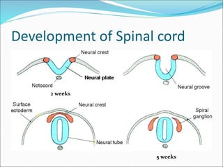
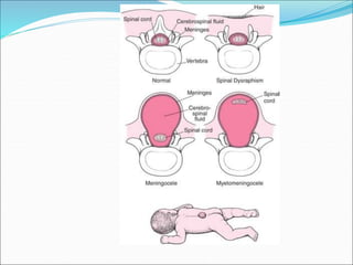
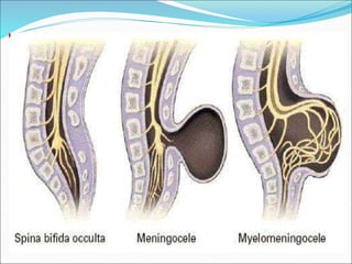
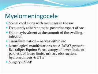
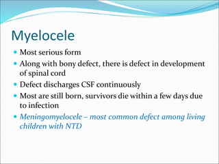
Subash Srivastav
Asst Professor
Definition
 Congenital defect in the posterior bony wall of the
spinal canal involving the laminae
 Most commonly seen in the lumbosacral region
Development of Spinal cord
2 weeks
5 weeks
Development of vertebral column
 Notochord – solid rod of cells in front of neural tube
 Vertebral bodies develop around Notochord
 From each vertebral body 2 projections grow around
the neural tube to form Vertebral arch OR Neural arch
 Fusion occurs first in the Thoracic region and extends
upwards and downwards
 Failure of fusion of these arches – SPINA BIFIDA
Incidence
 0.1% , excluding spina bifida occulta
 Myelocele – commonest type
 Most are still born; in survivors death occurs early due
to infection
Types
 Spina bifida occulta
 Meningocele
 Meningomyelocele
 Syringomyelocele
 Myelocele
Spina bifida occulta
 Common in lumbar/sacral region
 Small gap in one of vertebral arches (frequently only
one vertebra is involved)
 Gap is filled with fibrous tissue – no protrusion of cord
or membrane
 Indicators – patch of hair, lipoma, sinus, depression
 When cord lags behind vertebral column growth –
neurological deficiencies (nocturnal eneuresis,
backache)
 Many are symptomless
Meningocele
 Protrusion of meninges containing only CSF – cystic
swelling which is compressible
 Overlying skin remains intact
 Common in lumbosacral region; also occipital & root
of nose
 No neurological manifestations
 If associated with hydrocephalus (Arnold Chiari synd)
 Complications – infection, rupture
 Surgery required as early as possible
Myelomeningocele
 Spinal cord along with meninges in the sac
 Frequently adherent to the posterior aspect of sac
 Skin maybe absent at the summit of the swelling –
infection
 Transillumination – nerves within sac
 Neurological manifestations are ALWAYS present –
B/L talipes Equino Varus, atropy of lower limbs or
paralysis of lower limbs, urinary obstruction,
hydronephrosis & UTIs
 Surgery - ASAP
Syringomyelocele
 Rarest variety
 Central canal of spinal cord is dilated and the spinal
cord lies within the sac together with nerves
 Gross neurological deficits and paralytic
manifestations are seen
Myelocele
 Most serious form
 Along with bony defect, there is defect in development
of spinal cord
 Defect discharges CSF continuously
 Most are still born, survivors die within a few days due
to infection
 Meningomyelocele – most common defect among living
children with NTD

Spina bifida

  • 1.
  • 2.
    Definition  Congenital defectin the posterior bony wall of the spinal canal involving the laminae  Most commonly seen in the lumbosacral region
  • 3.
    Development of Spinalcord 2 weeks 5 weeks
  • 4.
    Development of vertebralcolumn  Notochord – solid rod of cells in front of neural tube  Vertebral bodies develop around Notochord  From each vertebral body 2 projections grow around the neural tube to form Vertebral arch OR Neural arch  Fusion occurs first in the Thoracic region and extends upwards and downwards  Failure of fusion of these arches – SPINA BIFIDA
  • 5.
    Incidence  0.1% ,excluding spina bifida occulta  Myelocele – commonest type  Most are still born; in survivors death occurs early due to infection
  • 6.
    Types  Spina bifidaocculta  Meningocele  Meningomyelocele  Syringomyelocele  Myelocele
  • 9.
    Spina bifida occulta Common in lumbar/sacral region  Small gap in one of vertebral arches (frequently only one vertebra is involved)  Gap is filled with fibrous tissue – no protrusion of cord or membrane  Indicators – patch of hair, lipoma, sinus, depression  When cord lags behind vertebral column growth – neurological deficiencies (nocturnal eneuresis, backache)  Many are symptomless
  • 10.
    Meningocele  Protrusion ofmeninges containing only CSF – cystic swelling which is compressible  Overlying skin remains intact  Common in lumbosacral region; also occipital & root of nose  No neurological manifestations  If associated with hydrocephalus (Arnold Chiari synd)  Complications – infection, rupture  Surgery required as early as possible
  • 11.
    Myelomeningocele  Spinal cordalong with meninges in the sac  Frequently adherent to the posterior aspect of sac  Skin maybe absent at the summit of the swelling – infection  Transillumination – nerves within sac  Neurological manifestations are ALWAYS present – B/L talipes Equino Varus, atropy of lower limbs or paralysis of lower limbs, urinary obstruction, hydronephrosis & UTIs  Surgery - ASAP
  • 12.
    Syringomyelocele  Rarest variety Central canal of spinal cord is dilated and the spinal cord lies within the sac together with nerves  Gross neurological deficits and paralytic manifestations are seen
  • 13.
    Myelocele  Most seriousform  Along with bony defect, there is defect in development of spinal cord  Defect discharges CSF continuously  Most are still born, survivors die within a few days due to infection  Meningomyelocele – most common defect among living children with NTD