Downloaded 235 times
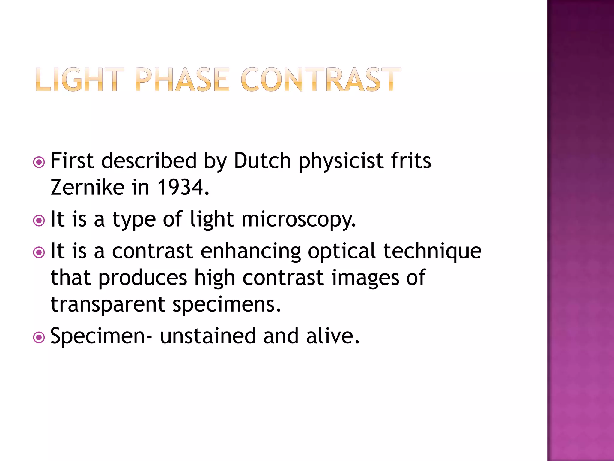
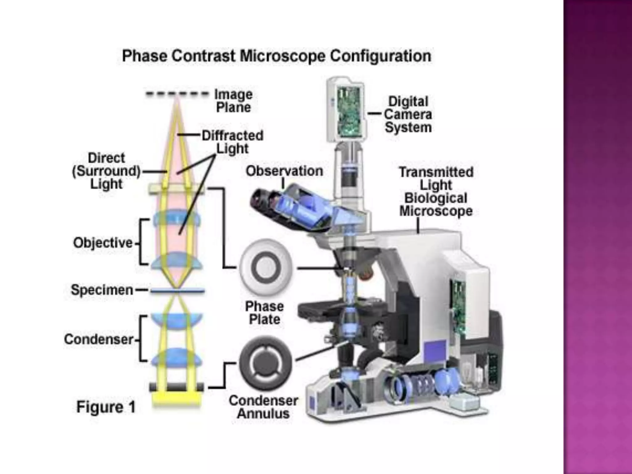
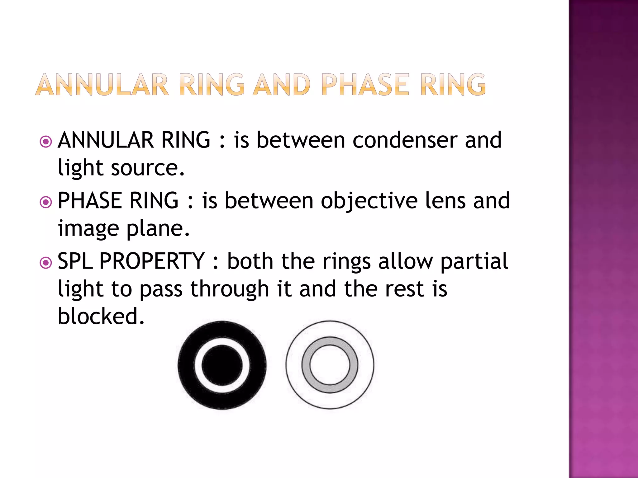
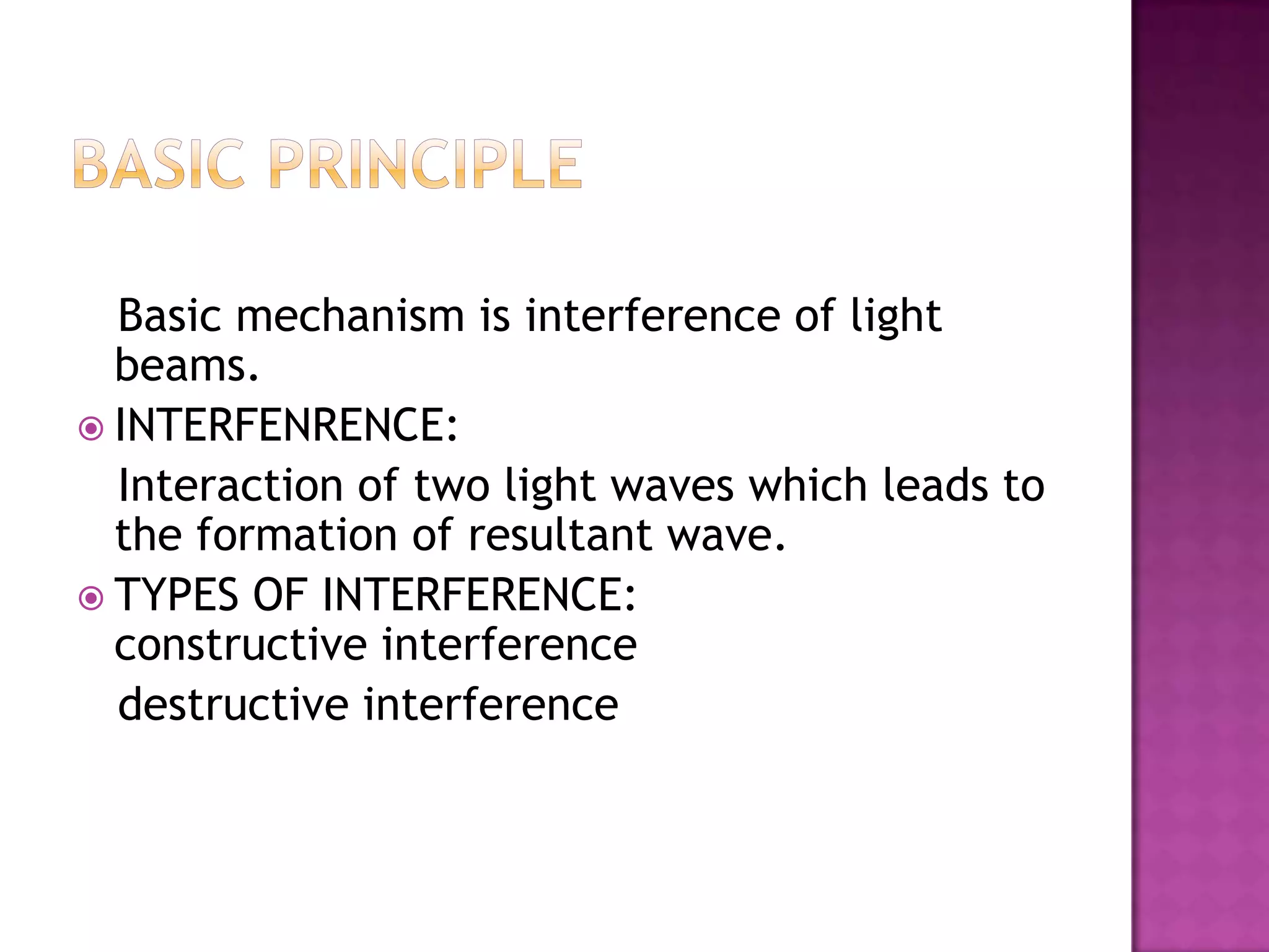
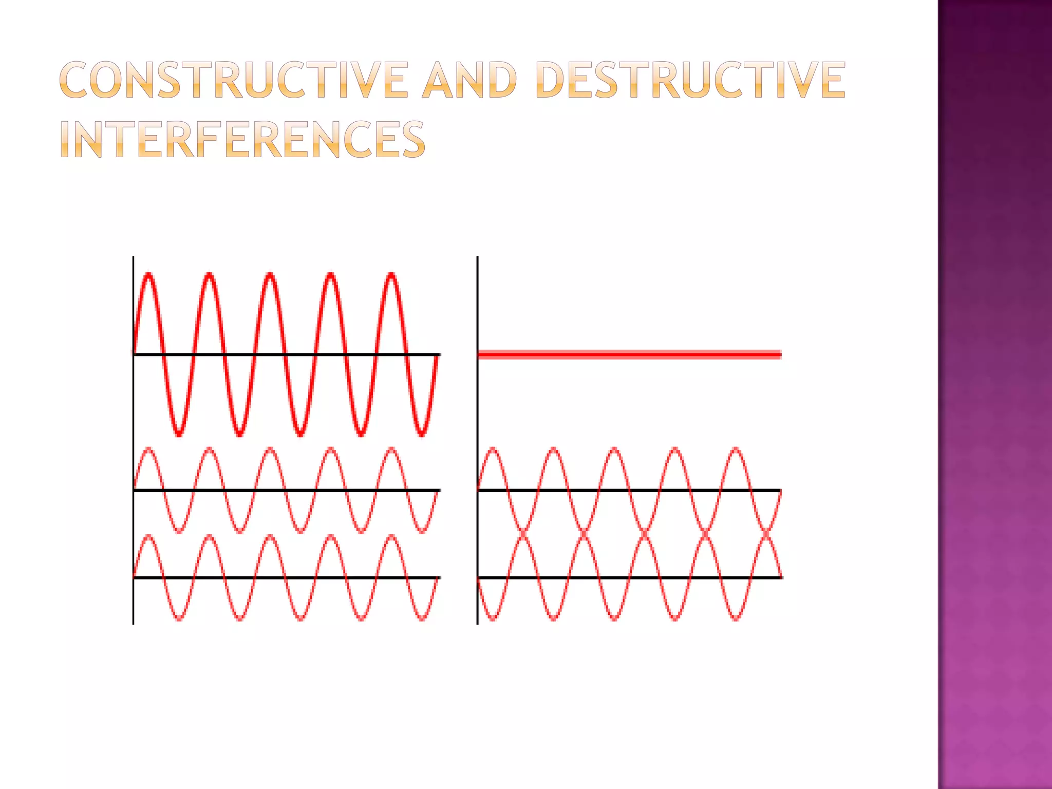
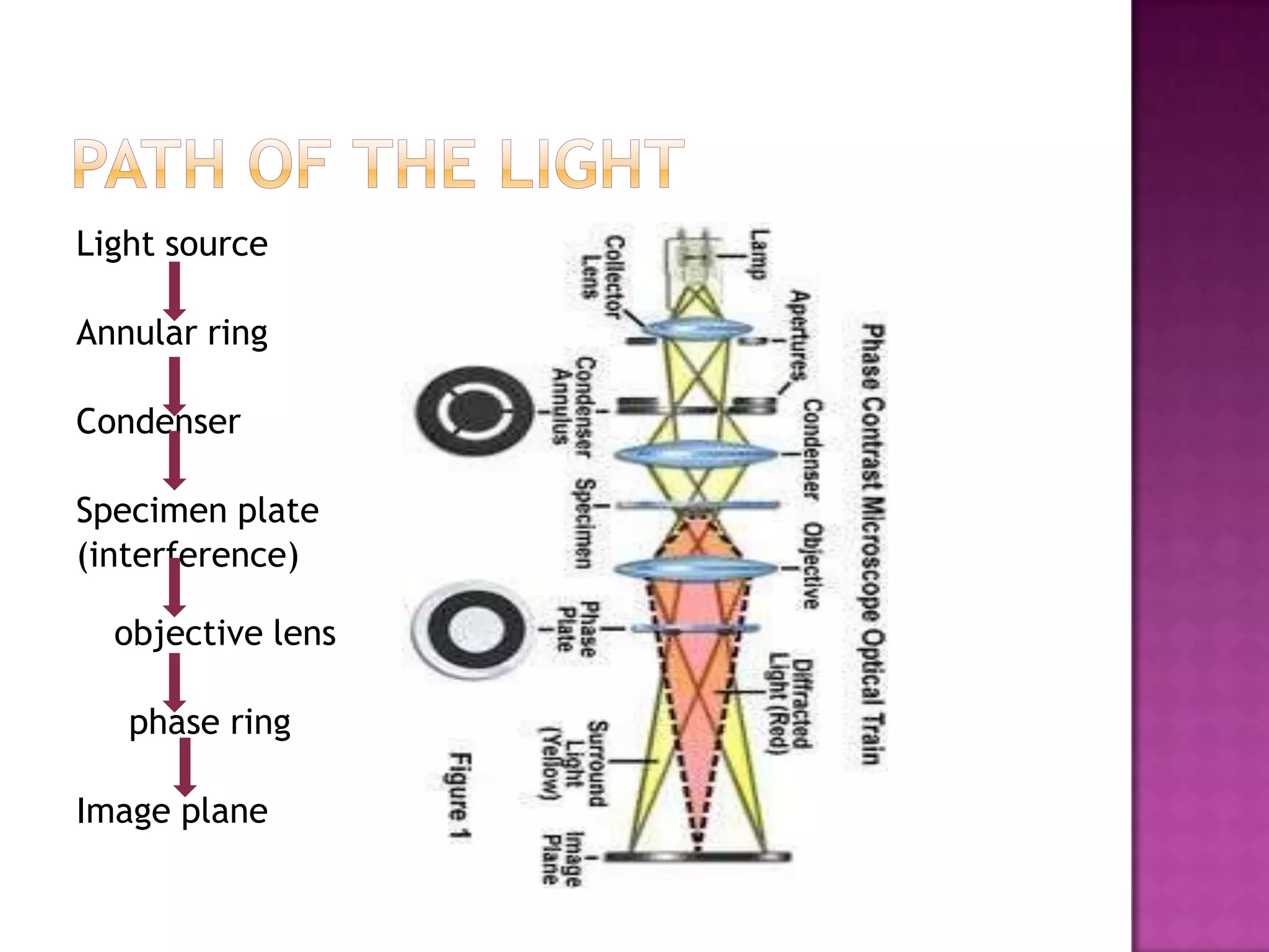
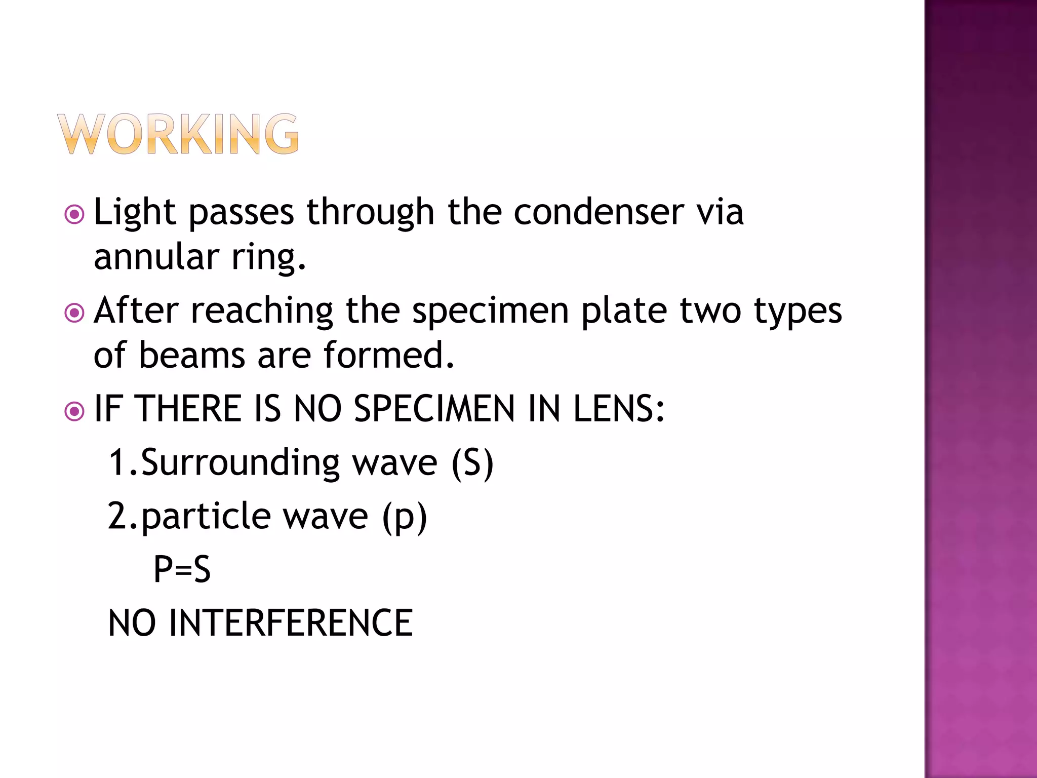
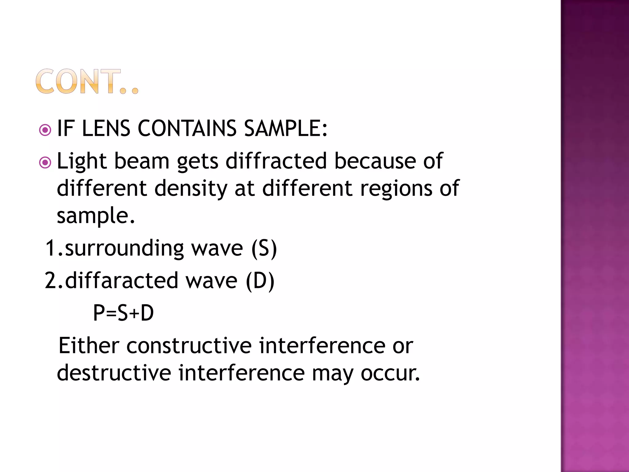
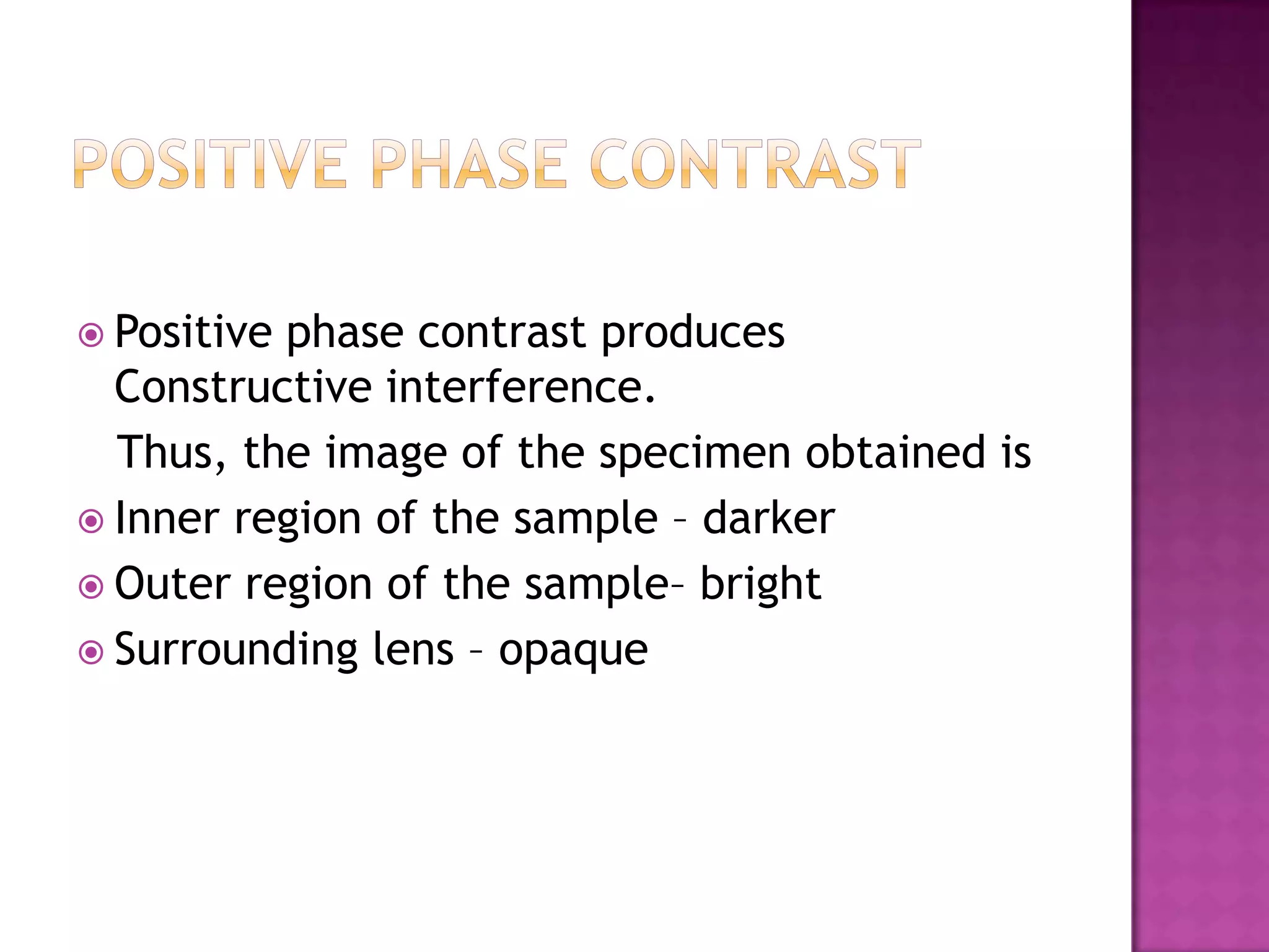
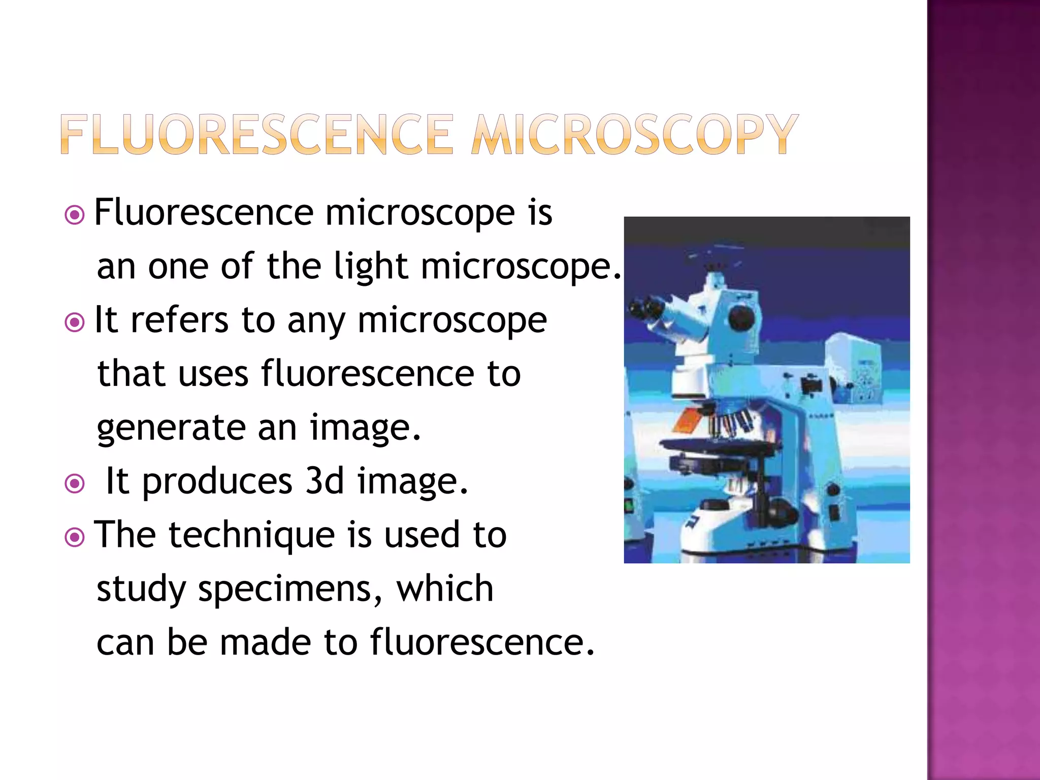
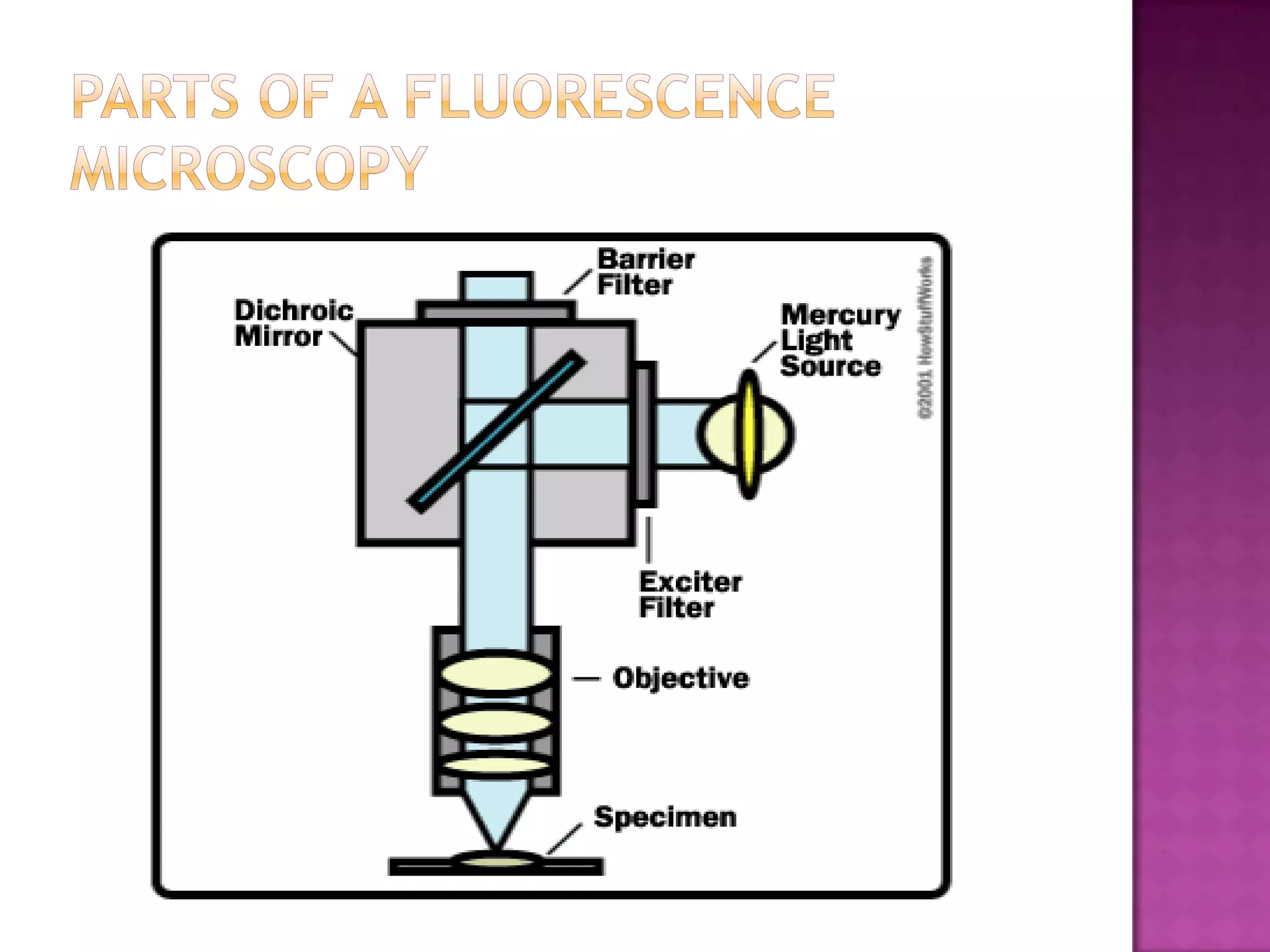
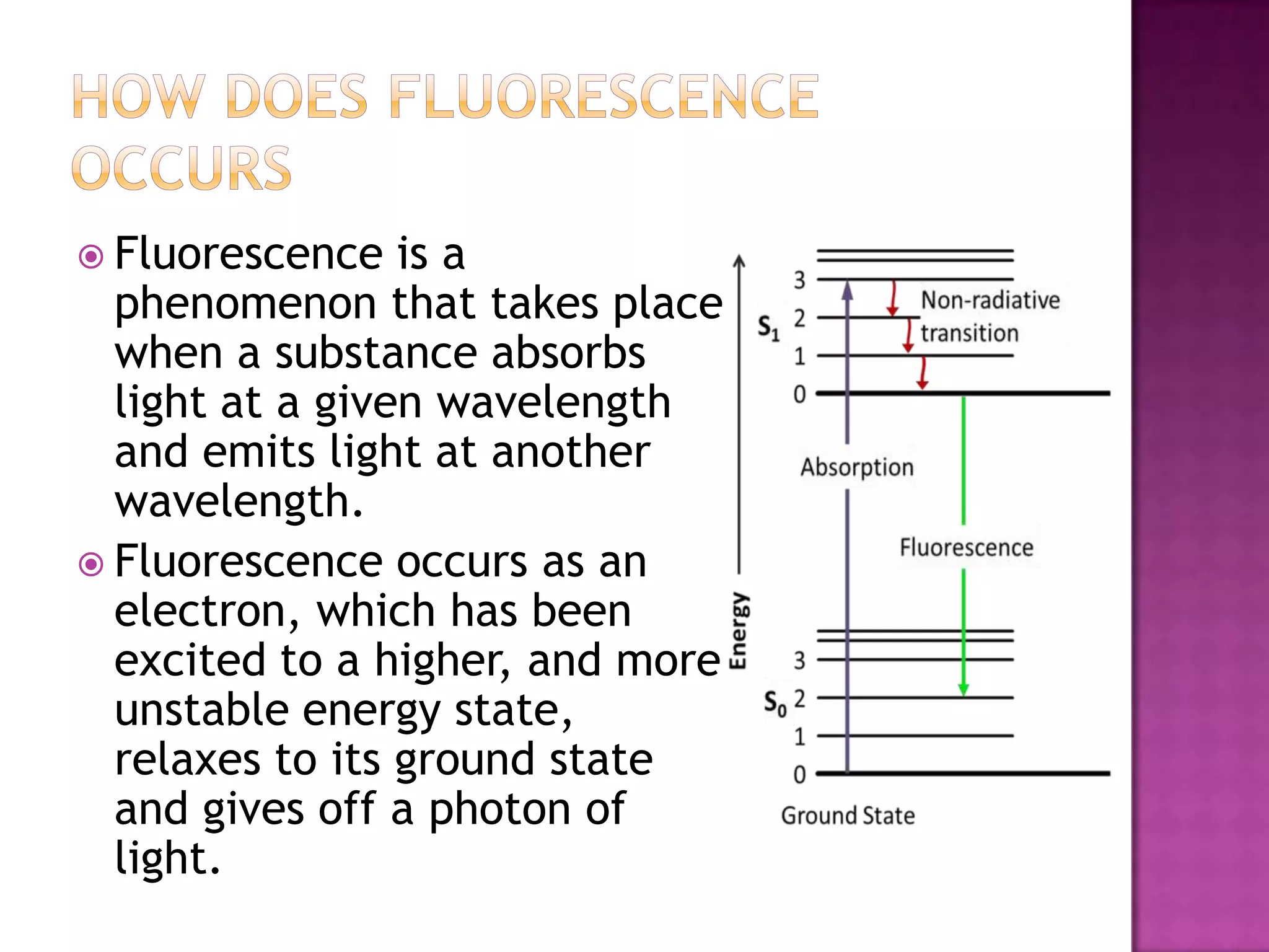
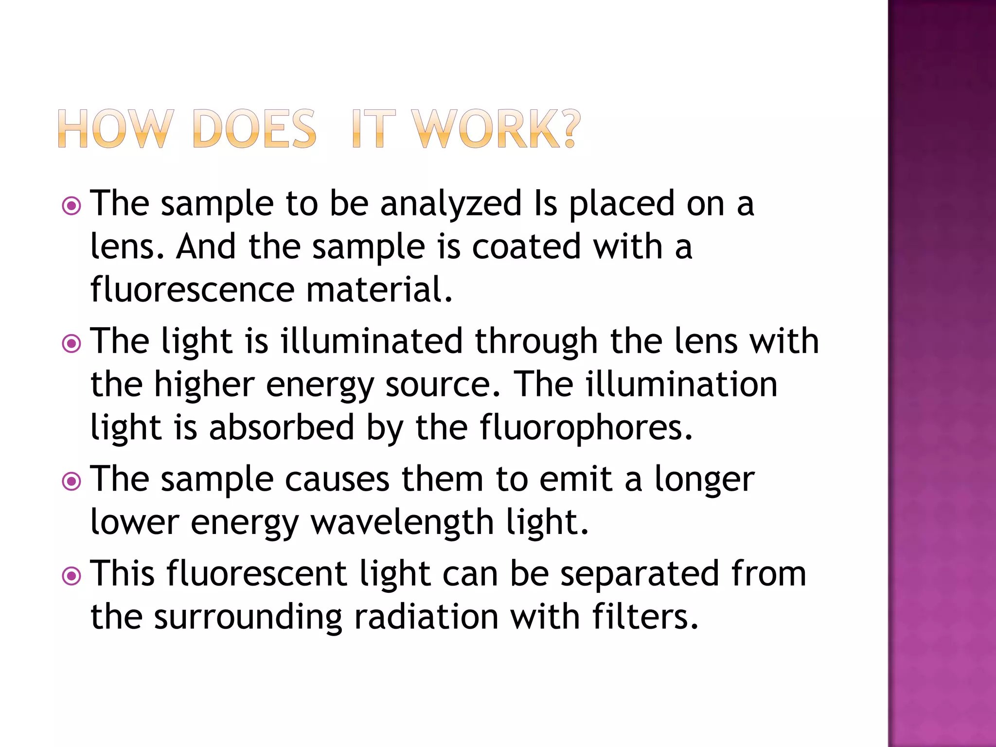
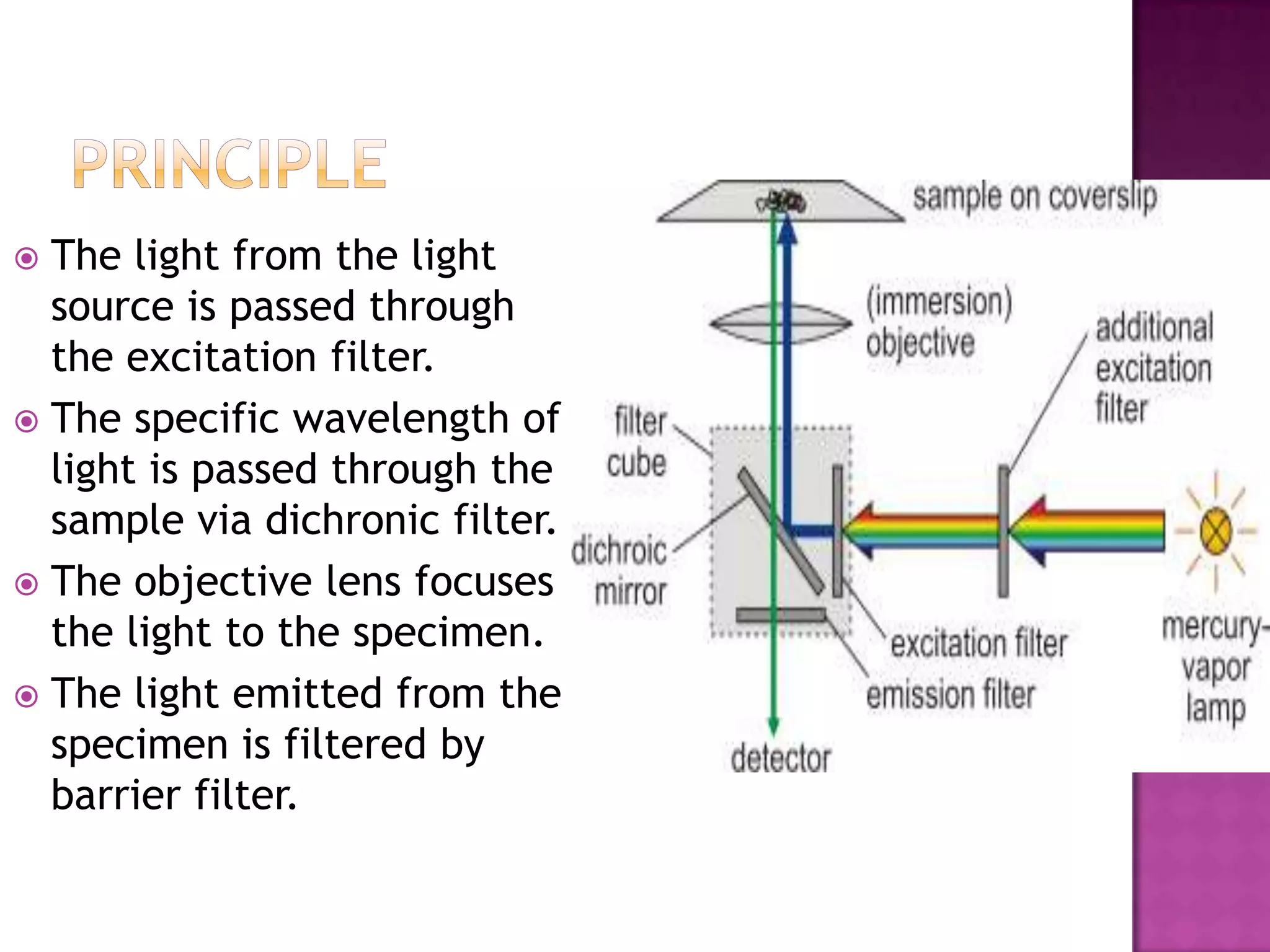


Phase contrast microscopy is a type of light microscopy technique that produces high contrast images of transparent specimens without staining. It works by interfering surrounding light waves and sample-diffracted waves to create an image with the inner regions of the sample appearing darker than the outer regions. Fluorescence microscopy uses fluorescent dyes excited by high-energy light to emit lower-energy light, allowing specific structures within samples to be imaged. It is used to study living cells and view genetic material.