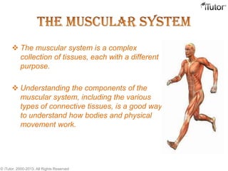
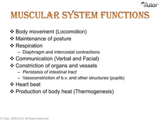
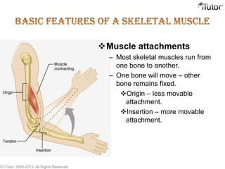
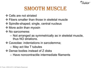
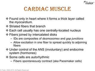
T- 1-855-694-8886
Email- info@iTutor.com
By iTutor.com
 The muscular system is a complex
collection of tissues, each with a different
purpose.
 Understanding the components of the
muscular system, including the various
types of connective tissues, is a good way
to understand how bodies and physical
movement work.
© iTutor. 2000-2013. All Rights Reserved
 Body movement (Locomotion)
 Maintenance of posture
 Respiration
– Diaphragm and intercostal contractions
 Communication (Verbal and Facial)
 Constriction of organs and vessels
– Peristalsis of intestinal tract
– Vasoconstriction of b.v. and other structures (pupils)
 Heart beat
 Production of body heat (Thermogenesis)
© iTutor. 2000-2013. All Rights Reserved
 Excitability: capacity of muscle to respond to a
stimulus.
 Contractility: ability of a muscle to shorten and
generate pulling force.
 Extensibility: muscle can be stretched back to its
original length.
 Elasticity: ability of muscle to recoil to original resting
length after stretched.
© iTutor. 2000-2013. All Rights Reserved
 Skeletal
– Attached to bones
– Makes up 40% of body weight
– Responsible for locomotion, facial expressions, posture, respiratory
movements, other types of body movement
– Voluntary in action; controlled by somatic motor neurons
 Smooth
– In the walls of hollow organs, blood vessels, eye, glands, uterus, skin
– Some functions: propel urine, mix food in digestive tract,
dilating/constricting pupils, regulating blood flow,
– In some locations, auto rhythmic
– Controlled involuntarily by endocrine and autonomic nervous systems
 Cardiac
– Heart: major source of movement of blood
– Auto rhythmic
– Controlled involuntarily by endocrine and autonomic nervous systems
© iTutor. 2000-2013. All Rights Reserved
Muscle attachments
– Most skeletal muscles run from
one bone to another.
– One bone will move – other
bone remains fixed.
Origin – less movable
attachment.
Insertion – more movable
attachment.
© iTutor. 2000-2013. All Rights Reserved
Muscle attachments (continued)
– Muscles attach to origins and insertions by
connective tissue
Fleshy attachments – connective tissue fibers are
short.
Indirect attachments – connective tissue forms a
tendon or aponeurosis.
– Bone markings present where tendons meet
bones
Tubercles, trochanters, and crests.
© iTutor. 2000-2013. All Rights Reserved
 Composed of muscle cells (fibers), connective tissue, blood
vessels, nerves.
 Fibers are long, cylindrical, and multinucleated.
 Tend to be smaller diameter in small muscles and larger in
large muscles. 1 mm- 4 cm in length.
 Develop from myoblasts; numbers remain constant.
 Striated appearance.
 Nuclei are peripherally located.
© iTutor. 2000-2013. All Rights Reserved
 SR is an elaborate, smooth endoplasmic reticulum
– runs longitudinally and surrounds each myofibril
– Form chambers called terminal cisternae on either side of the T-
tubules
 A single T-tubule and the 2 terminal cisternae form a triad
 SR stores Ca++ when muscle not contracting
– When stimulated, calcium released into sarcoplasm
– SR membrane has Ca++ pumps that function to pump Ca++ out of
the sarcoplasm back into the SR after contraction
© iTutor. 2000-2013. All Rights Reserved
 Many elongated myosin molecules shaped like golf clubs.
 Single filament contains roughly 300 myosin molecules.
 Molecule consists of two heavy myosin molecules wound together
to form a rod portion lying parallel to the myosin myofilament and
two heads that extend laterally.
 Myosin heads
1. Can bind to active sites on the actins molecules to form cross-bridges.
(Actin binding site)
2. Attached to the rod portion by a hinge region that can bend and
straighten during contraction.
3. Have ATPase activity: activity that breaks down adenosine
triphosphate (ATP), releasing energy. Part of the energy is used to
bend the hinge region of the myosin molecule during contraction
© iTutor. 2000-2013. All Rights Reserved
Tropomyosin Tropoin Actin
Myosin
© iTutor. 2000-2013. All Rights Reserved
 Thin Filament: composed of 3 major proteins
1. F (fibrous) actin
2. Tropomyosin
3. Troponin
 Two strands of fibrous (F) actin form a double helix extending the
length of the myofilament; attached at either end at sarcomere.
– Composed of G actin monomers each of which has a myosin-
binding site (see yellow dot)
– Actin site can bind myosin during muscle contraction.
 Tropomyosin: an elongated protein winds along the groove of the F
actin double helix.
 Troponin is composed of three subunits:
– Tn-A : binds to actin
– Tn-T :binds to tropomyosin,
– Tn-C :binds to calcium ions.
© iTutor. 2000-2013. All Rights Reserved
 Cells are not striated
 Fibers smaller than those in skeletal muscle
 Spindle-shaped; single, central nucleus
 More actin than myosin
 No sarcomeres
– Not arranged as symmetrically as in skeletal muscle,
thus NO striations.
 Caveolae: indentations in sarcolemma;
– May act like T tubules
 Dense bodies instead of Z disks
– Have noncontractile intermediate filaments
© iTutor. 2000-2013. All Rights Reserved
Grouped into sheets in walls of hollow organs.
 Longitudinal layer – muscle fibers run parallel to organ’s
long axis.
 Circular layer – muscle fibers run around circumference of
the organ.
 Both layers participate in peristalsis.
© iTutor. 2000-2013. All Rights Reserved
 Found only in heart where it forms a thick layer called
the myocardium.
 Striated fibers that branch
 Each cell usually has one centrally-located nucleus
 Fibers joined by intercalated disks
– IDs are composites of desmosomes and gap junctions
– Allow excitation in one fiber to spread quickly to adjoining
fibers
 Under control of the ANS (involuntary) and endocrine
system (hormones)
 Some cells are autorhythmic
– Fibers spontaneously contract (aka Pacemaker cells)
© iTutor. 2000-2013. All Rights Reserved
The end
Call us for more
Information:
www.iTutor.com
1-855-694-8886
Visit

The Muscular System

  • 1.
  • 2.
     The muscularsystem is a complex collection of tissues, each with a different purpose.  Understanding the components of the muscular system, including the various types of connective tissues, is a good way to understand how bodies and physical movement work. © iTutor. 2000-2013. All Rights Reserved
  • 3.
     Body movement(Locomotion)  Maintenance of posture  Respiration – Diaphragm and intercostal contractions  Communication (Verbal and Facial)  Constriction of organs and vessels – Peristalsis of intestinal tract – Vasoconstriction of b.v. and other structures (pupils)  Heart beat  Production of body heat (Thermogenesis) © iTutor. 2000-2013. All Rights Reserved
  • 4.
     Excitability: capacityof muscle to respond to a stimulus.  Contractility: ability of a muscle to shorten and generate pulling force.  Extensibility: muscle can be stretched back to its original length.  Elasticity: ability of muscle to recoil to original resting length after stretched. © iTutor. 2000-2013. All Rights Reserved
  • 5.
    © iTutor. 2000-2013.All Rights Reserved
  • 6.
     Skeletal – Attachedto bones – Makes up 40% of body weight – Responsible for locomotion, facial expressions, posture, respiratory movements, other types of body movement – Voluntary in action; controlled by somatic motor neurons  Smooth – In the walls of hollow organs, blood vessels, eye, glands, uterus, skin – Some functions: propel urine, mix food in digestive tract, dilating/constricting pupils, regulating blood flow, – In some locations, auto rhythmic – Controlled involuntarily by endocrine and autonomic nervous systems  Cardiac – Heart: major source of movement of blood – Auto rhythmic – Controlled involuntarily by endocrine and autonomic nervous systems © iTutor. 2000-2013. All Rights Reserved
  • 7.
    © iTutor. 2000-2013.All Rights Reserved
  • 8.
    Muscle attachments – Mostskeletal muscles run from one bone to another. – One bone will move – other bone remains fixed. Origin – less movable attachment. Insertion – more movable attachment. © iTutor. 2000-2013. All Rights Reserved
  • 9.
    Muscle attachments (continued) –Muscles attach to origins and insertions by connective tissue Fleshy attachments – connective tissue fibers are short. Indirect attachments – connective tissue forms a tendon or aponeurosis. – Bone markings present where tendons meet bones Tubercles, trochanters, and crests. © iTutor. 2000-2013. All Rights Reserved
  • 10.
     Composed ofmuscle cells (fibers), connective tissue, blood vessels, nerves.  Fibers are long, cylindrical, and multinucleated.  Tend to be smaller diameter in small muscles and larger in large muscles. 1 mm- 4 cm in length.  Develop from myoblasts; numbers remain constant.  Striated appearance.  Nuclei are peripherally located. © iTutor. 2000-2013. All Rights Reserved
  • 11.
    © iTutor. 2000-2013.All Rights Reserved
  • 12.
     SR isan elaborate, smooth endoplasmic reticulum – runs longitudinally and surrounds each myofibril – Form chambers called terminal cisternae on either side of the T- tubules  A single T-tubule and the 2 terminal cisternae form a triad  SR stores Ca++ when muscle not contracting – When stimulated, calcium released into sarcoplasm – SR membrane has Ca++ pumps that function to pump Ca++ out of the sarcoplasm back into the SR after contraction © iTutor. 2000-2013. All Rights Reserved
  • 13.
    © iTutor. 2000-2013.All Rights Reserved
  • 14.
     Many elongatedmyosin molecules shaped like golf clubs.  Single filament contains roughly 300 myosin molecules.  Molecule consists of two heavy myosin molecules wound together to form a rod portion lying parallel to the myosin myofilament and two heads that extend laterally.  Myosin heads 1. Can bind to active sites on the actins molecules to form cross-bridges. (Actin binding site) 2. Attached to the rod portion by a hinge region that can bend and straighten during contraction. 3. Have ATPase activity: activity that breaks down adenosine triphosphate (ATP), releasing energy. Part of the energy is used to bend the hinge region of the myosin molecule during contraction © iTutor. 2000-2013. All Rights Reserved
  • 15.
    Tropomyosin Tropoin Actin Myosin ©iTutor. 2000-2013. All Rights Reserved
  • 16.
     Thin Filament:composed of 3 major proteins 1. F (fibrous) actin 2. Tropomyosin 3. Troponin  Two strands of fibrous (F) actin form a double helix extending the length of the myofilament; attached at either end at sarcomere. – Composed of G actin monomers each of which has a myosin- binding site (see yellow dot) – Actin site can bind myosin during muscle contraction.  Tropomyosin: an elongated protein winds along the groove of the F actin double helix.  Troponin is composed of three subunits: – Tn-A : binds to actin – Tn-T :binds to tropomyosin, – Tn-C :binds to calcium ions. © iTutor. 2000-2013. All Rights Reserved
  • 17.
     Cells arenot striated  Fibers smaller than those in skeletal muscle  Spindle-shaped; single, central nucleus  More actin than myosin  No sarcomeres – Not arranged as symmetrically as in skeletal muscle, thus NO striations.  Caveolae: indentations in sarcolemma; – May act like T tubules  Dense bodies instead of Z disks – Have noncontractile intermediate filaments © iTutor. 2000-2013. All Rights Reserved
  • 18.
    © iTutor. 2000-2013.All Rights Reserved
  • 19.
    Grouped into sheetsin walls of hollow organs.  Longitudinal layer – muscle fibers run parallel to organ’s long axis.  Circular layer – muscle fibers run around circumference of the organ.  Both layers participate in peristalsis. © iTutor. 2000-2013. All Rights Reserved
  • 20.
     Found onlyin heart where it forms a thick layer called the myocardium.  Striated fibers that branch  Each cell usually has one centrally-located nucleus  Fibers joined by intercalated disks – IDs are composites of desmosomes and gap junctions – Allow excitation in one fiber to spread quickly to adjoining fibers  Under control of the ANS (involuntary) and endocrine system (hormones)  Some cells are autorhythmic – Fibers spontaneously contract (aka Pacemaker cells) © iTutor. 2000-2013. All Rights Reserved
  • 21.
    © iTutor. 2000-2013.All Rights Reserved
  • 22.
    The end Call usfor more Information: www.iTutor.com 1-855-694-8886 Visit