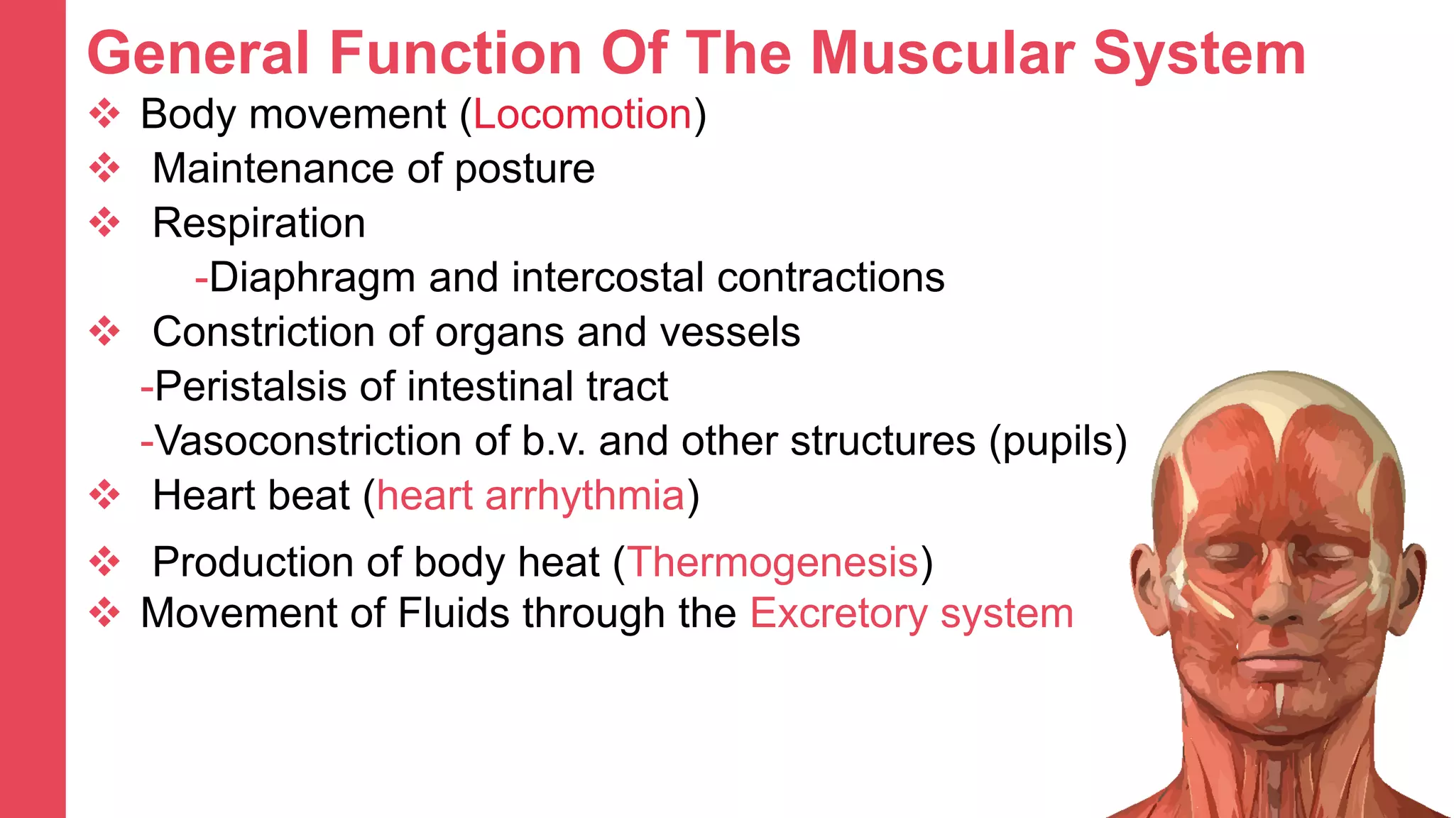
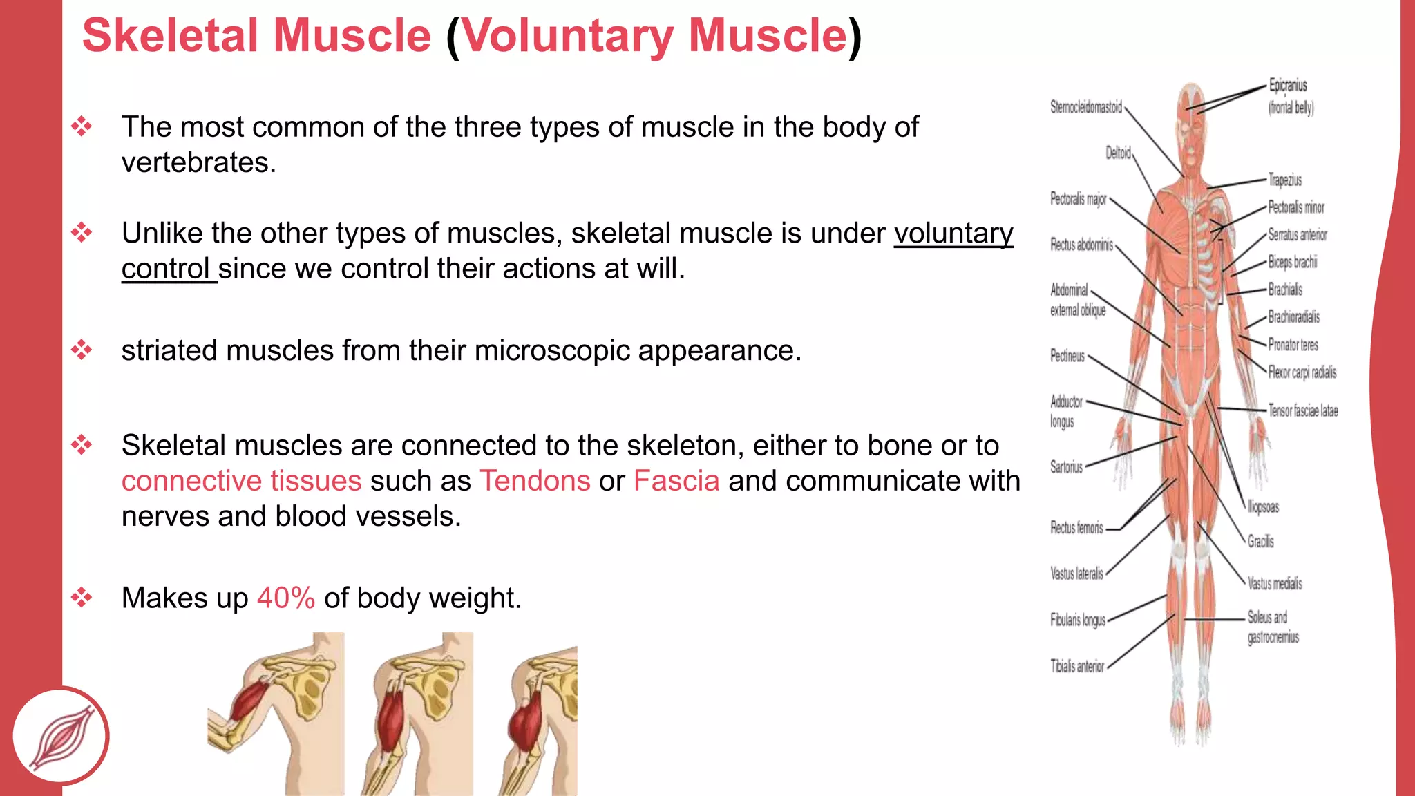
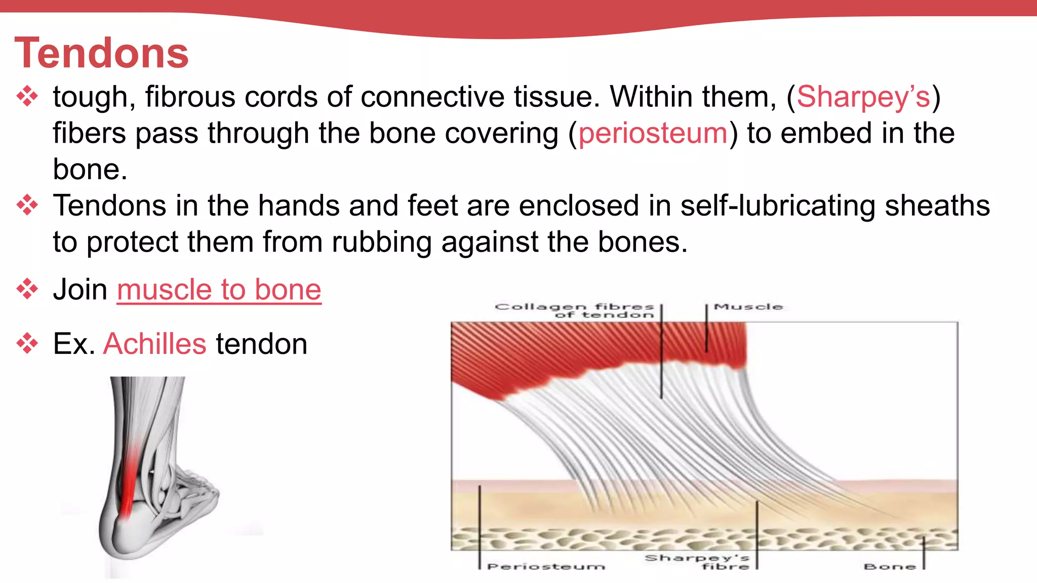
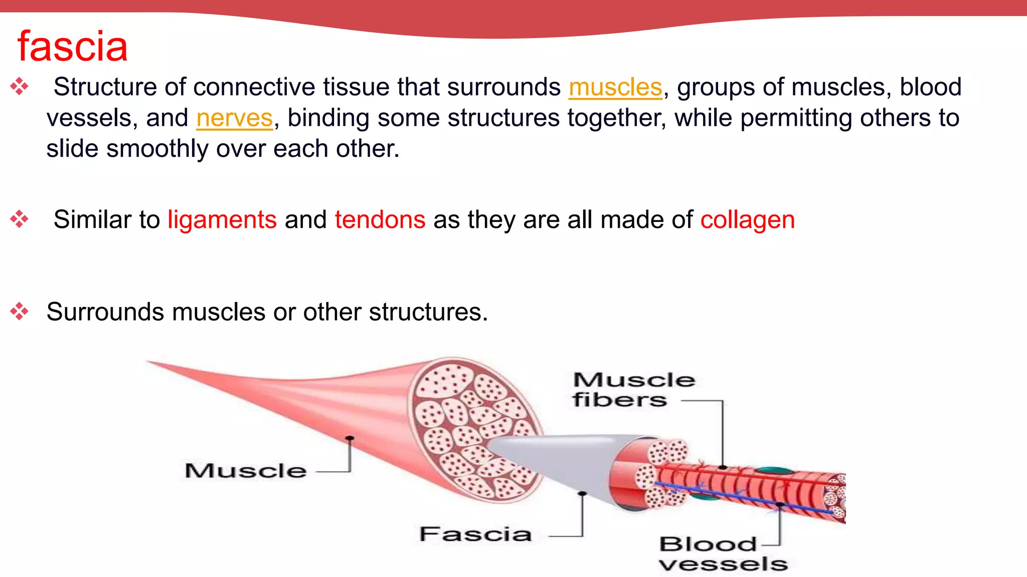
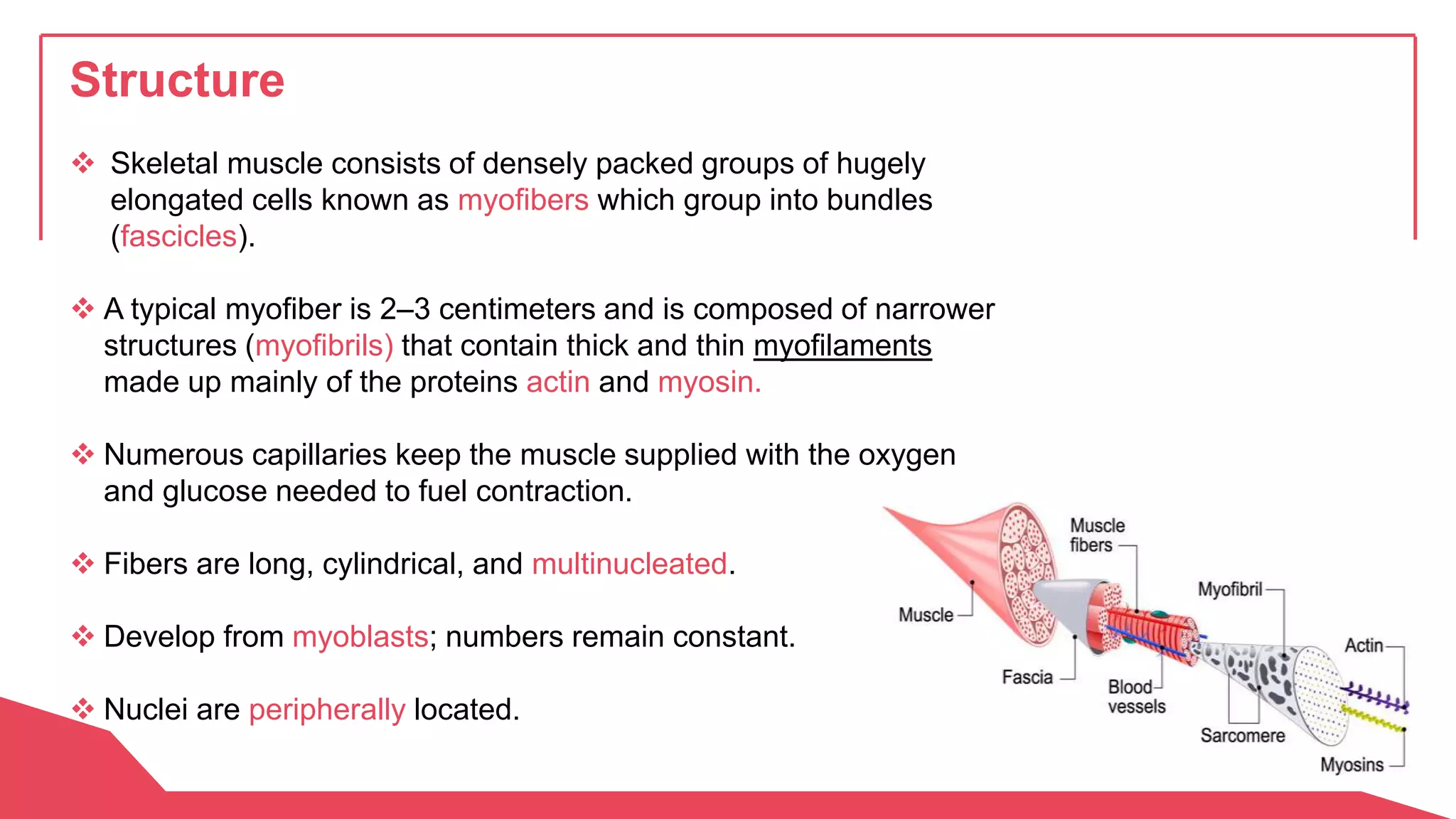
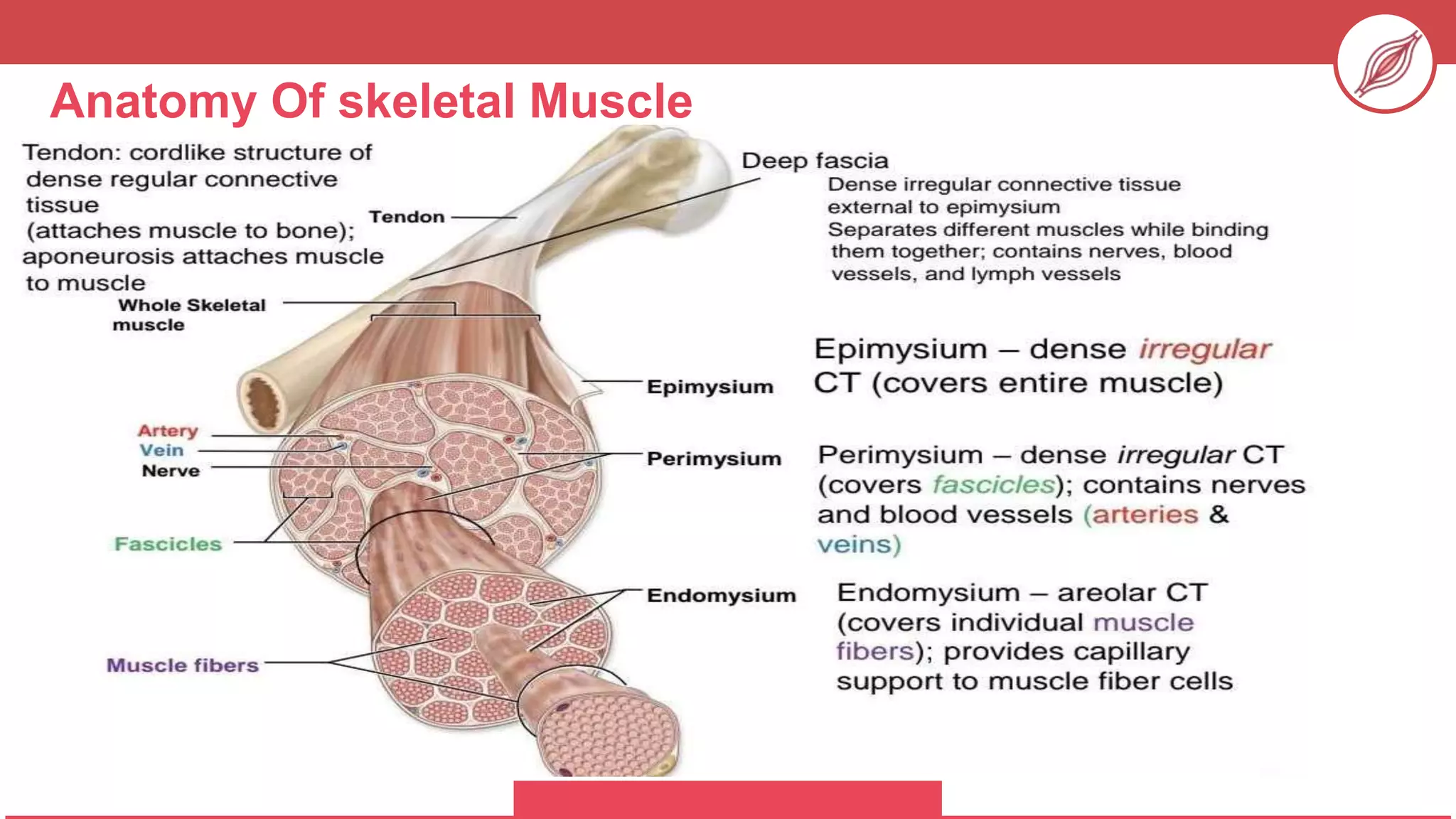
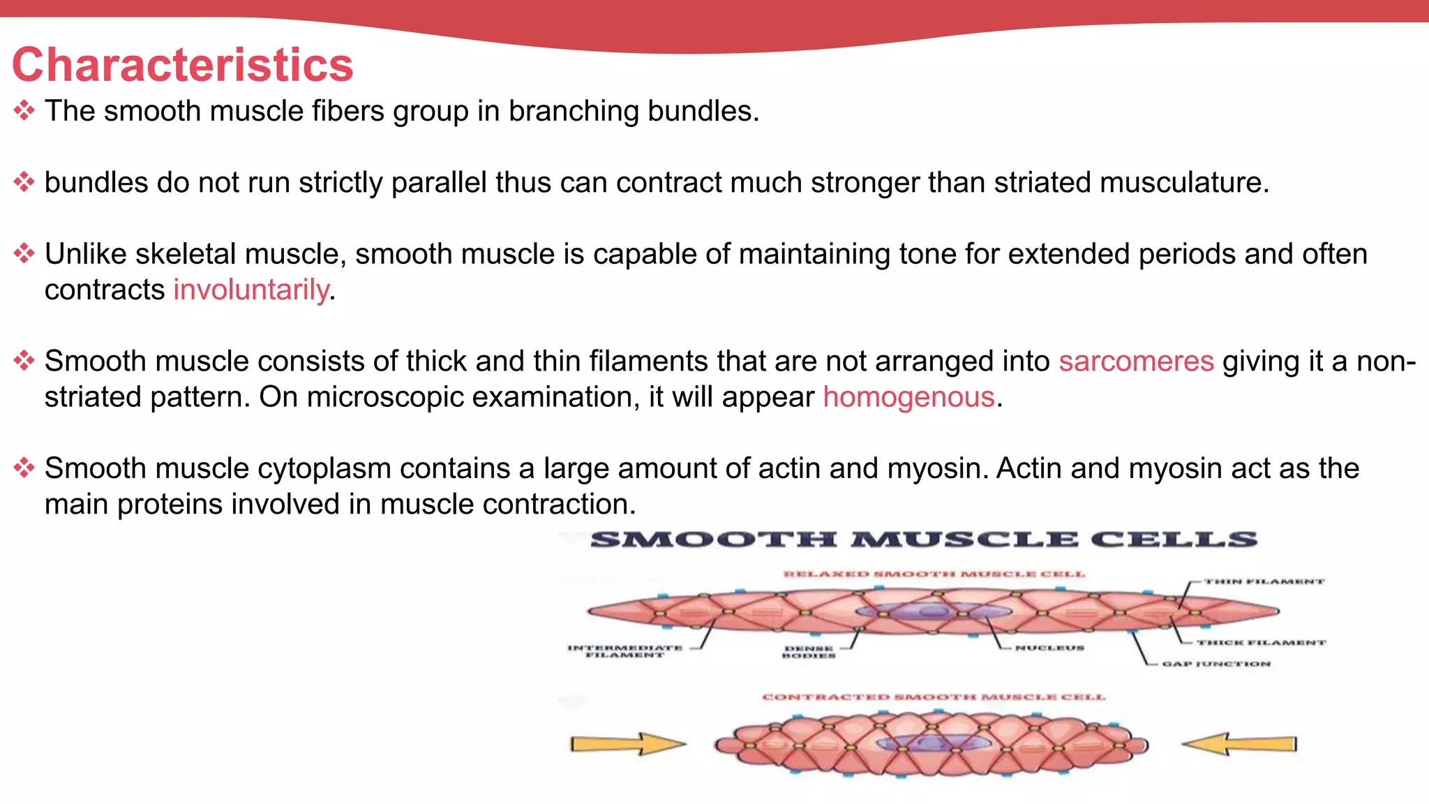
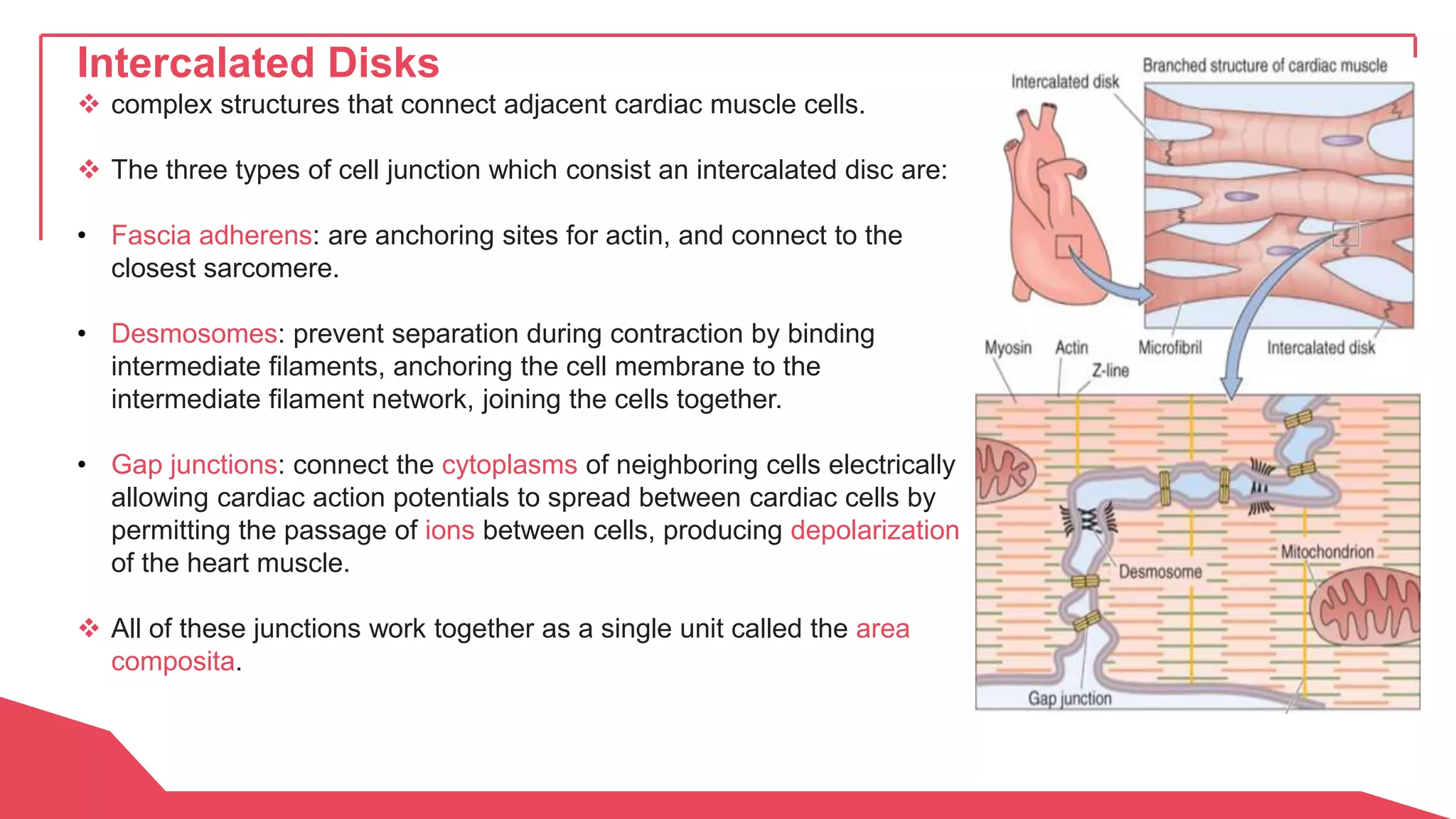
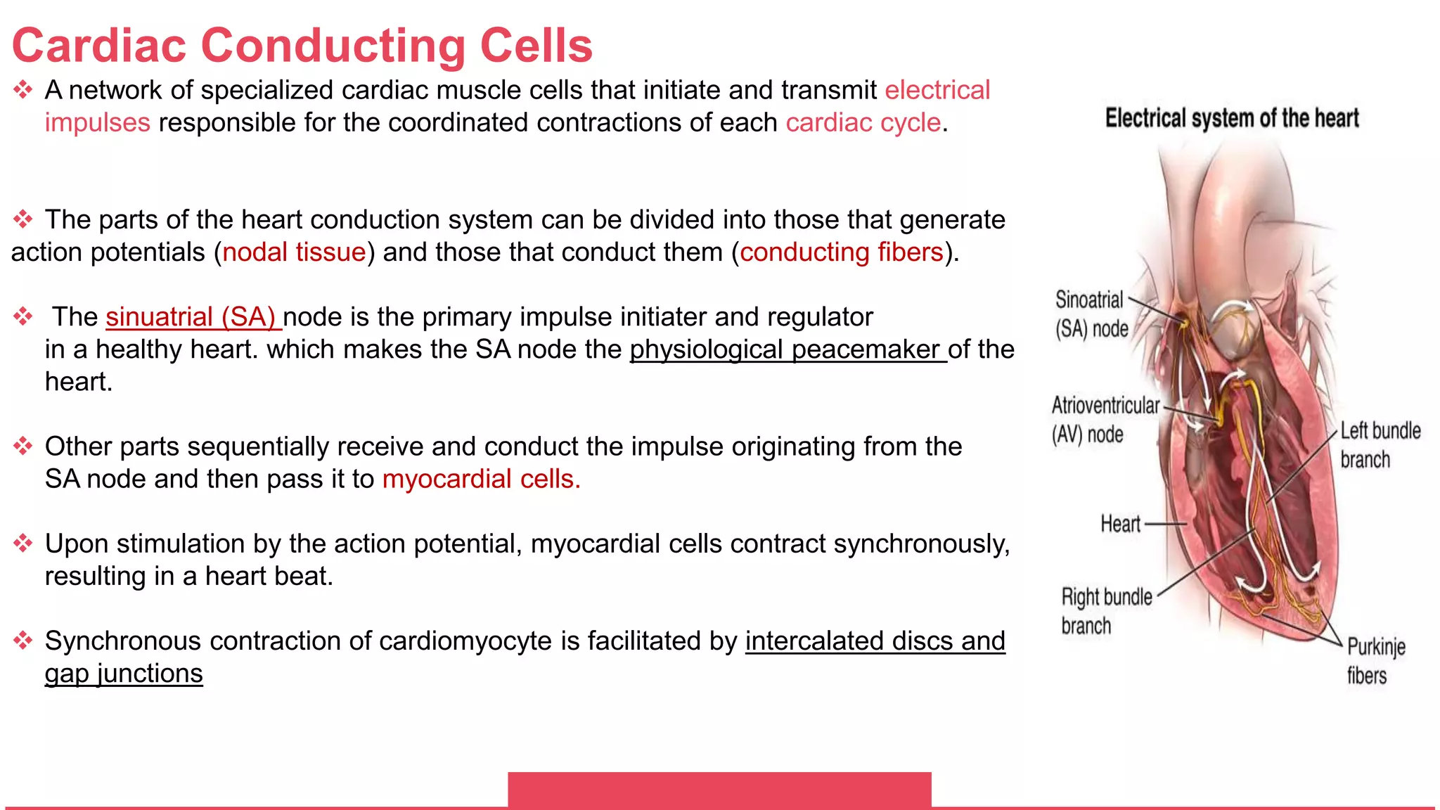
MUSCULAR
SYSTEM
Aland Bakr Shahid Jabar Examplary Highschool
Ahmed Azad 11-A
Yusf Rauf 2022-2023
The Muscular System: A complex collection of
tissues each with a different purpose. Understanding the
components of the muscular system, including the various
types of connective tissues, is a good way to understand
how bodies and physical movement work.
Respiratory
System
Urinary System
Circularory
System
Skeletal
System
Respiratory
System
Respiratory
System
General Function Of The Muscular System
 Body movement (Locomotion)
 Maintenance of posture
 Respiration
-Diaphragm and intercostal contractions
 Constriction of organs and vessels
-Peristalsis of intestinal tract
-Vasoconstriction of b.v. and other structures (pupils)
 Heart beat (heart arrhythmia)
 Production of body heat (Thermogenesis)
 Movement of Fluids through the Excretory system
PROPERTIES OF MUSCLE
 Excitability: capacity of muscle to respond to a stimulus.
Contractility: ability of a muscle to shorten and generate
pulling force.
Extensibility: muscles can be stretched back to its original
length.
Elasticity: ability of muscle to recoil to original resting
position.
Types Of Muscles
I. Skeletal Muscle
II. Smooth Muscle
III.Cardiac Muscle
Skeletal Muscle (Voluntary Muscle)
 The most common of the three types of muscle in the body of
vertebrates.
 Unlike the other types of muscles, skeletal muscle is under voluntary
control since we control their actions at will.
 striated muscles from their microscopic appearance.
 Skeletal muscles are connected to the skeleton, either to bone or to
connective tissues such as Tendons or Fascia and communicate with
nerves and blood vessels.
 Makes up 40% of body weight.
Tendons
 tough, fibrous cords of connective tissue. Within them, (Sharpey’s)
fibers pass through the bone covering (periosteum) to embed in the
bone.
 Tendons in the hands and feet are enclosed in self-lubricating sheaths
to protect them from rubbing against the bones.
 Join muscle to bone
 Ex. Achilles tendon
fascia
 Structure of connective tissue that surrounds muscles, groups of muscles, blood
vessels, and nerves, binding some structures together, while permitting others to
slide smoothly over each other.
 Similar to ligaments and tendons as they are all made of collagen
 Surrounds muscles or other structures.
Functions
 Chewing and swallowing, which are the first steps of digestion.
 Expanding and contracting your chest cavity so you can inhale and
exhale at will.
 Maintaining body posture.
 Moving the bones in different parts of your body.
 Protecting joints and holding them in place.
Muscle attachments
- Most skeletal muscles run from one bone to another.
- One bone will move, other bone remains fixed.
Origins less movable attachments.
Structure
 Skeletal muscle consists of densely packed groups of hugely
elongated cells known as myofibers which group into bundles
(fascicles).
 A typical myofiber is 2–3 centimeters and is composed of narrower
structures (myofibrils) that contain thick and thin myofilaments
made up mainly of the proteins actin and myosin.
 Numerous capillaries keep the muscle supplied with the oxygen
and glucose needed to fuel contraction.
 Fibers are long, cylindrical, and multinucleated.
 Develop from myoblasts; numbers remain constant.
 Nuclei are peripherally located.
Anatomy Of skeletal Muscle
Skeletal Muscles And Energy
Skeletal muscle cells are involved in
many processes relating to energy
such as:
Smooth Muscle
Smooth muscle is present throughout the body, where it serves a variety of functions, such
general functions include:
 Contraction (primary function)
 In the stomach and intestines, where it helps with digestion and nutrient collection.
 Throughout urinary system, helps rid the body of toxins and works in electrolyte balance.
 It is present throughout arteries and veins, where it plays a vital role in the regulation of blood pressure
and tissue oxygenation.
 Cardiovascular - regulation of blood flow and pressure via vascular resistance.
 Genital - contractions during pregnancy, propulsion of sperm.
 Respiratory tract - regulation of bronchiole diameter.
 Integument - raises hair with erector pili muscle.
 Sensory - dilation and constriction of the pupil as well as changing lens shape.
Characteristics
 The smooth muscle fibers group in branching bundles.
 bundles do not run strictly parallel thus can contract much stronger than striated musculature.
 Unlike skeletal muscle, smooth muscle is capable of maintaining tone for extended periods and often
contracts involuntarily.
 Smooth muscle consists of thick and thin filaments that are not arranged into sarcomeres giving it a non-
striated pattern. On microscopic examination, it will appear homogenous.
 Smooth muscle cytoplasm contains a large amount of actin and myosin. Actin and myosin act as the
main proteins involved in muscle contraction.
Cardiac Muscle (myocardium, Heart)
 is a specialized type of muscle tissue that forms the heart,
 has a very unique structure and could be described as intermediate or “in between” skeletal
and smooth muscle tissue.
 This muscle tissue contracts (Systole) and relaxes (Diastole) involuntary.
 responsible for blood pump and circulation through the blood vessels of the circulatory
system.
 contractility is the basis for its pumping action.
 The amount of blood pumped by the heart per minute (cardiac output) varies to meet the
metabolic needs. Is determined by the contractile force developed by the cardiac muscle
cells and by the frequency at which they are activated (rhythmicity).
Structure
 short, branching cell with one or two large, centrally located nuclei.
 averages about 25µm in diameter and 120µm in length.
 Adjacent cardiac muscle cells are joined together at their ends to form
cellular networks
 these branching networks of cardiac muscle cells are called cardiac fibers.
 The complex junctions that join cardiac muscle cells are
called intercalated discs
Components of the Cardiac Muscle Tissue
1. Cardiomyocytes
2. Intercalated discs
3. Cardiac conducting cells
Cardiomyocytes
 Individual cells that make up the cardiac muscle. The primary function is to contract, which generates the
pressure needed to pump blood through the circulatory system.
 These individual cells are tubular structures composed of chains of myofibrils that myofibrils consist of
repeating sections of sarcomeres.
 Sarcomeres are composed of long proteins that organize into thick and thin filaments, called myofilaments.
Which slide past each other as the muscle contracts and relaxes.
 Cardiomyocytes contain many mitochondria to produce large amounts of (ATP) and myoglobin to store oxygen
to meet the demands of muscle contraction.
 The outside of the cardiomyocyte is surrounded by a plasma membrane called the sarcolemma that acts as a
barrier between extracellular and intracellular contents
 Invaginations of the sarcolemma into the cytoplasm of the cardiomyocyte are called T-tubules.
Intercalated Disks
 complex structures that connect adjacent cardiac muscle cells.
 The three types of cell junction which consist an intercalated disc are:
• Fascia adherens: are anchoring sites for actin, and connect to the
closest sarcomere.
• Desmosomes: prevent separation during contraction by binding
intermediate filaments, anchoring the cell membrane to the
intermediate filament network, joining the cells together.
• Gap junctions: connect the cytoplasms of neighboring cells electrically
allowing cardiac action potentials to spread between cardiac cells by
permitting the passage of ions between cells, producing depolarization
of the heart muscle.
 All of these junctions work together as a single unit called the area
composita.
Cardiac Conducting Cells
 A network of specialized cardiac muscle cells that initiate and transmit electrical
impulses responsible for the coordinated contractions of each cardiac cycle.
 The parts of the heart conduction system can be divided into those that generate
action potentials (nodal tissue) and those that conduct them (conducting fibers).
 The sinuatrial (SA) node is the primary impulse initiater and regulator
in a healthy heart. which makes the SA node the physiological peacemaker of the
heart.
 Other parts sequentially receive and conduct the impulse originating from the
SA node and then pass it to myocardial cells.
 Upon stimulation by the action potential, myocardial cells contract synchronously,
resulting in a heart beat.
 Synchronous contraction of cardiomyocyte is facilitated by intercalated discs and
gap junctions
1) is triggered by calcium ions binding to the protein complex troponin. Which causes
the actin-binding sites to be exposed
2) The high-energy myosin head bridges the gap, forming a cross-bridge.
3) Once myosin binds to the actin, the Pi is released, and the myosin undergoes a
conformational change to a lower energy state. As myosin expends the energy, it
moves through the "power stroke," pulling the actin filament toward the M-line. When
the actin is pulled approximately 10 nm toward the M-line, the sarcomere shortens
and the muscle contracts. At the end of the power stroke, the myosin is in a low-
energy position. After the power stroke, ADP is released, but the cross-bridge formed
is still in place.
4) ATP then binds to myosin, moving the myosin to its high-energy state, releasing the
myosin head from the actin active site.
5) The ATP is hydrolyzed into ADP and inorganic phosphate (Pi) by the enzyme ATPase.
The energy released during ATP hydrolysis changes the angle of the myosin head
into a "cocked" position, which allows the cross-bridge cycle to start again; further
muscle contraction can occur. Therefore, without ATP, muscles would remain in their
contracted state, rather than their relaxed state.
Muscle Contraction
Muscles contract in a repeated pattern of binding and releasing between the two thin and thick
strands of the sarcoere. ATP is critical to prepare myosin for binding and to "recharge" the myosin.
 common complaint in clinical practice.
 In humans, defined as a decrease in maximal force or power production in response to
contractile activity.
 The causes of muscle fatigue is the result of relative depletion of ATP, when ATP is absent a
state of continuous contraction occurs, an example of muscle fatigue is when a marathon
runner collapses due to severe muscle cramps.
Muscle Fatigue Preventions
 Eating healthy or or light meals before exercise.
 Drinking plenty of beverages that contain electrolytes.
 Stretching your muscles before committing vigorous exertions may prevent you from injuries.
Muscle Fatigue
Muscular hypertrophy
An increase in the size of a muscle, or its cross-sectional area attributed to an
increase in the size and/or number of myofibrils (actin and myosin) within a
given muscle fiber.
Benefits of Muscle hypertrophy
There is a strong correlation between muscle cross-sectional area and
muscle strength. This means that more muscle mass creates greater
potential for developing maximal strength.
resistance training and muscle hypertrophy have a positive effect on body
composition, addressing two of the three factors that comprise energy
expenditure; resting metabolic rate (RMR), physical activity, and the thermic
effect of food.
Inducing Muscle Hypertrophy
The three primary factors that induce a hypertrophic response in the body include:
mechanical tension: The degree of mechanical tension from a resistance training
session is primarily determined by intensity (amount of weight lifted) and time under
tension (duration of the applied load).
muscle damage: Resistance training that creates an overload situation causes muscle
damage and an inflammatory response, potentiating the release of various growth
factors.
metabolic stress: Metabolic stress results from training programs that rely heavily on the
anaerobic energy production, decreasing the pH level and causing muscle fiber
degradation.
Sleep and nutrition each play a role in recovery and ultimately the body’s ability to undergo
supercompensation. The body uses sleep to repair damaged tissue and can play a vital
role in hypertrophy.

Muscular System

  • 1.
    MUSCULAR SYSTEM Aland Bakr ShahidJabar Examplary Highschool Ahmed Azad 11-A Yusf Rauf 2022-2023
  • 2.
    The Muscular System:A complex collection of tissues each with a different purpose. Understanding the components of the muscular system, including the various types of connective tissues, is a good way to understand how bodies and physical movement work.
  • 3.
    Respiratory System Urinary System Circularory System Skeletal System Respiratory System Respiratory System General FunctionOf The Muscular System  Body movement (Locomotion)  Maintenance of posture  Respiration -Diaphragm and intercostal contractions  Constriction of organs and vessels -Peristalsis of intestinal tract -Vasoconstriction of b.v. and other structures (pupils)  Heart beat (heart arrhythmia)  Production of body heat (Thermogenesis)  Movement of Fluids through the Excretory system
  • 4.
    PROPERTIES OF MUSCLE Excitability: capacity of muscle to respond to a stimulus. Contractility: ability of a muscle to shorten and generate pulling force. Extensibility: muscles can be stretched back to its original length. Elasticity: ability of muscle to recoil to original resting position.
  • 5.
    Types Of Muscles I.Skeletal Muscle II. Smooth Muscle III.Cardiac Muscle
  • 6.
    Skeletal Muscle (VoluntaryMuscle)  The most common of the three types of muscle in the body of vertebrates.  Unlike the other types of muscles, skeletal muscle is under voluntary control since we control their actions at will.  striated muscles from their microscopic appearance.  Skeletal muscles are connected to the skeleton, either to bone or to connective tissues such as Tendons or Fascia and communicate with nerves and blood vessels.  Makes up 40% of body weight.
  • 7.
    Tendons  tough, fibrouscords of connective tissue. Within them, (Sharpey’s) fibers pass through the bone covering (periosteum) to embed in the bone.  Tendons in the hands and feet are enclosed in self-lubricating sheaths to protect them from rubbing against the bones.  Join muscle to bone  Ex. Achilles tendon
  • 8.
    fascia  Structure ofconnective tissue that surrounds muscles, groups of muscles, blood vessels, and nerves, binding some structures together, while permitting others to slide smoothly over each other.  Similar to ligaments and tendons as they are all made of collagen  Surrounds muscles or other structures.
  • 9.
    Functions  Chewing andswallowing, which are the first steps of digestion.  Expanding and contracting your chest cavity so you can inhale and exhale at will.  Maintaining body posture.  Moving the bones in different parts of your body.  Protecting joints and holding them in place. Muscle attachments - Most skeletal muscles run from one bone to another. - One bone will move, other bone remains fixed. Origins less movable attachments.
  • 10.
    Structure  Skeletal muscleconsists of densely packed groups of hugely elongated cells known as myofibers which group into bundles (fascicles).  A typical myofiber is 2–3 centimeters and is composed of narrower structures (myofibrils) that contain thick and thin myofilaments made up mainly of the proteins actin and myosin.  Numerous capillaries keep the muscle supplied with the oxygen and glucose needed to fuel contraction.  Fibers are long, cylindrical, and multinucleated.  Develop from myoblasts; numbers remain constant.  Nuclei are peripherally located.
  • 11.
  • 12.
    Skeletal Muscles AndEnergy Skeletal muscle cells are involved in many processes relating to energy such as:
  • 13.
    Smooth Muscle Smooth muscleis present throughout the body, where it serves a variety of functions, such general functions include:  Contraction (primary function)  In the stomach and intestines, where it helps with digestion and nutrient collection.  Throughout urinary system, helps rid the body of toxins and works in electrolyte balance.  It is present throughout arteries and veins, where it plays a vital role in the regulation of blood pressure and tissue oxygenation.  Cardiovascular - regulation of blood flow and pressure via vascular resistance.  Genital - contractions during pregnancy, propulsion of sperm.  Respiratory tract - regulation of bronchiole diameter.  Integument - raises hair with erector pili muscle.  Sensory - dilation and constriction of the pupil as well as changing lens shape.
  • 14.
    Characteristics  The smoothmuscle fibers group in branching bundles.  bundles do not run strictly parallel thus can contract much stronger than striated musculature.  Unlike skeletal muscle, smooth muscle is capable of maintaining tone for extended periods and often contracts involuntarily.  Smooth muscle consists of thick and thin filaments that are not arranged into sarcomeres giving it a non- striated pattern. On microscopic examination, it will appear homogenous.  Smooth muscle cytoplasm contains a large amount of actin and myosin. Actin and myosin act as the main proteins involved in muscle contraction.
  • 15.
    Cardiac Muscle (myocardium,Heart)  is a specialized type of muscle tissue that forms the heart,  has a very unique structure and could be described as intermediate or “in between” skeletal and smooth muscle tissue.  This muscle tissue contracts (Systole) and relaxes (Diastole) involuntary.  responsible for blood pump and circulation through the blood vessels of the circulatory system.  contractility is the basis for its pumping action.  The amount of blood pumped by the heart per minute (cardiac output) varies to meet the metabolic needs. Is determined by the contractile force developed by the cardiac muscle cells and by the frequency at which they are activated (rhythmicity).
  • 16.
    Structure  short, branchingcell with one or two large, centrally located nuclei.  averages about 25µm in diameter and 120µm in length.  Adjacent cardiac muscle cells are joined together at their ends to form cellular networks  these branching networks of cardiac muscle cells are called cardiac fibers.  The complex junctions that join cardiac muscle cells are called intercalated discs Components of the Cardiac Muscle Tissue 1. Cardiomyocytes 2. Intercalated discs 3. Cardiac conducting cells
  • 17.
    Cardiomyocytes  Individual cellsthat make up the cardiac muscle. The primary function is to contract, which generates the pressure needed to pump blood through the circulatory system.  These individual cells are tubular structures composed of chains of myofibrils that myofibrils consist of repeating sections of sarcomeres.  Sarcomeres are composed of long proteins that organize into thick and thin filaments, called myofilaments. Which slide past each other as the muscle contracts and relaxes.  Cardiomyocytes contain many mitochondria to produce large amounts of (ATP) and myoglobin to store oxygen to meet the demands of muscle contraction.  The outside of the cardiomyocyte is surrounded by a plasma membrane called the sarcolemma that acts as a barrier between extracellular and intracellular contents  Invaginations of the sarcolemma into the cytoplasm of the cardiomyocyte are called T-tubules.
  • 18.
    Intercalated Disks  complexstructures that connect adjacent cardiac muscle cells.  The three types of cell junction which consist an intercalated disc are: • Fascia adherens: are anchoring sites for actin, and connect to the closest sarcomere. • Desmosomes: prevent separation during contraction by binding intermediate filaments, anchoring the cell membrane to the intermediate filament network, joining the cells together. • Gap junctions: connect the cytoplasms of neighboring cells electrically allowing cardiac action potentials to spread between cardiac cells by permitting the passage of ions between cells, producing depolarization of the heart muscle.  All of these junctions work together as a single unit called the area composita.
  • 19.
    Cardiac Conducting Cells A network of specialized cardiac muscle cells that initiate and transmit electrical impulses responsible for the coordinated contractions of each cardiac cycle.  The parts of the heart conduction system can be divided into those that generate action potentials (nodal tissue) and those that conduct them (conducting fibers).  The sinuatrial (SA) node is the primary impulse initiater and regulator in a healthy heart. which makes the SA node the physiological peacemaker of the heart.  Other parts sequentially receive and conduct the impulse originating from the SA node and then pass it to myocardial cells.  Upon stimulation by the action potential, myocardial cells contract synchronously, resulting in a heart beat.  Synchronous contraction of cardiomyocyte is facilitated by intercalated discs and gap junctions
  • 20.
    1) is triggeredby calcium ions binding to the protein complex troponin. Which causes the actin-binding sites to be exposed 2) The high-energy myosin head bridges the gap, forming a cross-bridge. 3) Once myosin binds to the actin, the Pi is released, and the myosin undergoes a conformational change to a lower energy state. As myosin expends the energy, it moves through the "power stroke," pulling the actin filament toward the M-line. When the actin is pulled approximately 10 nm toward the M-line, the sarcomere shortens and the muscle contracts. At the end of the power stroke, the myosin is in a low- energy position. After the power stroke, ADP is released, but the cross-bridge formed is still in place. 4) ATP then binds to myosin, moving the myosin to its high-energy state, releasing the myosin head from the actin active site. 5) The ATP is hydrolyzed into ADP and inorganic phosphate (Pi) by the enzyme ATPase. The energy released during ATP hydrolysis changes the angle of the myosin head into a "cocked" position, which allows the cross-bridge cycle to start again; further muscle contraction can occur. Therefore, without ATP, muscles would remain in their contracted state, rather than their relaxed state. Muscle Contraction Muscles contract in a repeated pattern of binding and releasing between the two thin and thick strands of the sarcoere. ATP is critical to prepare myosin for binding and to "recharge" the myosin.
  • 21.
     common complaintin clinical practice.  In humans, defined as a decrease in maximal force or power production in response to contractile activity.  The causes of muscle fatigue is the result of relative depletion of ATP, when ATP is absent a state of continuous contraction occurs, an example of muscle fatigue is when a marathon runner collapses due to severe muscle cramps. Muscle Fatigue Preventions  Eating healthy or or light meals before exercise.  Drinking plenty of beverages that contain electrolytes.  Stretching your muscles before committing vigorous exertions may prevent you from injuries. Muscle Fatigue
  • 22.
    Muscular hypertrophy An increasein the size of a muscle, or its cross-sectional area attributed to an increase in the size and/or number of myofibrils (actin and myosin) within a given muscle fiber. Benefits of Muscle hypertrophy There is a strong correlation between muscle cross-sectional area and muscle strength. This means that more muscle mass creates greater potential for developing maximal strength. resistance training and muscle hypertrophy have a positive effect on body composition, addressing two of the three factors that comprise energy expenditure; resting metabolic rate (RMR), physical activity, and the thermic effect of food.
  • 23.
    Inducing Muscle Hypertrophy Thethree primary factors that induce a hypertrophic response in the body include: mechanical tension: The degree of mechanical tension from a resistance training session is primarily determined by intensity (amount of weight lifted) and time under tension (duration of the applied load). muscle damage: Resistance training that creates an overload situation causes muscle damage and an inflammatory response, potentiating the release of various growth factors. metabolic stress: Metabolic stress results from training programs that rely heavily on the anaerobic energy production, decreasing the pH level and causing muscle fiber degradation. Sleep and nutrition each play a role in recovery and ultimately the body’s ability to undergo supercompensation. The body uses sleep to repair damaged tissue and can play a vital role in hypertrophy.

Editor's Notes