IRJET- Detection of Whitematter Ms-Lesion in Human Brain by Segmentation using Pso with Hpla Methods

This paper proposes a particle swarm optimization (PSO) algorithm combined with a hyper-parameter learning approach (HPLA) to improve segmentation accuracy of white matter MS lesions in human brain MRI scans. Existing methods produce inaccurate segmentations due to low accuracy and inability to find the global optimum. The proposed method represents segmentation parameters as hyper-parameters estimated using HPLA. A PSO algorithm then optimizes these hyper-parameters to maximize between-class variance and find the global optimal solution, achieving over 95% segmentation accuracy. This outperforms existing methods like GEREMA, MOPS, RMNMS and HPLA which have accuracies below 75-91%. The method provides an efficient way to accurately segment MS lesions

Recommended
More Related Content
What's hot
What's hot (8)
Similar to IRJET- Detection of Whitematter Ms-Lesion in Human Brain by Segmentation using Pso with Hpla Methods
Similar to IRJET- Detection of Whitematter Ms-Lesion in Human Brain by Segmentation using Pso with Hpla Methods (20)
More from IRJET Journal
More from IRJET Journal (20)
Recently uploaded
Recently uploaded (20)
IRJET- Detection of Whitematter Ms-Lesion in Human Brain by Segmentation using Pso with Hpla Methods
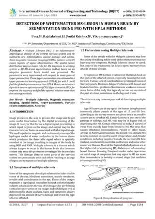
- 1. International Research Journal of Engineering and Technology (IRJET) e-ISSN: 2395-0056 Volume: 05 Issue: 04 | Apr-2018 www.irjet.net p-ISSN: 2395-0072 © 2018, IRJET | Impact Factor value: 6.171 | ISO 9001:2008 Certified Journal | Page 1544 DETECTION OF WHITEMATTER MS-LESION IN HUMAN BRAIN BY SEGMENTATION USING PSO WITH HPLA METHODS Uma.S1, Rajalakshmi.L2, Sruthi Krishna.N3, Vikramanarayanan.J4 1,2,3,4 8th Semester/Department of CSE/Dr. NGP Institute of Technology/Coimbatore/TN/India ---------------------------------------------------------------------***--------------------------------------------------------------------- Abstract - Multiple Sclerosis (MS) is an inflammatory neurological disease of the central nervous system and its hallmarks are, inflammation, axonal damage and edema. Brain magnetic resonance imaging (MRI) in patients with MS shows regions of signal abnormalities. The spatial lesion distribution plays a major role for MS diagnosis. In existing system, a 3D MS-lesion segmentation method based on an adaptive geometric brain model was presented. The parameters were represented with respect to more general hyper-parameters. These hyper-parametersareestimatedina hyper-parameter learning approach (HPLA), for which could find the global optimum more than HPLA. Hence,weintroduce a particle swarm optimization (PSO) algorithm with HPLAfor improve the accuracy and find the optimal solution morethan the existing methods. Key Words: Multiple Sclerosis, Magnetic resonance imaging, Spatial-lesion, Hyper-parameters, Particle swarm optimization, Accuracy 1. INTRODUCTION Image process is the way to process the image and to get some useful information by the digital processing of the image. It is a type that forms a digital signal processing in which input is given as the image and output may be the characteristicsor features associated with that input image. We need to polarize magnets and excitement process of the hydrogen nuclei of water molecules in the human tissue which produces the detectable signal which is spatially encoded in the image, resulting in images of the body by using MRI and NMR. Multiple sclerosis is a disease which always happen to occur in the human brain that immune system eats away the protective covering of the brain of the nerves. This damage disturbs some parts of the nervous system to communicate with each other resulting in a range of signs and symptoms of multiple sclerosis. 1.1 Symptoms of multiple sclerosis Some of the symptoms of multiple sclerosis includes double vision of the eye, blindness sometimes, muscle weakness, trouble with coordination in the eye. Those of the images maintains a topological equivalent subject with the healthy subjects which allows the use of techniques for performing cortical reconstruction of the images andunfoldingaswellas diffeomorphic shape analysis. Signs and symptoms always depend on the amount of nerve damage and which the type of nerve affected. 1.2 Factors increasing Multiple Sclerosis Some of the people with the Multiple Sclerosis may lose the abilityof walking,while someof the otherpeoplemaynot have any new symptoms. Multiple Sclerosis cannot be cured. But some of the treatments can help in speed recovery from attacks of those disease. Symptomsof MS: Certain treatmentof Electricalshockon the neck of the affected person, especially bending the neck forward Tumor, lack of coordination or prolonged vision of Slurred speech. Dizziness fatigue Problems with bowel and bladder functionsproblems.Numbnessorweaknessinoneor more limbs of the body that typically occurs on one side of the part at a time, sometimes on the legs and trunk These factors may increase your risk of developing multiple sclerosis: Age: MScan occur at any age of thehuman being butmost commonly affects people of the ages of 15 and 60. Sex: According to the survey women are about twice as possible as men are to develop MS. Family history: If any one of the parents or siblings had MS, you may be in higher risk of developing the MS. Certain infections in body: A variety of virus from outside have been linked to MS, the virus that causes infectious mononucleosis. People of other Asian, African or Native American have the lowest risk.Climate: MS ismore common in countrieswith temperaturesandclimatic conditions, including people of Canada, the northernUS,New Zealand, southeastern Australia and Europe and some other countries. Disease. Most of the thyroid affected person are at the higher risk of developing MS, diabetes or inflammatory bowel disease. Smoking: Frequent smokers who experience an initial stage of symptoms that may signal to MS are more than nonsmokers to develop a second stage that confirms relapsing-remitting MS. Figure1. Demyelination of progress
- 2. International Research Journal of Engineering and Technology (IRJET) e-ISSN: 2395-0056 Volume: 05 Issue: 04 | Apr-2018 www.irjet.net p-ISSN: 2395-0072 © 2018, IRJET | Impact Factor value: 6.171 | ISO 9001:2008 Certified Journal | Page 1545 2. Literature survey The Red nucleus of the midbrain structures of a sample interest on neuron imaging technique in manystudieswhich may benefited from the availability of automatic segmentation methodsof brain structures.Thehighcontrast in quantitative susceptibility mapping due to the high iron content thus called QSM images. This presents a segmentation method that leveragestheinformationofthese images to produce automated segmentations of the red nucleus (RN). By non-linearly registering a set of manually- traced training labels the algorithm builds a map of spatial priors for the structures to the midbrain of the nervous system. The main disadvantage of this is process speed is very slow. To inform a Gaussian mixture model of the image intensities, with smoothness constraints imposed to ensure anatomical plausibility of the brain, the priors are used. From a sample of 40 healthy younger and older subjects of human brain, the method was validated on manual segmentations. Average Dice scores in the left hemisphere were 0.81 (0.05) for the SN, 0.66 (0.14) for the STN and 0.88 (0.04) for the RN, and the similar values were obtained for the right hemisphere and the left hemisphereof the brain. In all structures, volumes of manually obtained segmentations and automatically obtained segmentations were correlated significantly. The algorithm showed lower accuracy on T2-weighted Fluid Attenuated Inversion Recovery (FLAIR) imagesand R2 *, which are also sensitiveto iron content in the human brain. To illustrate an application of the method, it shows that the manual segmentations were comparable to the automated ones regarding detection of age-related differences. In recent years, many brain structure of automatic segmentation methods have been proposed. However, the effect of lesions on their performance has not been evaluated and these methods are commonly tested with non-lesion brains. Here, we analyze the effect of multiple sclerosis (MS) lesions on three well-known brain structure of automatic segmentation methods, namely, FIRST, Free Surfer and multi-atlas fused by majority voting, which use deformable, learning-based and atlas-based strategies, respectively. To perform a quantitative analysis, the volume differences and the Dice similarity coefficient (DSC) differences between the simulated and the healthy images are calculated for the sub cortical structures and the brainstem. We observe that when lesions are present, the three strategies are affected. However, the lesions effects do not follow the same pattern; the lesions either make the segmentation accuracy augment surprisingly or the segmentation method underperform. On the other hand, when the lesions are overlaid or close to the structure of analysis, FIRST is more affected. The nucleus acumens(from - for the left hemisphere and high for the right hemisphere) is the most affected structure by the presence of lesions, whereas the structures that show less. Variation as a tool to measure the disease including the thalamus. The three segmentations are affected by the presence of MS lesions, which demonstratesthatthereexists a problem in the automatic brain structure segmentation methods of the deep gray matter (DGM) structures that has to be taken into account when using them progression commutative iron content. The influence of lesions on the segmentation on other disease is not investigated, is the drawback of this method. A work for the fully automatic segmentation and the robust segmentation of magnetic resonance imaging of the brain images called as the “Multi Atlas Label of Propagation with the Expectation and Maximization based refinement”. The present approach is based on highly performance label fusion, a robust registration approach (MAPER) and intensity-based label refinement using EM. The further adapt of framework to be applicable for the brain images segmentation with gross changesof the human brain in anatomy. To do for consistent process errors by relaxing anatomical values of priors of medical imaging obtained by multi atlas of the propagation and intensity refined by posterior probabilities and a weighting type to locally combine anatomical valuesatlasof priors. When compared to state of the art automatic labeling techniques employed MALP EM to segment 125 MR brain images were into 134 regions from subjects who had sustained traumatic brain injury, the MALP EM is competitive for the MR segmentation of the brain scans of healthy brain adults. If there were no manual reference labels available then protocol is employed to assess segmentation quality of the brain structures. Specifically to show are able to satisfy the TBI patients with favorable outputs compared from non-favorableoutcomeswith66.8% accuracy using the MRI images and 64.7% accuracy using acute-phase MRI images. Furthermore, then the images was able to be differentiated with the subjects with the presence of a midline in the brain system that may shift theMSlesionfrom those with diffusion in the brain injury with 76% accuracy. The thalamus, hippocampus, pitmen and palladium are affected particularly. TBI disease progressionispredictedby their involvement. The main drawback of this method islow accuracy. Magnetic resonance imaging is the primary imaging technique that may be the evaluation of the progression of the brain tumor of the human being before and after surgery. To exploit conventional MRI modalities in order to identify and segment of the brain images with neoplasm is the purpose of this approach. The main drawback of this method isthat it performslow accuracy and it is notusedfor all subject-related information.
- 3. International Research Journal of Engineering and Technology (IRJET) e-ISSN: 2395-0056 Volume: 05 Issue: 04 | Apr-2018 www.irjet.net p-ISSN: 2395-0072 © 2018, IRJET | Impact Factor value: 6.171 | ISO 9001:2008 Certified Journal | Page 1546 Figure 2.Lesion 2.1 Summary Based on the above survey,manyproblemswereobtained such that the classification steps process was low. The influence of lesions on the segmentation on the other diseases is not investigated.Low accuracywasalso obtained. And the above methods is not applicable for non-enhancing tumors and necrosis is included in the same class with all non-enhancing tissue. In experimental result, this method is not used all the subject-related information that is available. To overcome this, problem, we introduce a particle swarm optimization (PSO) algorithm with HPLA for improving the accuracy and find the optimal solution. 3. Proposed architecture Preprocess In a first stage, we align the image contrasts with a rigid registration,which is estimatedusingthe default parameters ofthe ITK libraries. Subsequently weremovethe skull in the images via the BET toolkit and perform a bias- field correction by extending to image contrasts. In a second stage, we compute a partial segmentation. Geremia process: This process describesthe adaptation oftherandomdecision forests to the segmentation of MS lesions and illustrates the visual features used. This process perform the following sequences, • Context-rich decision forest • Forest training • Prediction • Visual features Model of Population and subjects (MOPS): In this approach, we first recap the well-established global Gaussian Mixture Models (GMM) modelof brain tissue segmentation explored. This approach is used a local intensity prior probability and spatial tissue class prior estimated from a healthy reference population. Rotational invariant distance measure (RMNMS) This algorithm adapts the Non-Local Means (NLM) estimator to account for multi-modal images and rotation-invariant distance measure of the patches. The idea of the NLM is to reduce the noise of the image by averaging the vowel that would have a similar way Hyper Parameter Learning Approach (HPLA): In this approach, the hyper-parameter are estimated forfinding the global optimum. Particle swarmoptimization in this methoditprovidesmore accuracy. The proposed parameters were represented with respect to more general hyper-parameters. These hyper- parameters are estimated in a hyper-parameter learning approach (HPLA), for which could find the global optimum. Thus by combining the methods such as hyper-parameter Learning approach and [particle swarm optimization improves more accuracy and find the optimal solution. Fig 3.1 Process of PSO 4. Further enhancement Accuracy Under detected Over detected Properly detected GEREMIA 45 41.6667 66.667 MOPS 41.667 33.333 75 RMNMS 25 16.6667 83 HPLA 16.6667 12.500 91.667 PSO 13.45 10.56 95.2555
- 4. International Research Journal of Engineering and Technology (IRJET) e-ISSN: 2395-0056 Volume: 05 Issue: 04 | Apr-2018 www.irjet.net p-ISSN: 2395-0072 © 2018, IRJET | Impact Factor value: 6.171 | ISO 9001:2008 Certified Journal | Page 1547 5. CONCLUSION In this paper we present a particle swarm optimization (PSO) algorithm with HPLA is proposed for improve the accuracy and find the optimal solution. The proposed algorithm is used to segment brain magnetic resonance image. The solution correspondstomaximizingthebetween- class variance of the distribution of intensity levels in the image. Then, the proposed approach is very efficient global search algorithm. REFERENCES [1] Maddalena strumia,Frank R.schmidt, constantinos anastasopoulos, Cristina Granziera, Gunnar Krueger, Thomas Brox. White matter MS-Lesion segmentation using a geometric brain model. [2] Garzón, B., Sitnikov, R., Bäckman, L., & Kalpouzos, G. (2017). Automated segmentation of midbrain structures with high iron content. NeuroImage. [3] [9]MRI based medical image analysis: Survey on brain tumor grade classification Geethu Mohan., Monica Subashini(2017) [4] A new method of brain tissues segmentation from MRI with accuracy estimation Sudipta Ro., Samir Kumar Bandyodhyay(2016) [5] González-Villà, S., Valverde, S., Cabezas, M., Pareto, D., Vilanova, J. C., Ramió-Torrentà, L., ... & Lladó, X. (2017). Evaluating the effect of multiple sclerosis lesions on automatic brain structure segmentation. NeuroImage: Clinical, 15, 228-238 [6] Kanas, V. G., Zacharaki, E. I., Davatzikos, C., Sgarbas,K.N., & Megalooikonomou, V. (2015). A low cost approachfor brain tumor segmentation based on intensity modeling and 3D Random [7] Automated segmentation of cerebral deep gray matter from MRI scans: effect of field strength on sensitivity and reliability. Chu R, Hurwitz S,Tauhid S, BakshiR.BMC Neurol [8] Homeomorphic brain image segmentation with topological and statiscal atlases Pierre-Louis Bazin., Dzung L phan [9] Tumopr or abnormality identification from magnetic resonance images using statistical region fusion based segmentationBadri N arayan Subudhi., Veerakumar Thangaraj., Esakkirajan Sankaralingam., Ashish Ghosh(2015) [10] A multi-time-point modality-agnostic-basedmethodfor lesion filling in multiple sclerosisFerranPrados.,Manuel Jorge Cardoso.,Baris BIOGRAPHIES Uma. S pursuing BE computer science and engineering in Dr.NGP Institute of Technology.Memberof CSI standard. Vikramanarayan. J pursuing BE computer science and engineering in Dr. NGP Institute of Technology. And a member of CSI standard, Sruthi Krishna. N pursuing BE computer science and engineering in Dr. NGP Institute of Technology. And a member of CSI standard Rajalakshmi. L pursuing BE computer science and engineering in Dr. NGP Institute of Technology. And a member of CSI standard