Downloaded 296 times

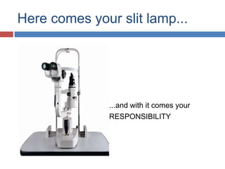
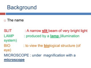
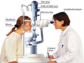
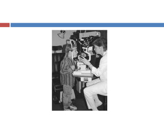
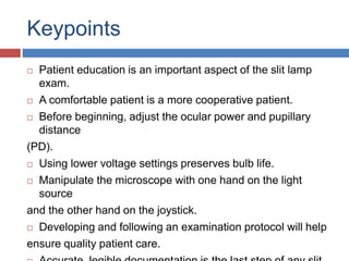
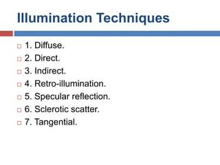


The document provides an overview of the slit lamp biomicroscope, a microscope used to examine the eye. It describes the history, development, parts, optics, and various illumination techniques of the slit lamp. The slit lamp allows a magnified three-dimensional view of the eye for documentation and quantitative measurements. Examination with the slit lamp involves using different illumination methods like diffuse, direct, indirect, and retro-illumination to view different structures of the eye. Proper technique and understanding of slit lamp use is important for quality eye examinations.


