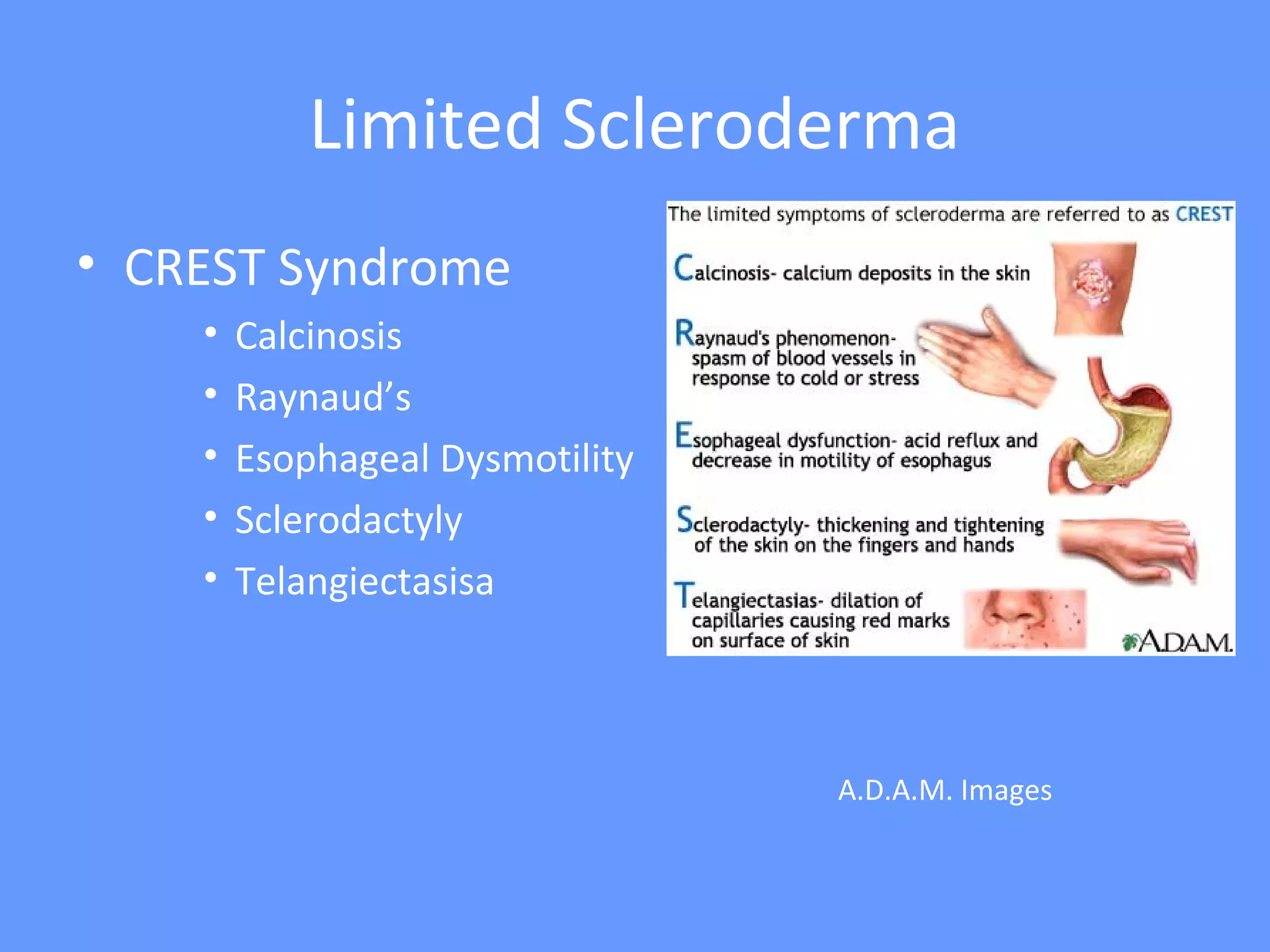
Systemic Sclerosis (definition)
• Multisystem disorder
• Unknown etiology
• Thickening of skin caused by accumulation of
connective tissue (collagen types I and III)
• Involvement of visceral organs
Epidemiology
• Peak age range: 35-64
• Younger age in women and with diffuse disease.
• Female:Male = 3:1
• 8:1 in child bearing years
• Incidence: 20/million per year in US
• Prevalence: 240/million in US.
Etiology
• Unknown
• Environmental Exposures
• Silica exposure in men conferred increased risk
• Silicone breast implants: no definite risk identified
• Aniline laced Contaminated rapseed oil in Spain
• Vinyl chloride exposure increased risk of SSc like
disorder: Eosinophilic Fasciitis
• bleomycin
• L-tryptophan: Eosinophilia Myalgia syndrome
Etiology
• Genetic Factors
• Familial Clustering: 1.5-2.5% of those with 1st
degree
relative
– Choctow Native Americans: prevalence 4720/million.
• HLA-haplotypes: there are higher risk haplotypes in
certain populations
Pathogenesis: general principles
• Endogenous or exogenous pathogen stimulates antigen
presenting cells.
• Antigen presenting cells stimulate CD4+ T cells
• Cytokines are produced by both of these cells.
• Cytokines stimulate growth factors to stimulate fibroblasts
to produce collagen
• Vascular damage occurs with thickened intima and
narrowing of the lumen.
• Narrowing of the lumen leads to ischemia.
• Ischemia leads to prostacyclin production which is a
platelet aggregant and platelets bind to endothelium and
release PDGF which is chemotactic and mitogenic for
fibroblasts.
Pathogenesis
Pathogenesis of Scleroderma
Up to Date
Forms of Systemic Sclerosis
• Limited Scleroderma
• Skin thickening is distal to elbows and knees, not involving
trunk
• Can involve perioral skin thickening (pursing of lips)
• Less organ involvement
• Seen in CREST syndrome
• Isolated pulmonary hypertension can occur
• Diffuse Scleroderma
• Skin thickening proximal to elbows and knees, involving the
trunk
• More likely to have organ involvement
• Pulmonary fibrosis and Renal Crisis are more common.
2013 ACR Diagnostic Criteria
Limited Scleroderma
• More gradual process
• Can have Raynaud’s for years (even up to decade)
• Skin involvement distal to elbows and knees
• Often with perioral involvement (pursing of lips)
• Capillaroscopy
• with dilated capillary loops but without dropout.
• Less organ involvement
• though 10-15% with isolated pulmonary hypertension.
• Renal involvement is rare.
• Anti-centromere Ab in 70-80%
Limited Scleroderma
• CREST Syndrome
• Calcinosis
• Raynaud’s
• Esophageal Dysmotility
• Sclerodactyly
• Telangiectasisa
A.D.A.M. Images
CREST Syndrome
ACR and Mayo Foundation
Calcinosis on x-ray
Gupta E., et al. Malaysian Family
Physician. 2008;3(3):xx-xx ACR
Nailfold Capillaroscopy
Diffuse Scleroderma
• More Rapid Process
• Often with onset of skin thickening within a year of
Raynaud’s symptoms
• Skin involvement proximal to elbows and knees
• Often can involve the trunk
• Capillaroscopy reveals dropout
• With capillary dilatation and dropout.
• Early organ involvement
• Renal, interstitial lung disease, myocardial, diffuse
gastrointestinal – often within the first 3 years.
• Antibodies
• Anti-Scl-70, anti-RNA Polymerase III.
Diffuse Scleroderma
ACR
American Osteopathic College of Dermatology, Grand
Rounds
Netter
Organs Involved
• Skin
• Musculoskeletal
• Pulmonary
• Renal
• Gastrointestinal
• Cardiac
Skin Involvement
• Early stages:
• Perivascular infiltrate which are primarily T cells.
• Skin swelling which eventually becomes skin thickening.
• Involves the hands and/or feet (distal).
• Late Stages:
• Finger-like projections of collagen extend from the dermis to
the subcutaneous tissue to anchor skin deeper.
• Skin becomes firm, thick and tight.
• Skin thickening moves proximally.
• Fibroblasts and collagen deposition.
• Hair and wrinkles overlying area of skin thickening
disappears.
Skin involvement in Scleroderma
• May regress on its own over years
• reverse pattern (ie, starting with regression of skin
thickening in the trunk, then proximal extremities, then
more distal).
• Digital Ulcers:
• on extensor surface of PIP’s and elbows; may become
secondarily infected.
• Digital ischemia:
• with pits in the distal aspect of the digits related to
prolonged Raynaud’s.
• Thinning of the lips, beak-like nose.
Skin Manifestations
Kahaleh B. Rheum Dis Clin N Amer 2008:57-71
Sclero.org
International
Scleroderma Network
ACR
Musculoskeletal
• Arthritis
• in > 50% with swelling, stiffness, and pain in the joints
of the hands.
• Carpal Tunnel Syndrome.
• Contractures
• related to skin thickening.
• Polymyositis
• may occur as part of mixed connective tissue disease or
overlap.
Pulmonary
• leading cause of death
• since we are better at control of renal disease.
• Symptoms:
• exertional dyspnea
• Types of lung Involvement:
• Interstitial lung disease.
• Isolated pulmonary hypertension.
Interstitial Lung Disease
• Inflammatory phase
• with ground glass opacities and linear infiltrates
• lower 2/3 of the lung fields on CT scan.
• Fibrosis:
• Late phase with honeycombing.
• Diagnosis
– Pulmonary function tests
• restrictive pattern with low FVC, low residual volume, low DLCO.
– High Resolution CT Scan
– BAL: often not required
– Lung biopsy: often not required
• ILD is most commonly associated with diffuse scleroderma.
• Anti-Scl-70
Interstitial Lung Disease
Up to Date 2005 Up to Date 2005
Primary Pulmonary Hypertension
• Symptoms:
• exertional dyspnea.
• Frequency
• 10-15% of patients with systemic sclerosis
• Definition:
• Mean PA blood pressure >25mmHg at rest or >30mmHg
with exercise on right heart catheterization.
• Estimated systolic pulmonary artery pressure of >35mmHg
on Echocardiogram
• Pathogenesis
• Intimal fibrosis and medial hypertrophy of the pulmonary
arterioles and arteries.
Pulmonary Hypertension
Up to Date 2005
Doppler Echocardiogram to estimate
pulmonary artery pressure.
Roberts JD. Pulm Circ 2011;1:160-181.
Other Pulmonary Associations
• Pneumonia:
• due to aspiration secondary to GERD; skin thickening of
chest may reduce effectiveness of cough.
• Alveolar carcinoma: increased incidence
• Bronchogenic carcinoma: increased incidence.
Renal Manifestations of Systemic
Sclerosis
• Scleroderma Renal Crisis
• Abruptly developing severe hypertension
– Rise in SBP by > 30 mmHg, DBP by > 20 mm Hg
• One of the following:
– Increase in serum creatinine by 50% over baseline or creatinine > 120%
of upper limit.
– Proteinuria > 2+ by dipstick.
– Hematuria > 2+ by dipstick or > 10 RBC/HPF
– Thrombocytopenia < 100
– Hemolysis (schisctocytes, low platelets, increased reticulocyte count).
• Can cause headache, encephalopathy, seizures, LV failure.
• 90% with blood pressure > 150/90.
• Can occur also with lower blood pressures < 140/90 and this
confers worse prognosis.
Steen et al., ClinExp. Rheumatol. 2003
Scleroderma Renal Crisis
Up to Date 2012
Risk Factors for Renal Crisis
• Rapidly progressive skin thickening within the
first 2-3 years.
• Steroid use (prednisone > 15 mg)
• Anti-polymerase III Ab.
• Pericardial Effusion.
Treatment of Scleroderma Renal Crisis
• Medical Emergency: generally with admission.
• Initiation of ACE inhibitors such as captopril;
lifelong treatment with ACE inhibitors.
• Dose escalation of captopril.
• ACE-inhibitors do not prevent SRC.
Treatment of Scleroderma Renal Crisis
Steen, Clinics in Dermatology, 1994
Without
Renal Crisis - Prognosis
• Improved overall with ACE-inhibitors.
• Even with ACE-inhibitors 20-50% will progress
to ESRD.
• Among patients who required dialysis during
the acute phase, an appreciable proportion
(40-50%) will be able to discontinue dialysis.
Gastrointestinal Manifestations
• Esophageal dysmotility: in up to 90%.
• Pathophysiology:
– reduced tone of gastroesophageal sphincter and distal dilatation of the
esophagus.
– Lamina propia and submucosal tissue with Inflammatory changes and
increased collagen on pathology.
• Symptoms
– Dysphagia, GERD; many asymptomatic.
• Diagnosis:
– Esophageal manometry, Esophagram, CT scan.
• Treatment
– Proton Pump Inhibitors
– Elevation of head of the bed.
• Complications:
– Barret’s Esophagus.
Gastrointestinal Manifestations
• Gastric Involvement:
• Symptoms: Early satiety.
• Diagnosis: Nuclear Gastric Emptying Test.
• Treatment: promotility agents
• Watermelon Stomach: dilated vessel which can cause bleeding.
• Small Intestinal involvement
• Symptoms: distension, pain, bloating, steatorrhea
• nutritional deficiencies secondary to bacterial overgrowth.
» Vitamin B6/B12/folate/25-OH Vit D, low albumin
• Diagnosis:
– glucose hydrogen breath test
– Low D-xylose absorption test
– small bowel aspiration (only if resistance to rotating antibiotics)
• Treatment: Rotating antibiotics, Reglan, Erythromycin
Image of Watermelon Stomach: University of Michigan Rheumatology Website
Gastrointestinal Manifestations
• Colon Involvement:
• Can cause symptoms of constipation due to decreased
peristalsis.
• Fecal incontinence can occur due to alterations of
internal and external sphincter.
Cardiac Manifestations
• Forms of cardiac involvement
• Pericardial Effusion
– symptomatic pericarditis in 20%
• Microvascular CAD:
– recurrent vasospasm of coronary arteries
– Necrosis
– patchy myocardial fibrosis; leads to diastolic > systolic
dysfunction.
• Myocarditis
– Inflammation which leads to fibrosis
• Arrhythmias and conduction abnormalities
– Fibrosis of cardiac conduction system.
– AV conduction defects and arrhythmias.
Cardiac Involvement
Adapted from Desai, et al; Curr Opin Rheumatol 2011m 23:545-554
Cardiac Manifestation Prevalence Diagnosis Treatment
Myocarditis Rare Cardiac MRI, Biopsy Cytoxan + steroids
Pericardial effusion 5-16% Echocardiogram None; NSAIDs if
symptomatic
Microvascular CAD > 60% MRI/nuclear medicine Calcium channel
blockers
Macrovascular CAD 25% Coronary Angiogram Stenting/medical tx
Bradyarrhythmias Rare EKG/Holter Pacemaker
Tachyarrhythmias 15% EKG/Holter Diltiazem, ablation,
defibrillator
Scleroderma Autoantibodies
Antigen ANA
Pattern
Frequency Clinical
Associations
Organs Involved
Scl-70
(topoisomerase 1)
Speckled 10-40 dcSSC Lung fibrosis
RNA Polymerase III Speck/Nuc 4-25 dcSSC Renal,
Pulmonary HTN
Centromere Centromere 15-40 lcSSc, CREST Pulmonary HTN
Esophageal
U1-RNP Speckled 5-35 lcSSC, MCTD Muscle
U3 RNP (fibrillarin) Nucleolar 1-5 dcSSC, poor prognosis Muscle
Pulmonary HTN
PM-SCL Nucleolar 3-6 Overlap, mixed Muscle
Th/To Nucleolar 1-7 lcSSc Pulmonary HTN,
Lung fibrosis,
Small bowel
Anti U11/U12 Nucleolar 1-5 lcSSc & dcSSC Lung Fibrosis
Anti-Ku 1-3 Overlap Ssc Muscle, Joint,
SLE overlap
Adapted from: Nihtyanova SI, Denton CP. Nat Rev Rheumatol 2010; 6:112
Scleroderma Treatment
• Depends on clinical manifestations
• Aggressive disease versus stable disease
• Reversible inflammation vs Vasoconstriction.
• Organ Involvement
• Treatment is directed at organ involved.
Raynaud’s
• Calcium Channel Blockers: nifedipine
• Nitroglycerin patches
• Sildenafil (Viagra) (but not in combination
with nitroglycerine) –usually for refractory
Raynaud’s.
• Parental vasodilators (iloprost) – for severe
disease with impending digital ischemia.
Gastrointestinal Involvement
• GERD
• Proton pump inhibitor.
• Delayed Gastric Emptying and peristalis
disorders
• Supportive
• Promotilants are sometimes used.
Pulmonary Involvement
• Interstitial Lung Disease: with active
inflammation
• Mycophenolate
• Azithioprine
• Cytoxan - IV
• plus lower dose of steroids if RNA Poly III neg (ie 10 mg
daily); avoid steroids if RNA Poly III positive.
• Pulmonary Hypertension
• Vasodilators: bosentan, sildenafil, epoprostenol,
treprostinil, iloprost.
• Lung Heart Transplant
Myositis
• Polymyositis overlap or MCTD
• Similarly to myositis alone with methotrexate,
azathioprine in combination of low dose steroids.
• Tend to keep prednisone dose at around 10 mg or less
to avoid risk of renal crisis.
Cardiac Involvement
• Pericarditis:
• NSAIDs
• Drainage of effusion if tamponade
• Myocarditis with elevated CK-MB & troponin
• If CAD is excluded, MRI and biopsy confirms, then
treatment would generally be with low dose
prednisone (10 mg/day) and cytoxan; nifedipine may
also be helpful.
Skin Disease
• Stable disease: no treatment
• Advancing diffuse skin involvement:
• Methotrexate
• Mycophenolate
• Current trial with Tocilizumab (Actemra)
• D-penicillamine 125 mg/day.
• Research on various anti-fibrosis therapies is being
performed (imatinib, Gleevac).
Differential Diagnosis
• Scleredema
• No Raynauds, negative antibodies, seen in IDDM
• Proximal skin thickening (trunk, shoulders, back)
• Scleromyxedema
• Skin thickening/induration on head, neck, arms, trunk
• Monoclonal gammopathy (multiple myeloma/AL amyloid)
• Skin biopsy differentiates.
• Endocrinologic: diabetes and hypothyroid myxedema
• Can be associated with skin induration.
• In diabetes can have sclerodactyly (Diabetic Cheiroarthropathy) - dorsal
• POEMS (polyneuropathy, organomegaly, endocrinopathy, monoclonal gammopathy, skin thickening).
• Nephrogenic Systemic fibrosis
• Chronic kidney disease and gadolinium MRI contrast
• Can involve hands and feet.
• Eosinophilic fasciitis:
• Hands and feet are spared, peripheral blood eosinophilia, peau de orange appearance
• Diagnosis is via skin biopsy.
• Graft versus Host disease
• History of bone marrow transplant, no Raynaud’s symptoms.
• Diagnosis is via skin biopsy.
Cases
Case 1
• 50 year old female who has CREST syndrome with anti-
centromere antibody:
• Raynaud’s controlled with nifedipine
• only digital skin thickening of the hands which is unchanged
• GERD on omeprazole
• telangiectasia.
• She currently has no complaints.
• Labs:
• CMP, CBC, ESR, CRP, total CK all normal, anti-centromere Ab positivity.
• Echocardiogram and PFT’s 1 month ago:
• Echo: normal with normal estimated PA pressures.
• PFT’s: normal lung volumes, normal DLCO.
• What is next step:
Case 1
• Renew medications
• Nifedipine and omeprazole
• This case highlights the most typical case seen
in clinics with stable disease.
• Things to watch for:
• Change in skin disease
• Periodic echocardiogram and PFT’s.
• General exam
Case 2
• 60 year old male with Raynaud’s for 4 months prior to onset of skin
involvement
• Skin thickening has ascended to involve proximal extremities, chest, and abdomen
within 1 year.
• The patient reports mild shortness of breath recently.
• Exam:
• Vitals: T 98.9, BP 124/73, pulse 80, resp rate 18
• Raynaud’s is noted without digital ulcer.
• Cardiovascular exam normal.
• Gastrointestinal exam is normal.
• Dry crackles noted at both bases.
• Extremities: no edema.
• Labs:
• CBC, CMP, total CK are all normal
• ESR 35, CRP 1.8 (upper limit of normal is 1.0).
• Anti-Scl-70 Ab positive, RNA Pol III negative.
• What is next step?
Case 2
• PFT’s: TLC decreased 80% to 55%, VC decreased 85% to 50%, RV decreased 83% to 62%, DLCO
decreased 75% to 45%.
• Bronchoscopy performed: all cultures & cytology negative (neutrophils and eosinophils are
present).
• Echocardiogram: no pulmonary hypertension.
• Lung Biopsy shown on right.
• What is the diagnosis? What is the treatment?
Learningradiology.com Oikonomou A, Prassopoulos P - Insights Imaging (2012)
Strek, ME. Amer Col Chest
Physicians 2012
Case 2
• Interstitial lung disease associated with
scleroderma with active inflammation.
• Mycophenolate, Cytoxan, or Azathioprine
• Prednisone (low dose) 10 mg daily; gradual
taper
Case 3
• 50 year old female presents with
• onset of Raynaud’s for 1 year,
• developed skin thickening from the digits of the hands to just distal to the
elbows.
• She has noticed difficulty getting out of chairs and lifting objects overhead.
• Exam:
• VS: Temp 98.2, BP 124/72, pulse 78, respiratory rate 16
• Cardiovascular and pulmonary exams normal.
• Gastrointestinal exam is normal.
• Muscle weakness of thighs and shoulder regions is noted.
• No skin lesions other than skin thickening.
• Labs:
• CBC, chem-7, ESR, CRP all normal, PM-SCL Ab positivity
• Total CK 3000 (mostly CKMM), AST 158, ALT 105, GGT normal.
• What is the next step?
Case 3
• MRI of the thigh
• Biopsy of thigh musculature
• What is the diagnosis? What is the treatment?
EMG, Nerve Conduction Studies
Olsen NJ, et al. Rheum Dis Clin N. Amer 1996;22(4):783-796
Seidman, RJ. Medscape
Case 3
• Scleroderma/Myositis overlap.
• Methotrexate or Azathioprine
• Low dose prednisone: 10 mg daily
• Over the next few months, CK levels normalize
and prednisone dose is gradually tapered, and
the patient’s strength improves.
Case 4
• 35 year old female with
• limited scleroderma for 3 years, anti-centromere Ab
positive.
• with stable skin disease involving the digits of the hands
only; new “rash” appeared 1 month ago, gradually
worsening, no change in last week.
• Raynaud’s have been quite severe, but not on therapy.
• Exam
• VS: Temp 97.9, BP 123/76, pulse 82, RR 16
• Cardiac, pulmonary, gastrointestinal exams normal, no
edema
• Skin: see next slide
Case 4
• Labs:
• CBC, CMP, ESR, CRP all normal; anti-centromere Ab
positive, anti-phospholipid Ab neg, echo with bubble
study negative
• What is the diagnosis? What is next step?
Sclero.org
International Scleroderma Network
Case 4
• Digital Ischemia due to Raynaud’s
• Start calcium channel blocker
• Nifedipine 30 mg PO daily.
• Close follow-up and increase dose of nifedipine as
blood pressure tolerates.
• If not responding:
• Can start nitroglycerin patch or can start sildenafil (not
both).
Case 5
• 58 year old male with:
• Rapid onset scleroderma with Raynaud’s for 6 months then
skin thickening that spread to proximal arm, proximal thigh,
chest, and abdomen within 1.5 years.
• Blood pressure generally runs 110/70
• has mild headache, and has noticed some swelling of the
legs.
• Exam:
• VS: Temp 98.4, BP 160/105, pulse 70, RR 16.
• Cardiac, pulmonary, gastrointestinal exam all normal;
neurologic exam is non-focal.
• There is only mild bilateral lower extremity edema.
Case 5
• Labs
• Creatinine 2.0 (baseline is 0.6), CBC normal, ESR and
CRP normal, urine with 1+ protein, no RBC or WBC;
known to be RNA Pol III positive.
• What is the diagnosis? What is the next step.
Case 5
• Scleroderma Renal Crisis
• Treatment:
• Hospitalization
• Start ACE-inhibitor: captopril with dose escalation.
References
• Medscape
• Up To Date
• Desai, et al; Curr Opin Rheumatol 2011; 23:545-554
• Curr Opin Rheumatol 23;505-510
• Fischer A; CHEST 2006; 130:976 –981
• Rheum Dis Clin N Am;2003;29:293–313
• Arthritis Rheum 2006;54:3962-3970
• Rheumatology 2009;48:iii32–iii35
• Steen VD; Rheum Dis Clin N Am 2003;29:315–333
• Hudson M, et al; Medicine 2010;89:976-981
• Bon LV; Curr Opin Rheumatol 2011;23:505–510
• Barnes J; Curr Opin Rheumatol 2012, 24:165–170
Definition of Criteria
Skin Scoring

Systemic sclerosis

  • 1.
    Systemic Sclerosis (definition) •Multisystem disorder • Unknown etiology • Thickening of skin caused by accumulation of connective tissue (collagen types I and III) • Involvement of visceral organs
  • 2.
    Epidemiology • Peak agerange: 35-64 • Younger age in women and with diffuse disease. • Female:Male = 3:1 • 8:1 in child bearing years • Incidence: 20/million per year in US • Prevalence: 240/million in US.
  • 3.
    Etiology • Unknown • EnvironmentalExposures • Silica exposure in men conferred increased risk • Silicone breast implants: no definite risk identified • Aniline laced Contaminated rapseed oil in Spain • Vinyl chloride exposure increased risk of SSc like disorder: Eosinophilic Fasciitis • bleomycin • L-tryptophan: Eosinophilia Myalgia syndrome
  • 4.
    Etiology • Genetic Factors •Familial Clustering: 1.5-2.5% of those with 1st degree relative – Choctow Native Americans: prevalence 4720/million. • HLA-haplotypes: there are higher risk haplotypes in certain populations
  • 5.
    Pathogenesis: general principles •Endogenous or exogenous pathogen stimulates antigen presenting cells. • Antigen presenting cells stimulate CD4+ T cells • Cytokines are produced by both of these cells. • Cytokines stimulate growth factors to stimulate fibroblasts to produce collagen • Vascular damage occurs with thickened intima and narrowing of the lumen. • Narrowing of the lumen leads to ischemia. • Ischemia leads to prostacyclin production which is a platelet aggregant and platelets bind to endothelium and release PDGF which is chemotactic and mitogenic for fibroblasts.
  • 6.
  • 7.
  • 8.
    Forms of SystemicSclerosis • Limited Scleroderma • Skin thickening is distal to elbows and knees, not involving trunk • Can involve perioral skin thickening (pursing of lips) • Less organ involvement • Seen in CREST syndrome • Isolated pulmonary hypertension can occur • Diffuse Scleroderma • Skin thickening proximal to elbows and knees, involving the trunk • More likely to have organ involvement • Pulmonary fibrosis and Renal Crisis are more common.
  • 9.
  • 10.
    Limited Scleroderma • Moregradual process • Can have Raynaud’s for years (even up to decade) • Skin involvement distal to elbows and knees • Often with perioral involvement (pursing of lips) • Capillaroscopy • with dilated capillary loops but without dropout. • Less organ involvement • though 10-15% with isolated pulmonary hypertension. • Renal involvement is rare. • Anti-centromere Ab in 70-80%
  • 11.
    Limited Scleroderma • CRESTSyndrome • Calcinosis • Raynaud’s • Esophageal Dysmotility • Sclerodactyly • Telangiectasisa A.D.A.M. Images
  • 12.
    CREST Syndrome ACR andMayo Foundation
  • 13.
    Calcinosis on x-ray GuptaE., et al. Malaysian Family Physician. 2008;3(3):xx-xx ACR
  • 14.
  • 15.
    Diffuse Scleroderma • MoreRapid Process • Often with onset of skin thickening within a year of Raynaud’s symptoms • Skin involvement proximal to elbows and knees • Often can involve the trunk • Capillaroscopy reveals dropout • With capillary dilatation and dropout. • Early organ involvement • Renal, interstitial lung disease, myocardial, diffuse gastrointestinal – often within the first 3 years. • Antibodies • Anti-Scl-70, anti-RNA Polymerase III.
  • 16.
    Diffuse Scleroderma ACR American OsteopathicCollege of Dermatology, Grand Rounds Netter
  • 17.
    Organs Involved • Skin •Musculoskeletal • Pulmonary • Renal • Gastrointestinal • Cardiac
  • 18.
    Skin Involvement • Earlystages: • Perivascular infiltrate which are primarily T cells. • Skin swelling which eventually becomes skin thickening. • Involves the hands and/or feet (distal). • Late Stages: • Finger-like projections of collagen extend from the dermis to the subcutaneous tissue to anchor skin deeper. • Skin becomes firm, thick and tight. • Skin thickening moves proximally. • Fibroblasts and collagen deposition. • Hair and wrinkles overlying area of skin thickening disappears.
  • 19.
    Skin involvement inScleroderma • May regress on its own over years • reverse pattern (ie, starting with regression of skin thickening in the trunk, then proximal extremities, then more distal). • Digital Ulcers: • on extensor surface of PIP’s and elbows; may become secondarily infected. • Digital ischemia: • with pits in the distal aspect of the digits related to prolonged Raynaud’s. • Thinning of the lips, beak-like nose.
  • 20.
    Skin Manifestations Kahaleh B.Rheum Dis Clin N Amer 2008:57-71 Sclero.org International Scleroderma Network ACR
  • 21.
    Musculoskeletal • Arthritis • in> 50% with swelling, stiffness, and pain in the joints of the hands. • Carpal Tunnel Syndrome. • Contractures • related to skin thickening. • Polymyositis • may occur as part of mixed connective tissue disease or overlap.
  • 22.
    Pulmonary • leading causeof death • since we are better at control of renal disease. • Symptoms: • exertional dyspnea • Types of lung Involvement: • Interstitial lung disease. • Isolated pulmonary hypertension.
  • 23.
    Interstitial Lung Disease •Inflammatory phase • with ground glass opacities and linear infiltrates • lower 2/3 of the lung fields on CT scan. • Fibrosis: • Late phase with honeycombing. • Diagnosis – Pulmonary function tests • restrictive pattern with low FVC, low residual volume, low DLCO. – High Resolution CT Scan – BAL: often not required – Lung biopsy: often not required • ILD is most commonly associated with diffuse scleroderma. • Anti-Scl-70
  • 24.
    Interstitial Lung Disease Upto Date 2005 Up to Date 2005
  • 25.
    Primary Pulmonary Hypertension •Symptoms: • exertional dyspnea. • Frequency • 10-15% of patients with systemic sclerosis • Definition: • Mean PA blood pressure >25mmHg at rest or >30mmHg with exercise on right heart catheterization. • Estimated systolic pulmonary artery pressure of >35mmHg on Echocardiogram • Pathogenesis • Intimal fibrosis and medial hypertrophy of the pulmonary arterioles and arteries.
  • 26.
    Pulmonary Hypertension Up toDate 2005 Doppler Echocardiogram to estimate pulmonary artery pressure. Roberts JD. Pulm Circ 2011;1:160-181.
  • 27.
    Other Pulmonary Associations •Pneumonia: • due to aspiration secondary to GERD; skin thickening of chest may reduce effectiveness of cough. • Alveolar carcinoma: increased incidence • Bronchogenic carcinoma: increased incidence.
  • 28.
    Renal Manifestations ofSystemic Sclerosis • Scleroderma Renal Crisis • Abruptly developing severe hypertension – Rise in SBP by > 30 mmHg, DBP by > 20 mm Hg • One of the following: – Increase in serum creatinine by 50% over baseline or creatinine > 120% of upper limit. – Proteinuria > 2+ by dipstick. – Hematuria > 2+ by dipstick or > 10 RBC/HPF – Thrombocytopenia < 100 – Hemolysis (schisctocytes, low platelets, increased reticulocyte count). • Can cause headache, encephalopathy, seizures, LV failure. • 90% with blood pressure > 150/90. • Can occur also with lower blood pressures < 140/90 and this confers worse prognosis. Steen et al., ClinExp. Rheumatol. 2003
  • 29.
  • 30.
    Risk Factors forRenal Crisis • Rapidly progressive skin thickening within the first 2-3 years. • Steroid use (prednisone > 15 mg) • Anti-polymerase III Ab. • Pericardial Effusion.
  • 31.
    Treatment of SclerodermaRenal Crisis • Medical Emergency: generally with admission. • Initiation of ACE inhibitors such as captopril; lifelong treatment with ACE inhibitors. • Dose escalation of captopril. • ACE-inhibitors do not prevent SRC.
  • 32.
    Treatment of SclerodermaRenal Crisis Steen, Clinics in Dermatology, 1994 Without
  • 33.
    Renal Crisis -Prognosis • Improved overall with ACE-inhibitors. • Even with ACE-inhibitors 20-50% will progress to ESRD. • Among patients who required dialysis during the acute phase, an appreciable proportion (40-50%) will be able to discontinue dialysis.
  • 34.
    Gastrointestinal Manifestations • Esophagealdysmotility: in up to 90%. • Pathophysiology: – reduced tone of gastroesophageal sphincter and distal dilatation of the esophagus. – Lamina propia and submucosal tissue with Inflammatory changes and increased collagen on pathology. • Symptoms – Dysphagia, GERD; many asymptomatic. • Diagnosis: – Esophageal manometry, Esophagram, CT scan. • Treatment – Proton Pump Inhibitors – Elevation of head of the bed. • Complications: – Barret’s Esophagus.
  • 35.
    Gastrointestinal Manifestations • GastricInvolvement: • Symptoms: Early satiety. • Diagnosis: Nuclear Gastric Emptying Test. • Treatment: promotility agents • Watermelon Stomach: dilated vessel which can cause bleeding. • Small Intestinal involvement • Symptoms: distension, pain, bloating, steatorrhea • nutritional deficiencies secondary to bacterial overgrowth. » Vitamin B6/B12/folate/25-OH Vit D, low albumin • Diagnosis: – glucose hydrogen breath test – Low D-xylose absorption test – small bowel aspiration (only if resistance to rotating antibiotics) • Treatment: Rotating antibiotics, Reglan, Erythromycin Image of Watermelon Stomach: University of Michigan Rheumatology Website
  • 36.
    Gastrointestinal Manifestations • ColonInvolvement: • Can cause symptoms of constipation due to decreased peristalsis. • Fecal incontinence can occur due to alterations of internal and external sphincter.
  • 37.
    Cardiac Manifestations • Formsof cardiac involvement • Pericardial Effusion – symptomatic pericarditis in 20% • Microvascular CAD: – recurrent vasospasm of coronary arteries – Necrosis – patchy myocardial fibrosis; leads to diastolic > systolic dysfunction. • Myocarditis – Inflammation which leads to fibrosis • Arrhythmias and conduction abnormalities – Fibrosis of cardiac conduction system. – AV conduction defects and arrhythmias.
  • 38.
    Cardiac Involvement Adapted fromDesai, et al; Curr Opin Rheumatol 2011m 23:545-554 Cardiac Manifestation Prevalence Diagnosis Treatment Myocarditis Rare Cardiac MRI, Biopsy Cytoxan + steroids Pericardial effusion 5-16% Echocardiogram None; NSAIDs if symptomatic Microvascular CAD > 60% MRI/nuclear medicine Calcium channel blockers Macrovascular CAD 25% Coronary Angiogram Stenting/medical tx Bradyarrhythmias Rare EKG/Holter Pacemaker Tachyarrhythmias 15% EKG/Holter Diltiazem, ablation, defibrillator
  • 39.
    Scleroderma Autoantibodies Antigen ANA Pattern FrequencyClinical Associations Organs Involved Scl-70 (topoisomerase 1) Speckled 10-40 dcSSC Lung fibrosis RNA Polymerase III Speck/Nuc 4-25 dcSSC Renal, Pulmonary HTN Centromere Centromere 15-40 lcSSc, CREST Pulmonary HTN Esophageal U1-RNP Speckled 5-35 lcSSC, MCTD Muscle U3 RNP (fibrillarin) Nucleolar 1-5 dcSSC, poor prognosis Muscle Pulmonary HTN PM-SCL Nucleolar 3-6 Overlap, mixed Muscle Th/To Nucleolar 1-7 lcSSc Pulmonary HTN, Lung fibrosis, Small bowel Anti U11/U12 Nucleolar 1-5 lcSSc & dcSSC Lung Fibrosis Anti-Ku 1-3 Overlap Ssc Muscle, Joint, SLE overlap Adapted from: Nihtyanova SI, Denton CP. Nat Rev Rheumatol 2010; 6:112
  • 40.
    Scleroderma Treatment • Dependson clinical manifestations • Aggressive disease versus stable disease • Reversible inflammation vs Vasoconstriction. • Organ Involvement • Treatment is directed at organ involved.
  • 41.
    Raynaud’s • Calcium ChannelBlockers: nifedipine • Nitroglycerin patches • Sildenafil (Viagra) (but not in combination with nitroglycerine) –usually for refractory Raynaud’s. • Parental vasodilators (iloprost) – for severe disease with impending digital ischemia.
  • 42.
    Gastrointestinal Involvement • GERD •Proton pump inhibitor. • Delayed Gastric Emptying and peristalis disorders • Supportive • Promotilants are sometimes used.
  • 43.
    Pulmonary Involvement • InterstitialLung Disease: with active inflammation • Mycophenolate • Azithioprine • Cytoxan - IV • plus lower dose of steroids if RNA Poly III neg (ie 10 mg daily); avoid steroids if RNA Poly III positive. • Pulmonary Hypertension • Vasodilators: bosentan, sildenafil, epoprostenol, treprostinil, iloprost. • Lung Heart Transplant
  • 44.
    Myositis • Polymyositis overlapor MCTD • Similarly to myositis alone with methotrexate, azathioprine in combination of low dose steroids. • Tend to keep prednisone dose at around 10 mg or less to avoid risk of renal crisis.
  • 45.
    Cardiac Involvement • Pericarditis: •NSAIDs • Drainage of effusion if tamponade • Myocarditis with elevated CK-MB & troponin • If CAD is excluded, MRI and biopsy confirms, then treatment would generally be with low dose prednisone (10 mg/day) and cytoxan; nifedipine may also be helpful.
  • 46.
    Skin Disease • Stabledisease: no treatment • Advancing diffuse skin involvement: • Methotrexate • Mycophenolate • Current trial with Tocilizumab (Actemra) • D-penicillamine 125 mg/day. • Research on various anti-fibrosis therapies is being performed (imatinib, Gleevac).
  • 47.
    Differential Diagnosis • Scleredema •No Raynauds, negative antibodies, seen in IDDM • Proximal skin thickening (trunk, shoulders, back) • Scleromyxedema • Skin thickening/induration on head, neck, arms, trunk • Monoclonal gammopathy (multiple myeloma/AL amyloid) • Skin biopsy differentiates. • Endocrinologic: diabetes and hypothyroid myxedema • Can be associated with skin induration. • In diabetes can have sclerodactyly (Diabetic Cheiroarthropathy) - dorsal • POEMS (polyneuropathy, organomegaly, endocrinopathy, monoclonal gammopathy, skin thickening). • Nephrogenic Systemic fibrosis • Chronic kidney disease and gadolinium MRI contrast • Can involve hands and feet. • Eosinophilic fasciitis: • Hands and feet are spared, peripheral blood eosinophilia, peau de orange appearance • Diagnosis is via skin biopsy. • Graft versus Host disease • History of bone marrow transplant, no Raynaud’s symptoms. • Diagnosis is via skin biopsy.
  • 48.
  • 49.
    Case 1 • 50year old female who has CREST syndrome with anti- centromere antibody: • Raynaud’s controlled with nifedipine • only digital skin thickening of the hands which is unchanged • GERD on omeprazole • telangiectasia. • She currently has no complaints. • Labs: • CMP, CBC, ESR, CRP, total CK all normal, anti-centromere Ab positivity. • Echocardiogram and PFT’s 1 month ago: • Echo: normal with normal estimated PA pressures. • PFT’s: normal lung volumes, normal DLCO. • What is next step:
  • 50.
    Case 1 • Renewmedications • Nifedipine and omeprazole • This case highlights the most typical case seen in clinics with stable disease. • Things to watch for: • Change in skin disease • Periodic echocardiogram and PFT’s. • General exam
  • 51.
    Case 2 • 60year old male with Raynaud’s for 4 months prior to onset of skin involvement • Skin thickening has ascended to involve proximal extremities, chest, and abdomen within 1 year. • The patient reports mild shortness of breath recently. • Exam: • Vitals: T 98.9, BP 124/73, pulse 80, resp rate 18 • Raynaud’s is noted without digital ulcer. • Cardiovascular exam normal. • Gastrointestinal exam is normal. • Dry crackles noted at both bases. • Extremities: no edema. • Labs: • CBC, CMP, total CK are all normal • ESR 35, CRP 1.8 (upper limit of normal is 1.0). • Anti-Scl-70 Ab positive, RNA Pol III negative. • What is next step?
  • 52.
    Case 2 • PFT’s:TLC decreased 80% to 55%, VC decreased 85% to 50%, RV decreased 83% to 62%, DLCO decreased 75% to 45%. • Bronchoscopy performed: all cultures & cytology negative (neutrophils and eosinophils are present). • Echocardiogram: no pulmonary hypertension. • Lung Biopsy shown on right. • What is the diagnosis? What is the treatment? Learningradiology.com Oikonomou A, Prassopoulos P - Insights Imaging (2012) Strek, ME. Amer Col Chest Physicians 2012
  • 53.
    Case 2 • Interstitiallung disease associated with scleroderma with active inflammation. • Mycophenolate, Cytoxan, or Azathioprine • Prednisone (low dose) 10 mg daily; gradual taper
  • 54.
    Case 3 • 50year old female presents with • onset of Raynaud’s for 1 year, • developed skin thickening from the digits of the hands to just distal to the elbows. • She has noticed difficulty getting out of chairs and lifting objects overhead. • Exam: • VS: Temp 98.2, BP 124/72, pulse 78, respiratory rate 16 • Cardiovascular and pulmonary exams normal. • Gastrointestinal exam is normal. • Muscle weakness of thighs and shoulder regions is noted. • No skin lesions other than skin thickening. • Labs: • CBC, chem-7, ESR, CRP all normal, PM-SCL Ab positivity • Total CK 3000 (mostly CKMM), AST 158, ALT 105, GGT normal. • What is the next step?
  • 55.
    Case 3 • MRIof the thigh • Biopsy of thigh musculature • What is the diagnosis? What is the treatment? EMG, Nerve Conduction Studies Olsen NJ, et al. Rheum Dis Clin N. Amer 1996;22(4):783-796 Seidman, RJ. Medscape
  • 56.
    Case 3 • Scleroderma/Myositisoverlap. • Methotrexate or Azathioprine • Low dose prednisone: 10 mg daily • Over the next few months, CK levels normalize and prednisone dose is gradually tapered, and the patient’s strength improves.
  • 57.
    Case 4 • 35year old female with • limited scleroderma for 3 years, anti-centromere Ab positive. • with stable skin disease involving the digits of the hands only; new “rash” appeared 1 month ago, gradually worsening, no change in last week. • Raynaud’s have been quite severe, but not on therapy. • Exam • VS: Temp 97.9, BP 123/76, pulse 82, RR 16 • Cardiac, pulmonary, gastrointestinal exams normal, no edema • Skin: see next slide
  • 58.
    Case 4 • Labs: •CBC, CMP, ESR, CRP all normal; anti-centromere Ab positive, anti-phospholipid Ab neg, echo with bubble study negative • What is the diagnosis? What is next step? Sclero.org International Scleroderma Network
  • 59.
    Case 4 • DigitalIschemia due to Raynaud’s • Start calcium channel blocker • Nifedipine 30 mg PO daily. • Close follow-up and increase dose of nifedipine as blood pressure tolerates. • If not responding: • Can start nitroglycerin patch or can start sildenafil (not both).
  • 60.
    Case 5 • 58year old male with: • Rapid onset scleroderma with Raynaud’s for 6 months then skin thickening that spread to proximal arm, proximal thigh, chest, and abdomen within 1.5 years. • Blood pressure generally runs 110/70 • has mild headache, and has noticed some swelling of the legs. • Exam: • VS: Temp 98.4, BP 160/105, pulse 70, RR 16. • Cardiac, pulmonary, gastrointestinal exam all normal; neurologic exam is non-focal. • There is only mild bilateral lower extremity edema.
  • 61.
    Case 5 • Labs •Creatinine 2.0 (baseline is 0.6), CBC normal, ESR and CRP normal, urine with 1+ protein, no RBC or WBC; known to be RNA Pol III positive. • What is the diagnosis? What is the next step.
  • 62.
    Case 5 • SclerodermaRenal Crisis • Treatment: • Hospitalization • Start ACE-inhibitor: captopril with dose escalation.
  • 63.
    References • Medscape • UpTo Date • Desai, et al; Curr Opin Rheumatol 2011; 23:545-554 • Curr Opin Rheumatol 23;505-510 • Fischer A; CHEST 2006; 130:976 –981 • Rheum Dis Clin N Am;2003;29:293–313 • Arthritis Rheum 2006;54:3962-3970 • Rheumatology 2009;48:iii32–iii35 • Steen VD; Rheum Dis Clin N Am 2003;29:315–333 • Hudson M, et al; Medicine 2010;89:976-981 • Bon LV; Curr Opin Rheumatol 2011;23:505–510 • Barnes J; Curr Opin Rheumatol 2012, 24:165–170
  • 64.
  • 65.