Download as PDF, PPTX

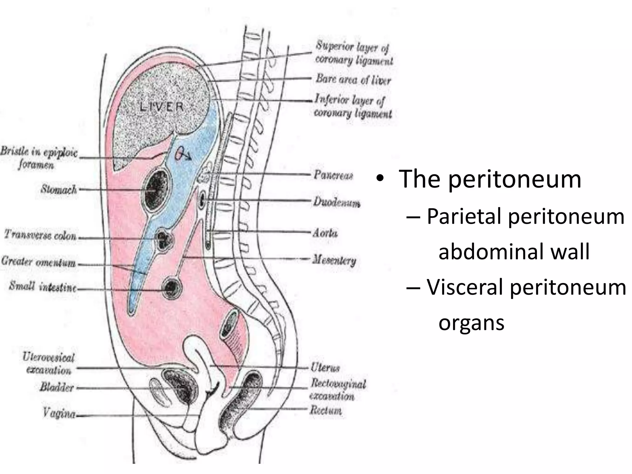
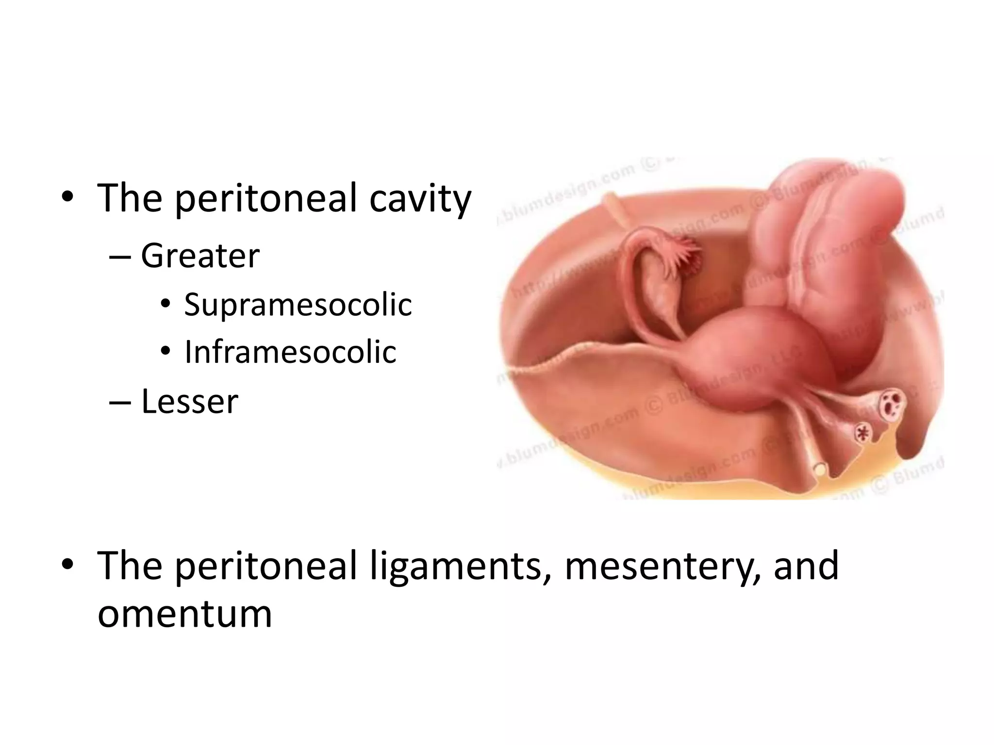
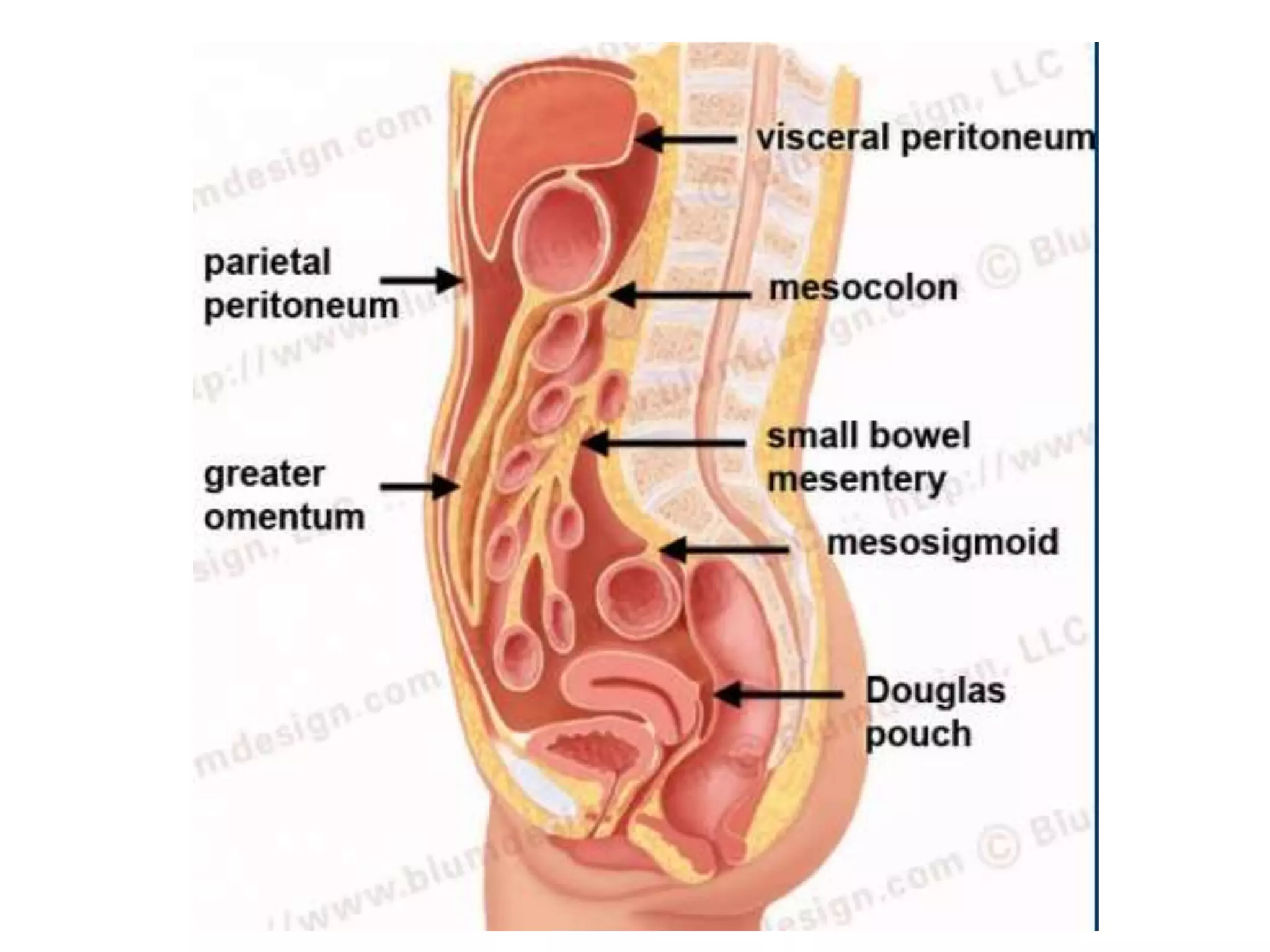
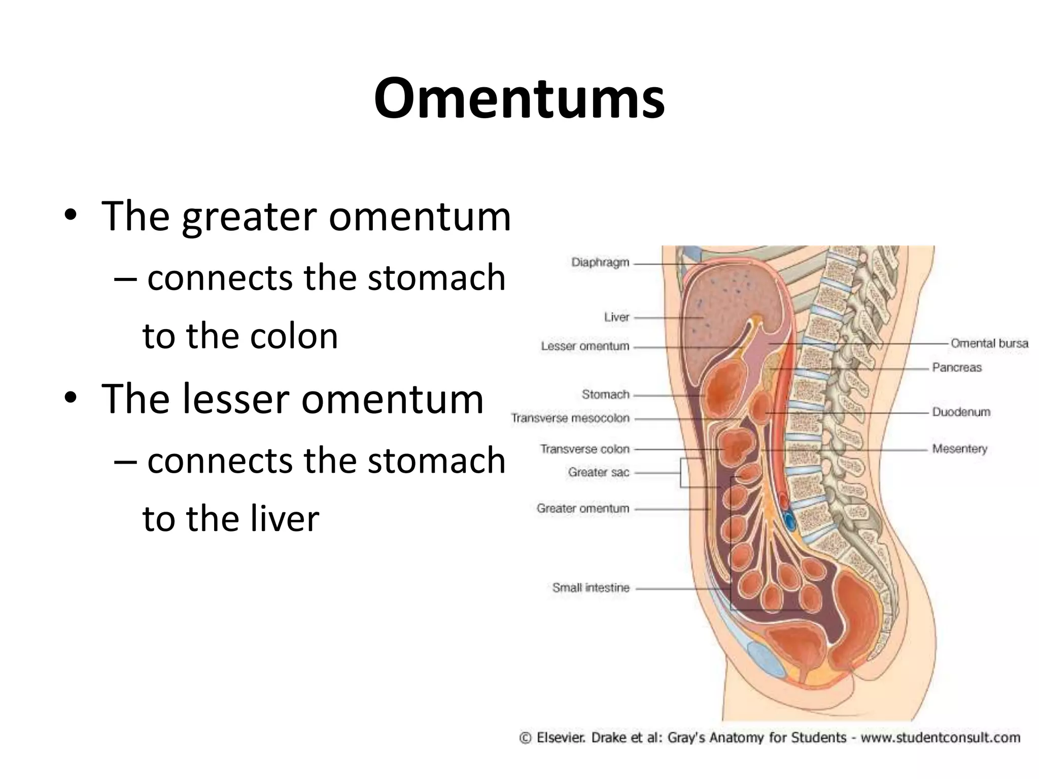
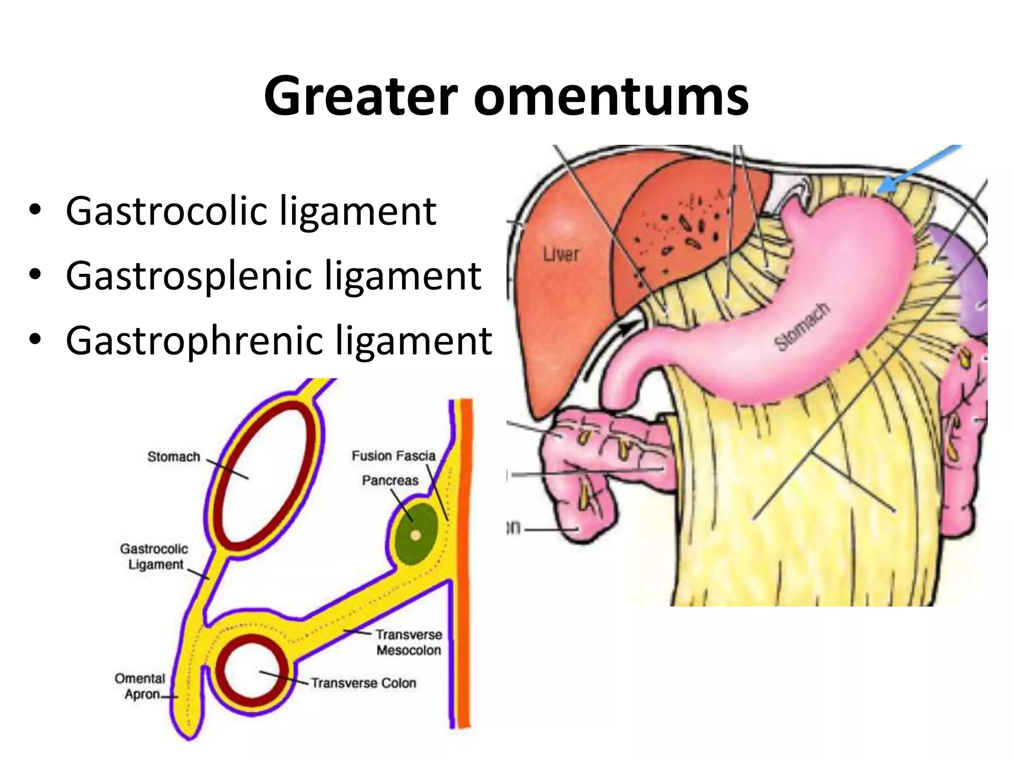
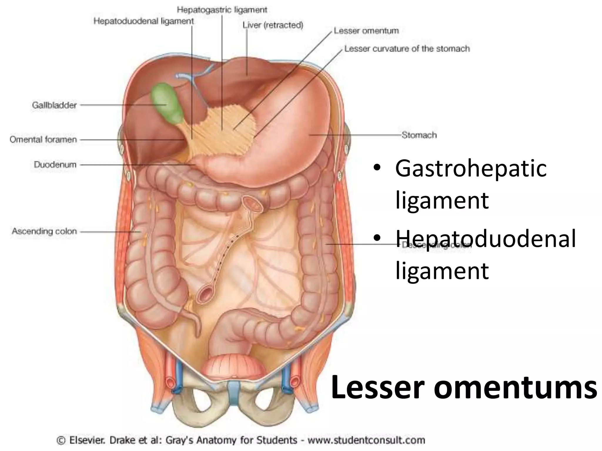

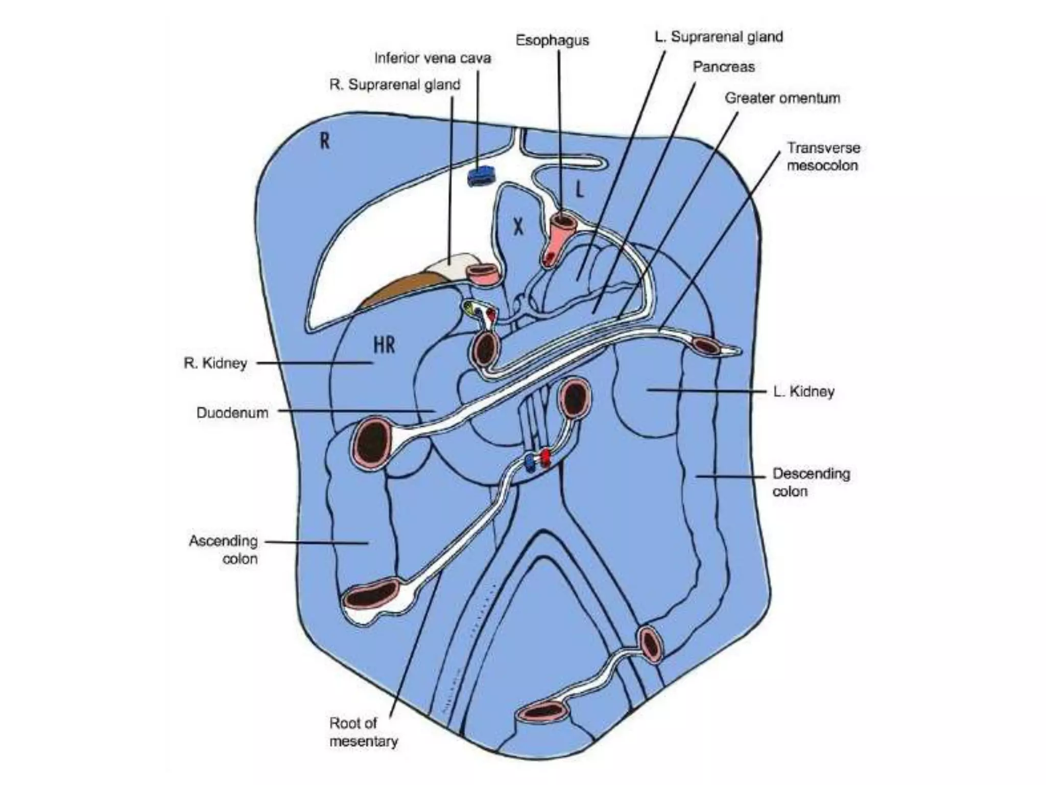
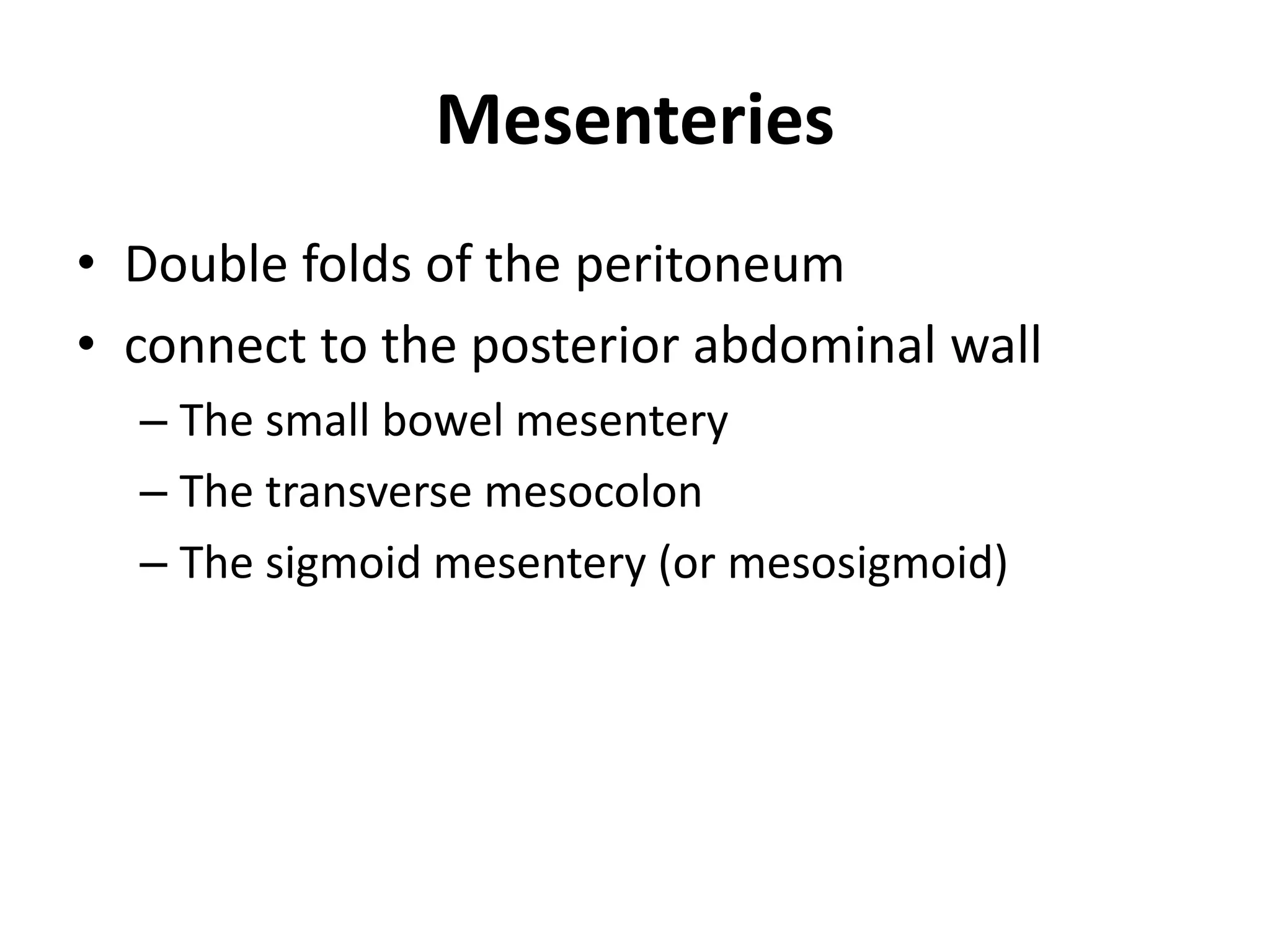
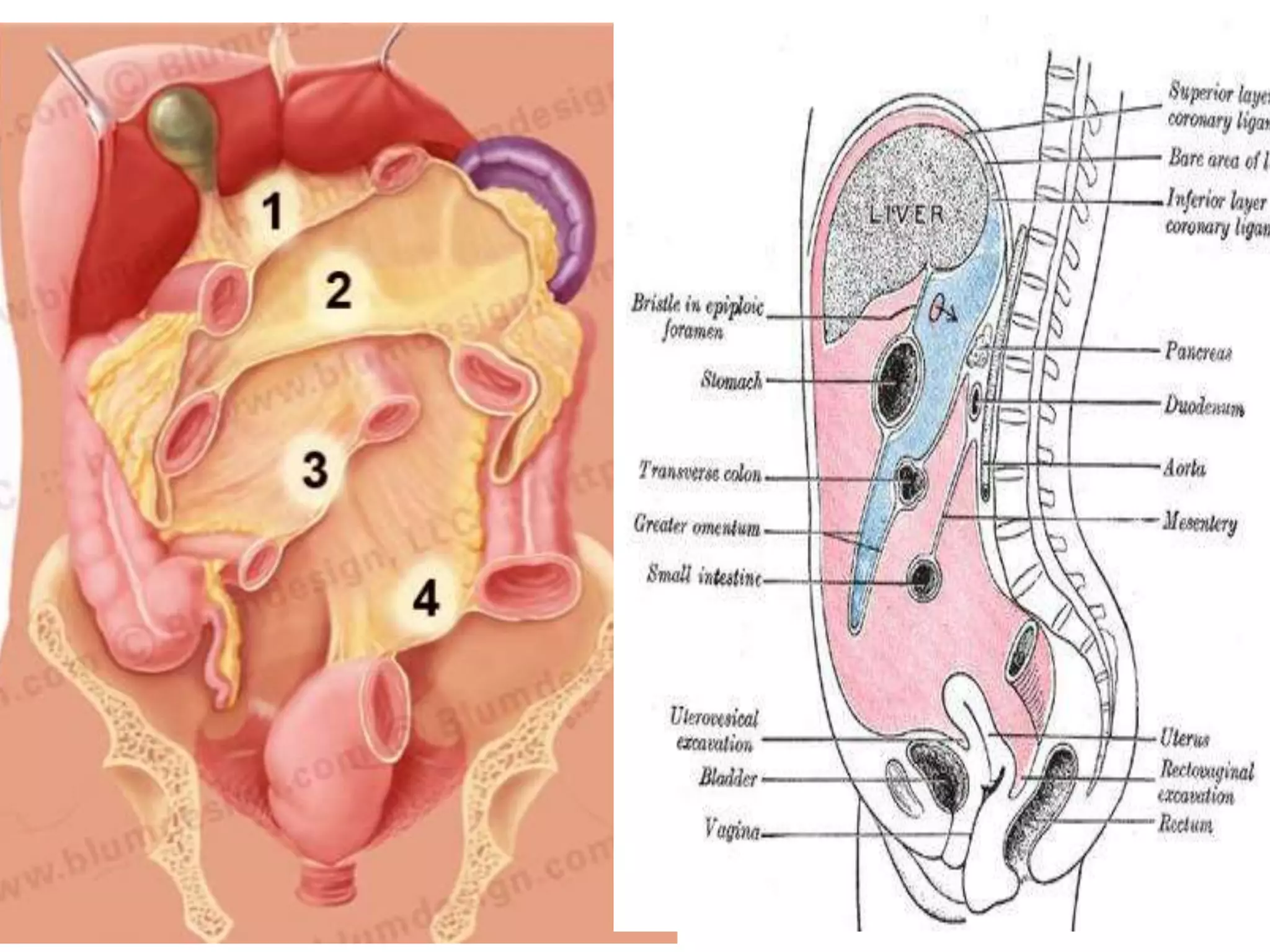
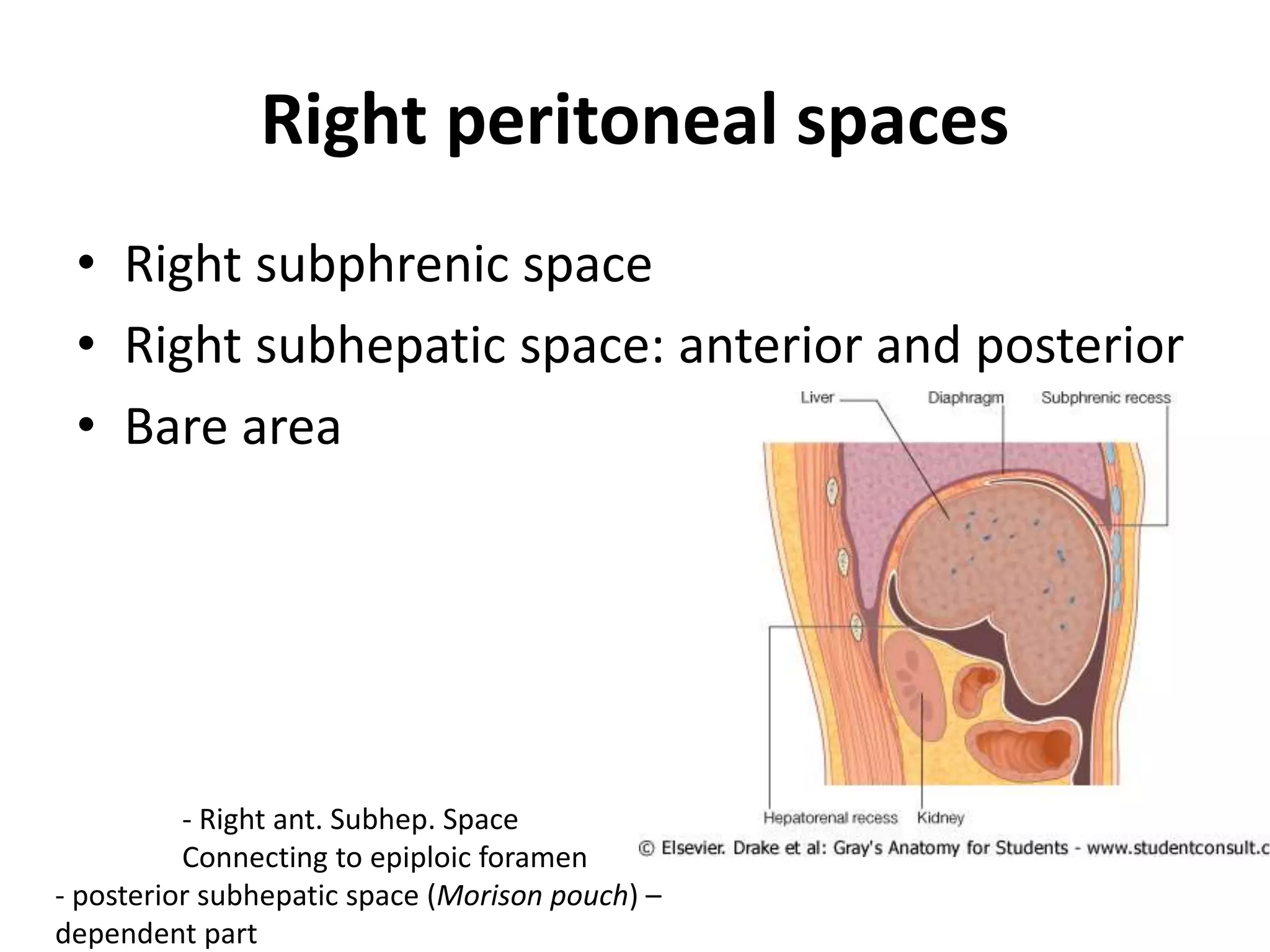
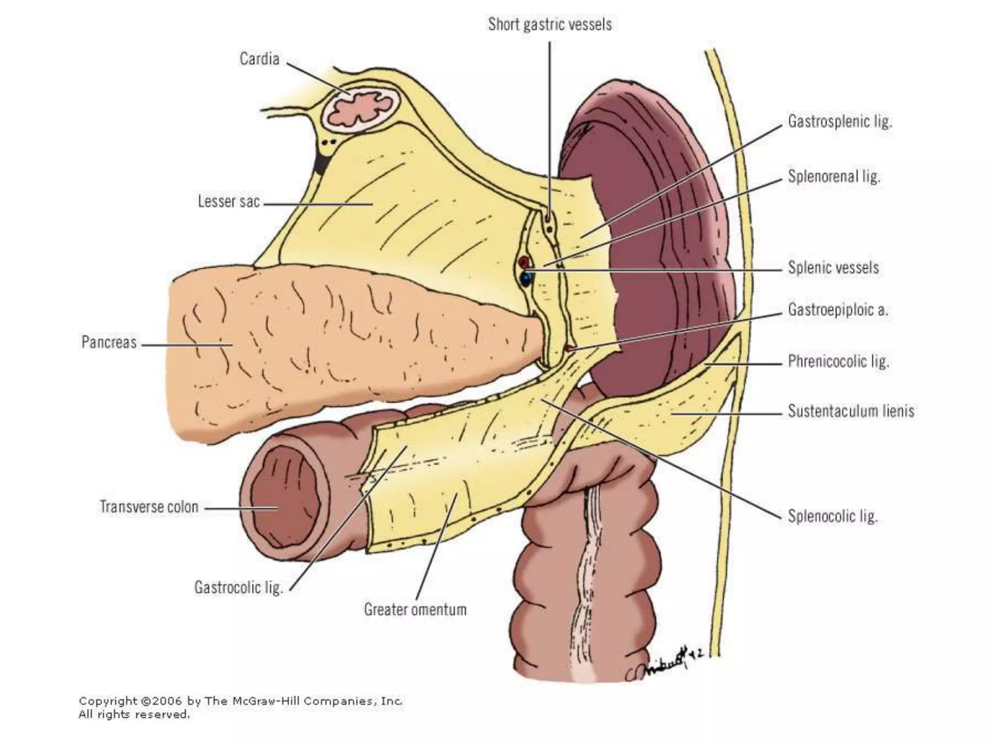
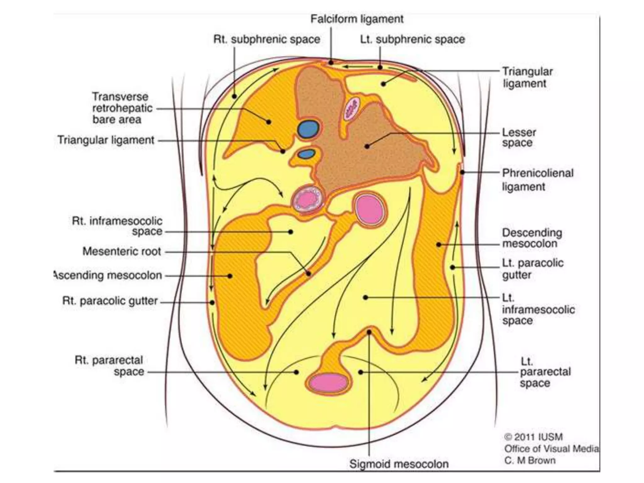
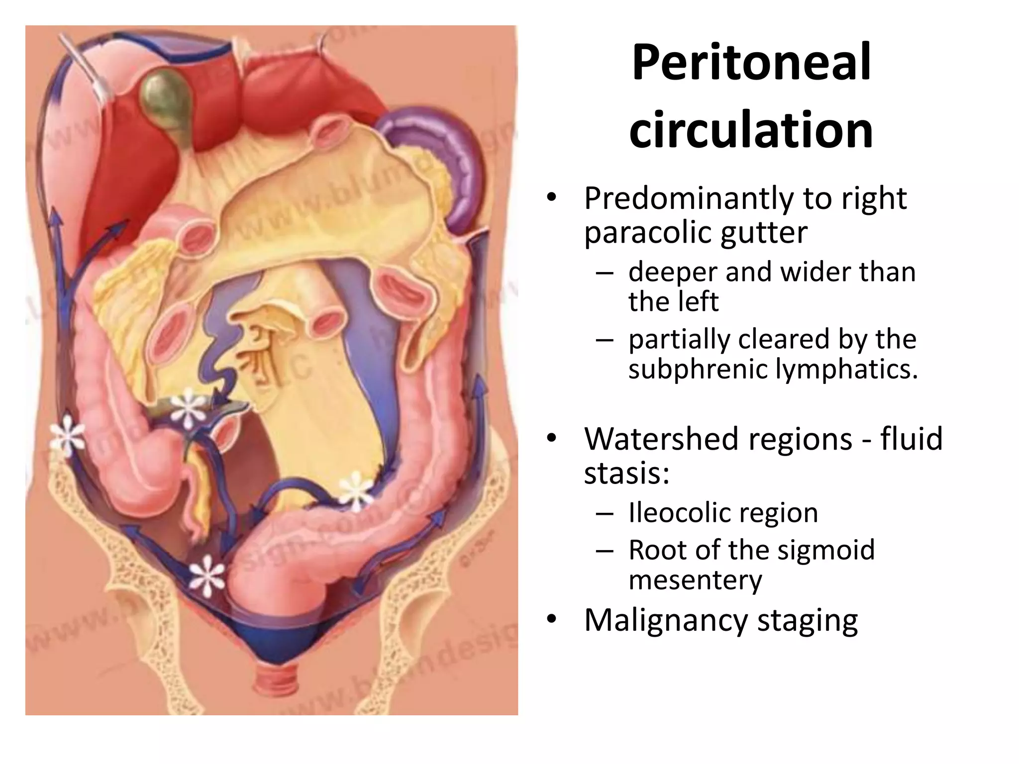
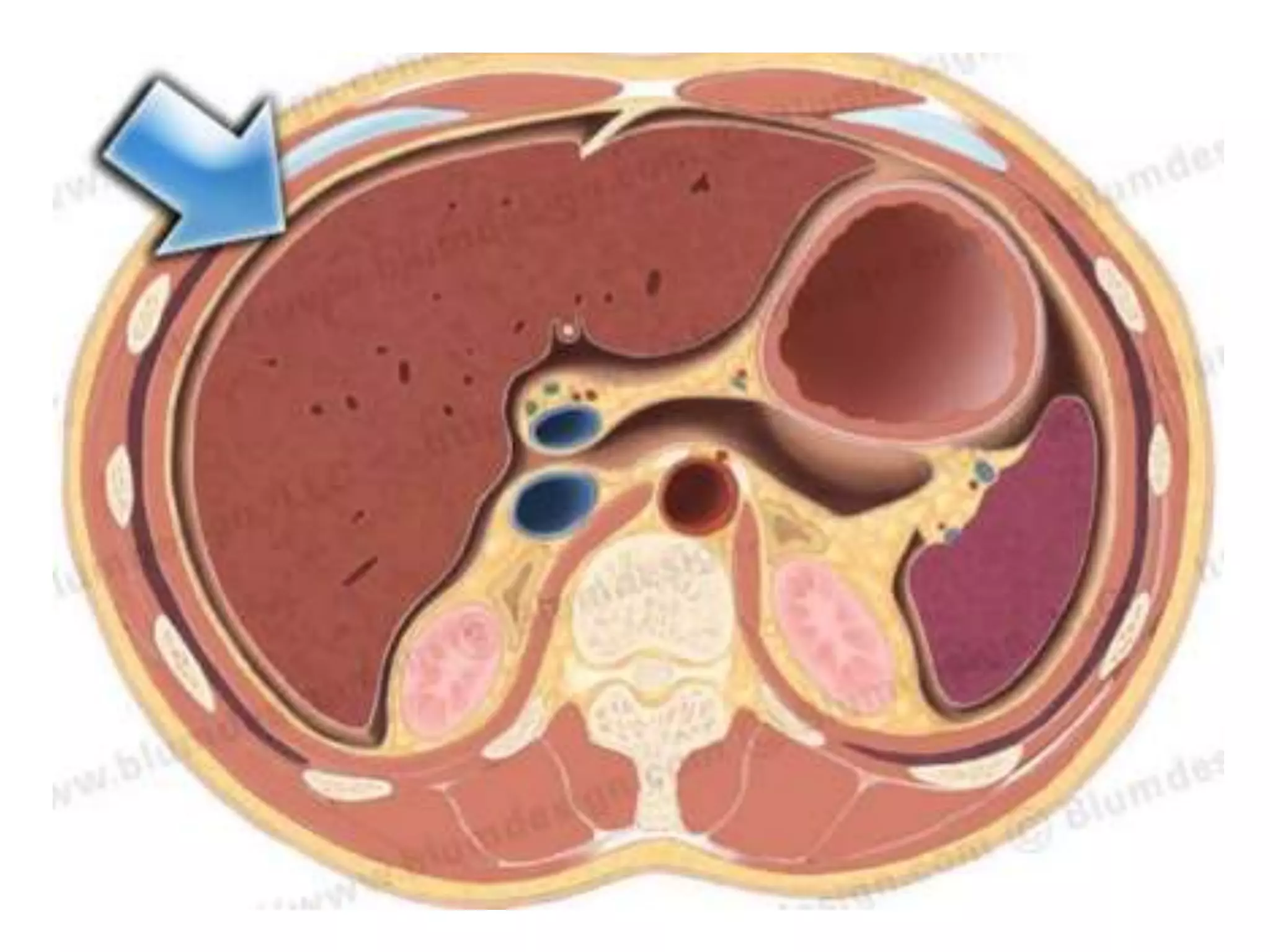
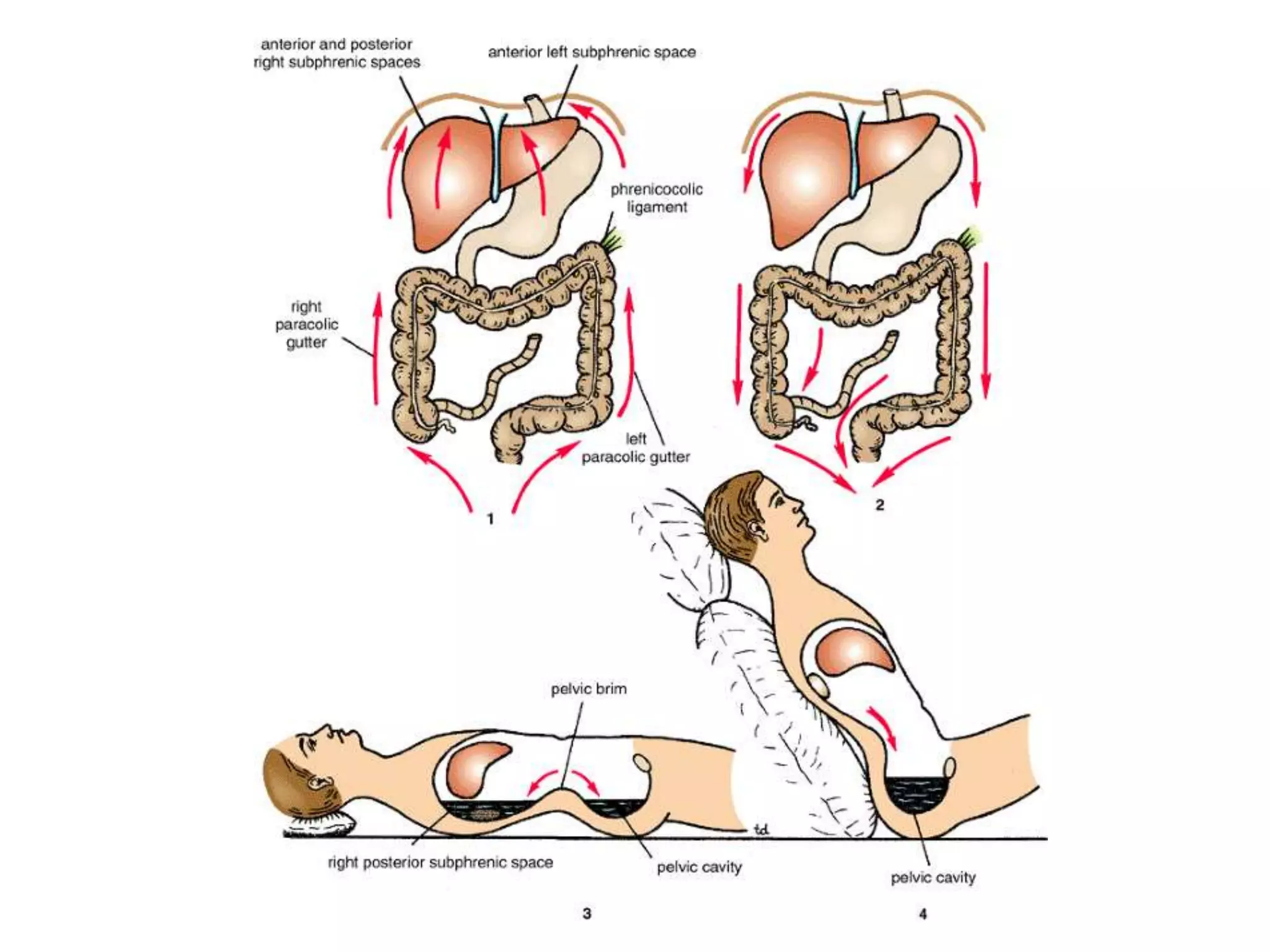

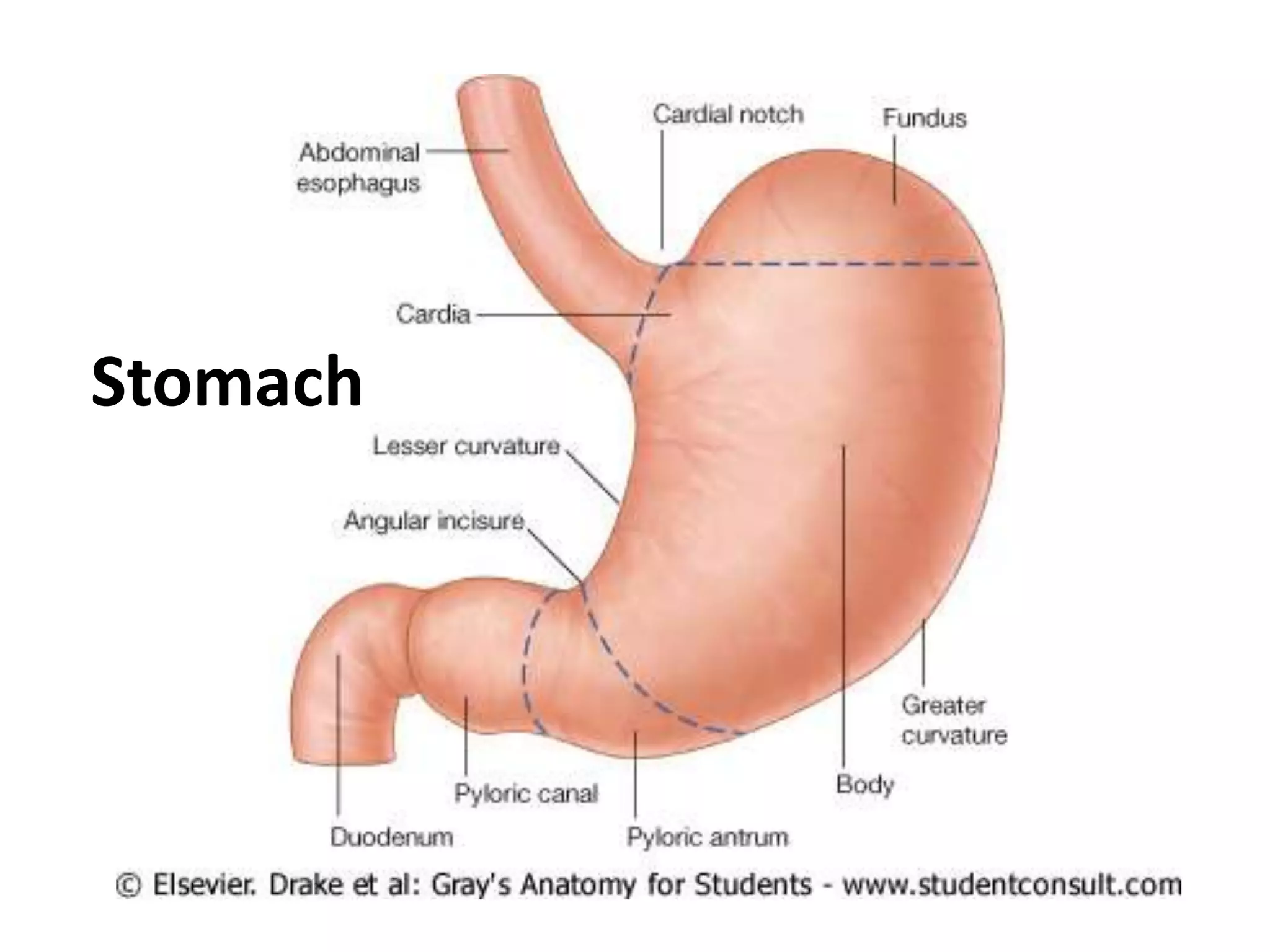
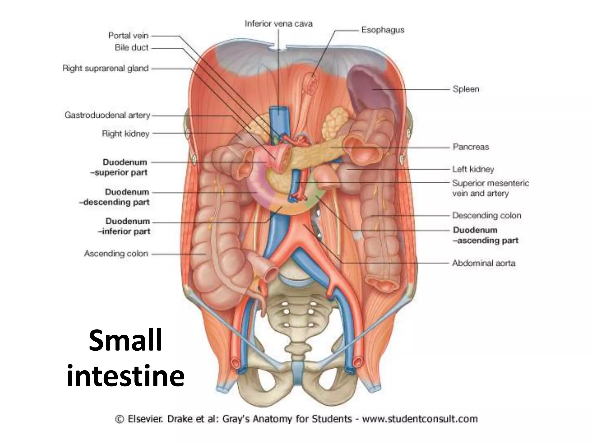
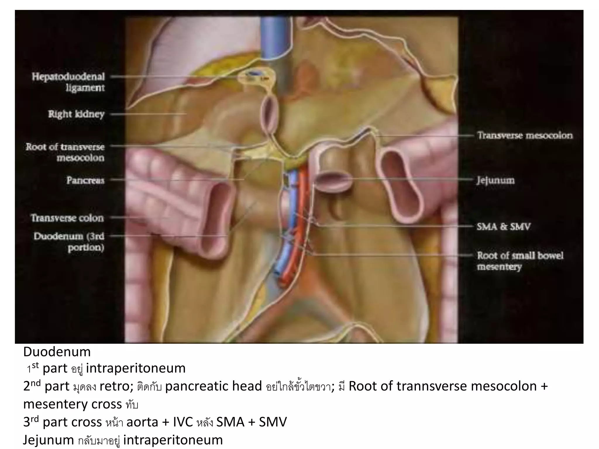
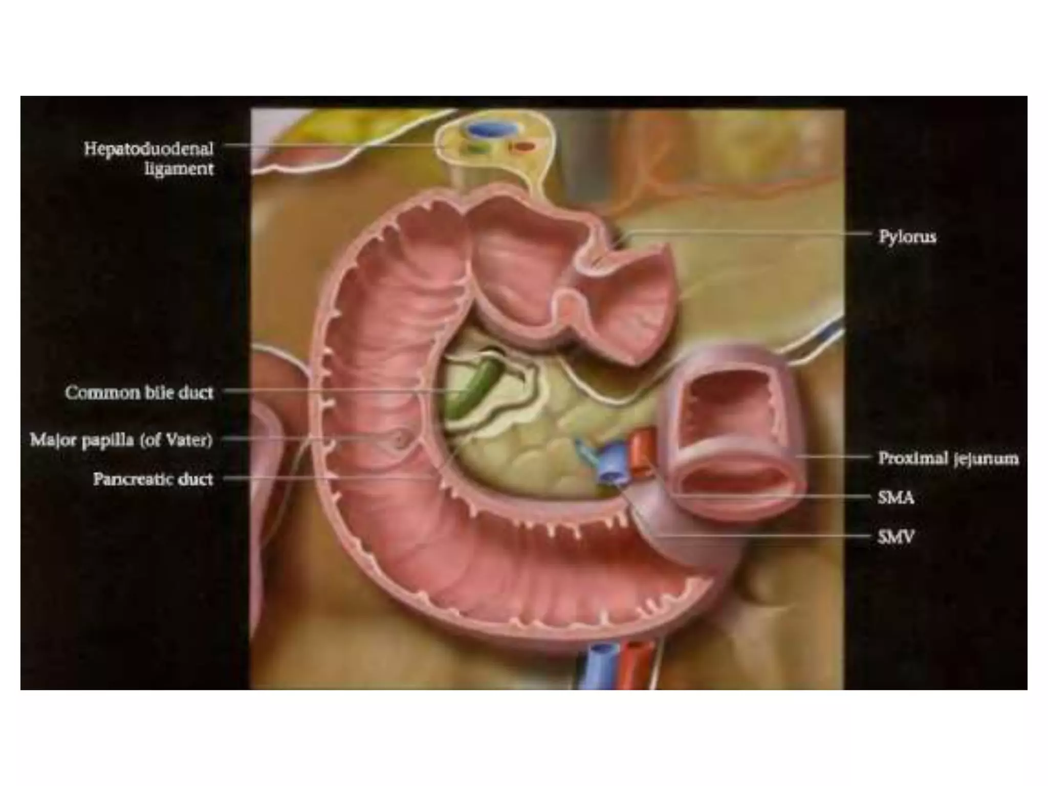
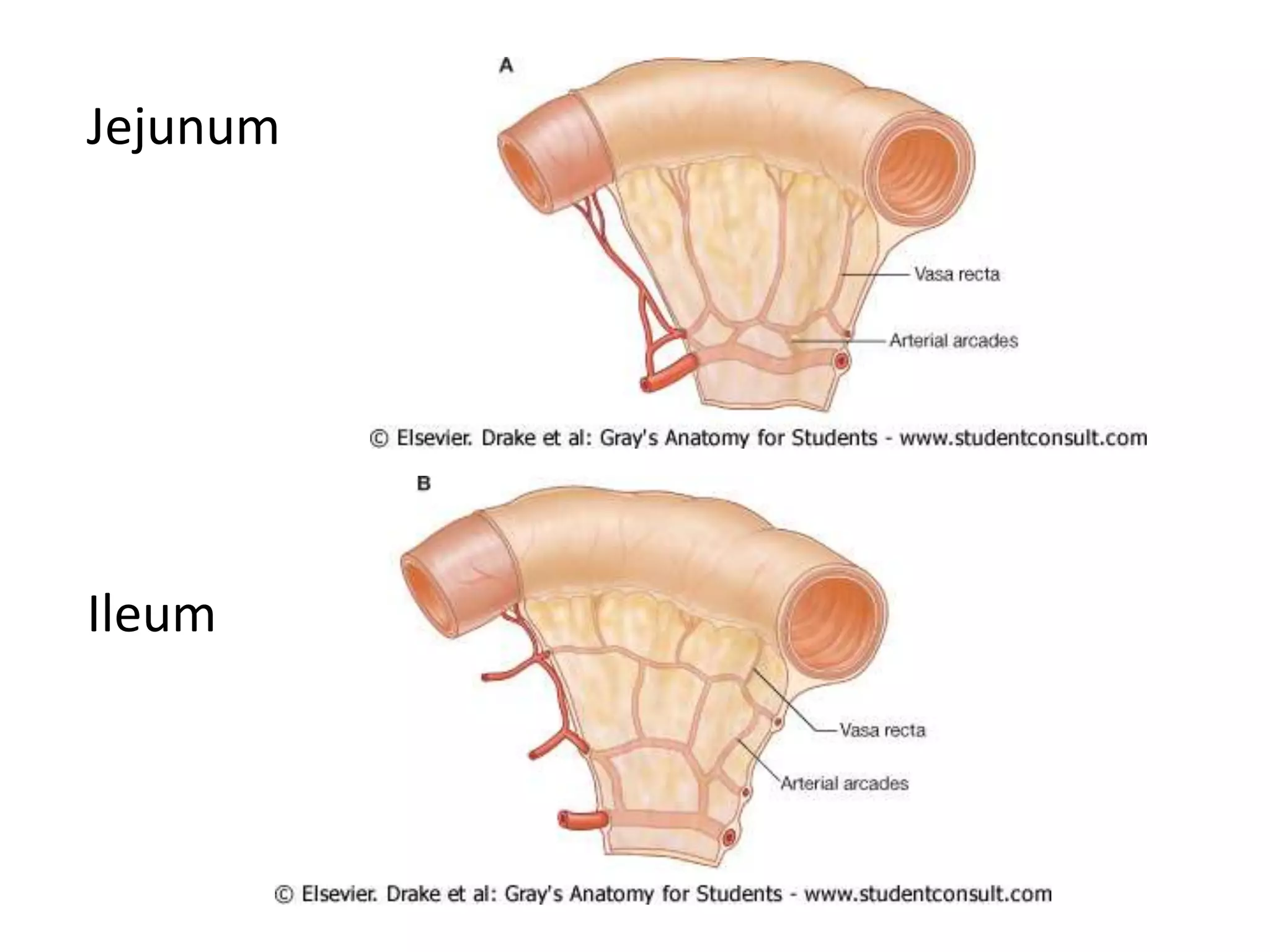
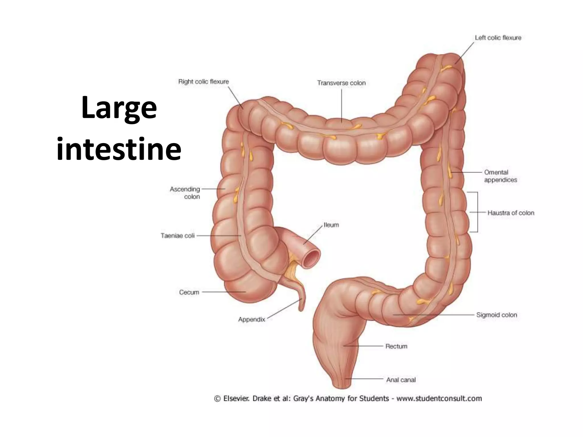
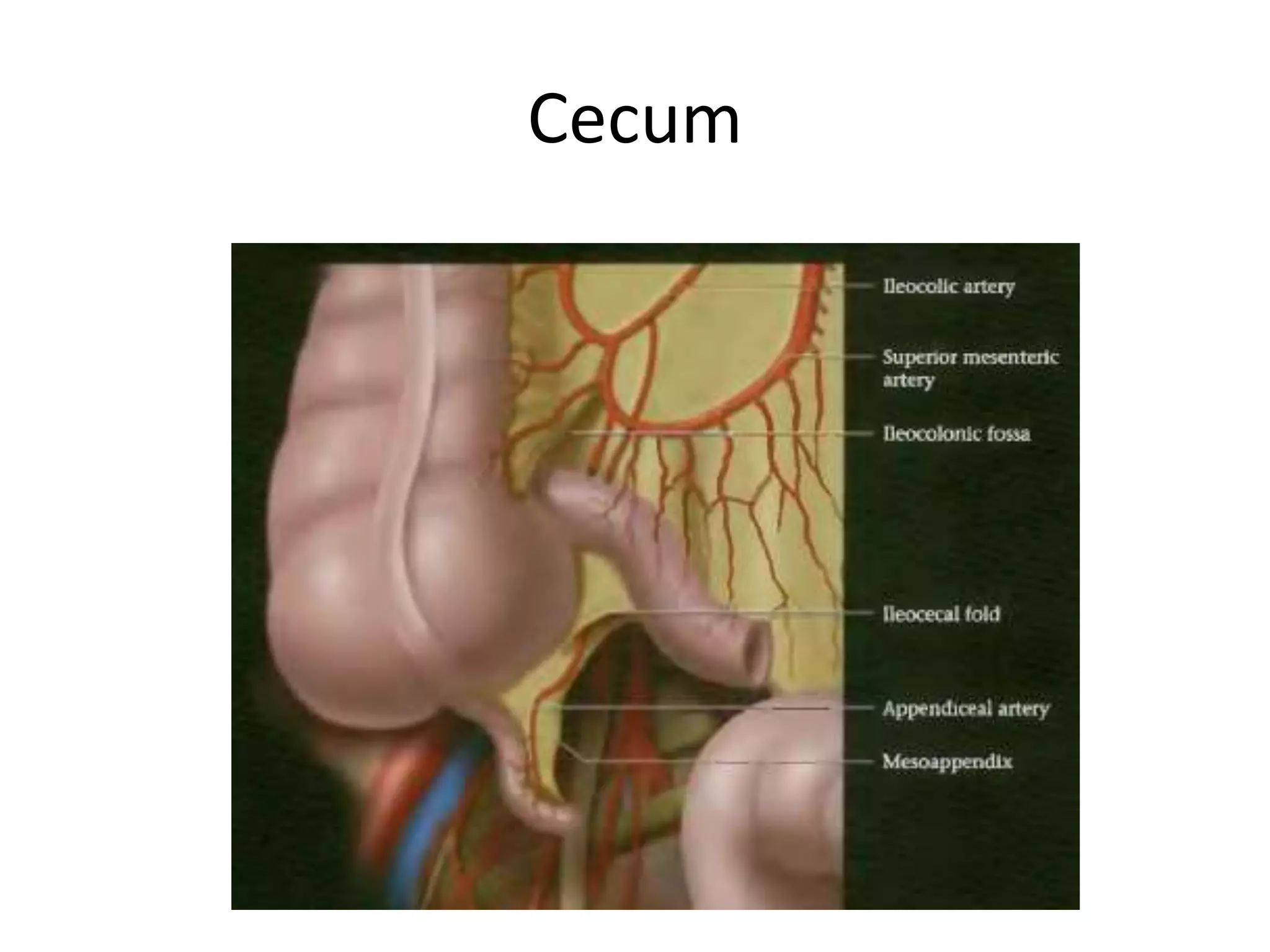

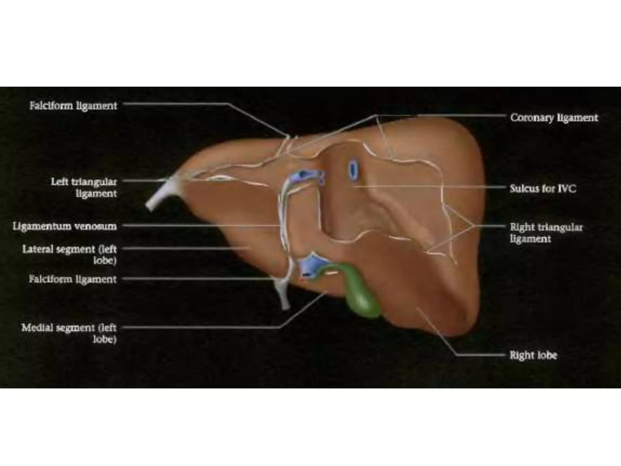
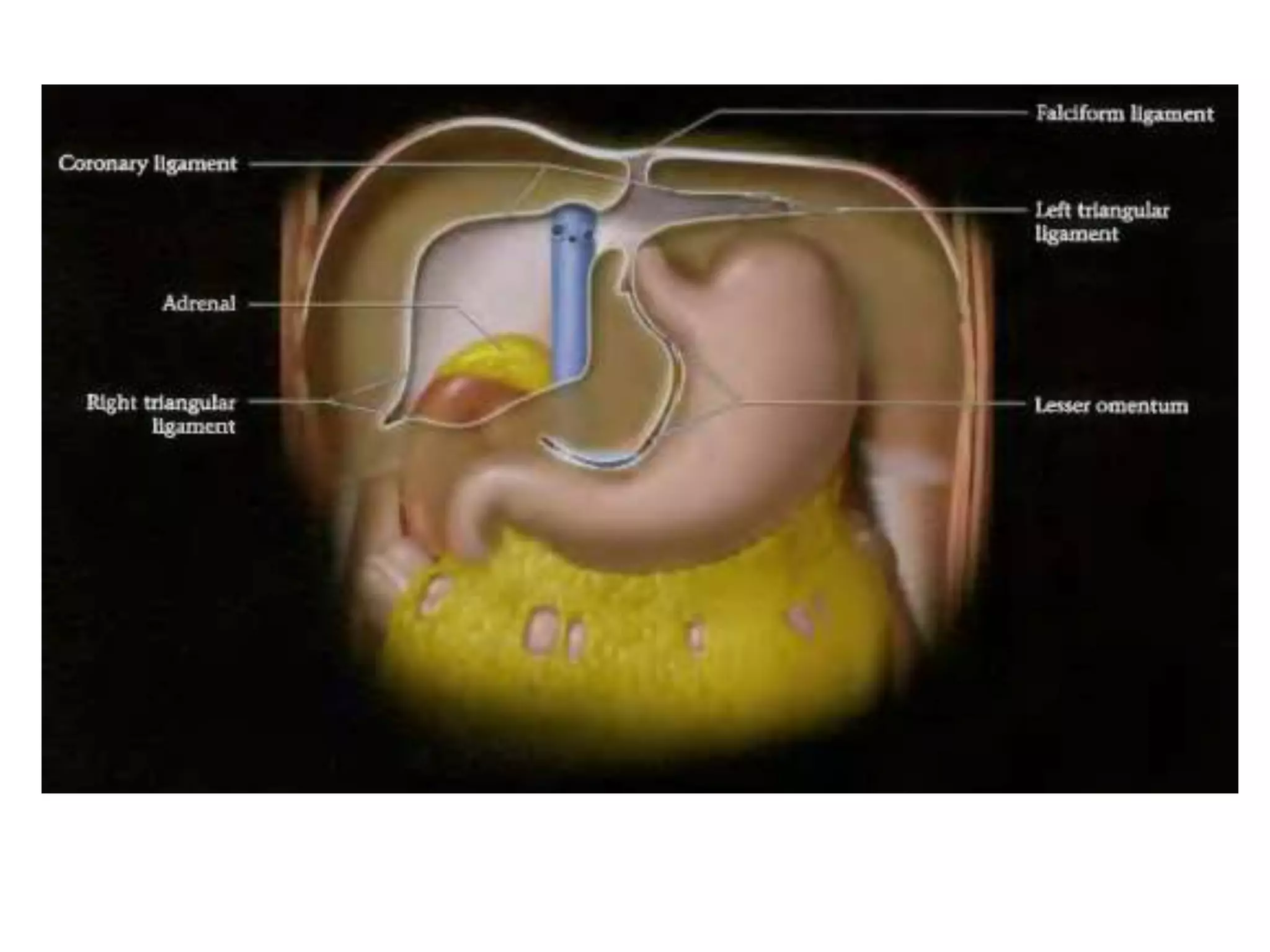

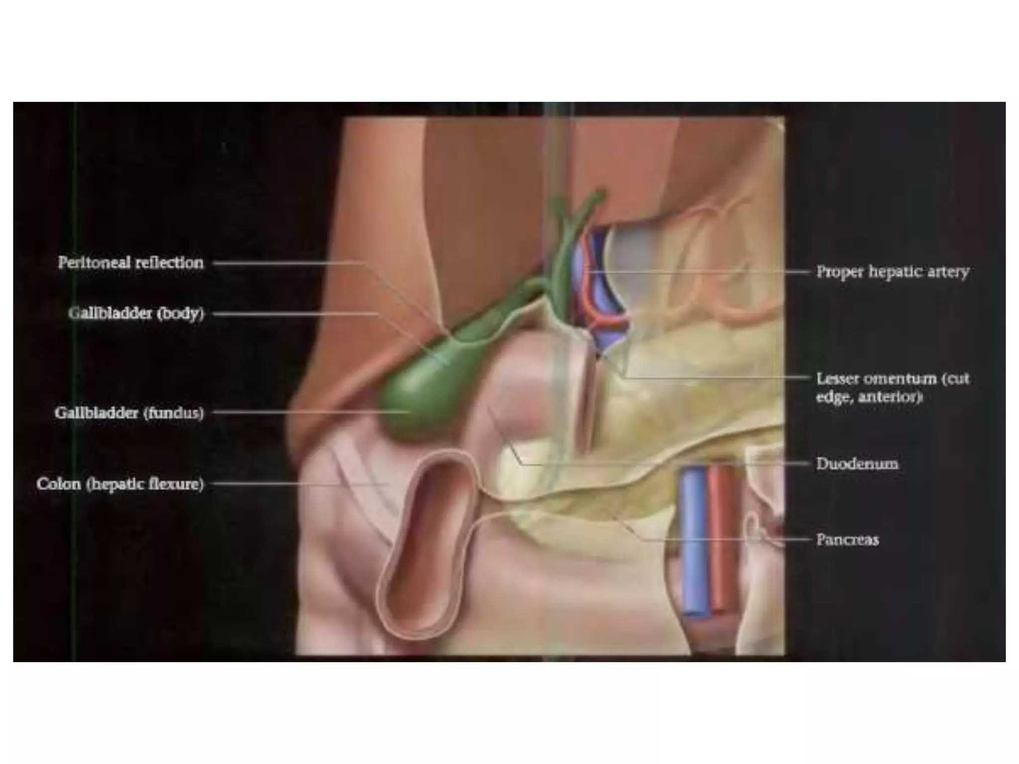
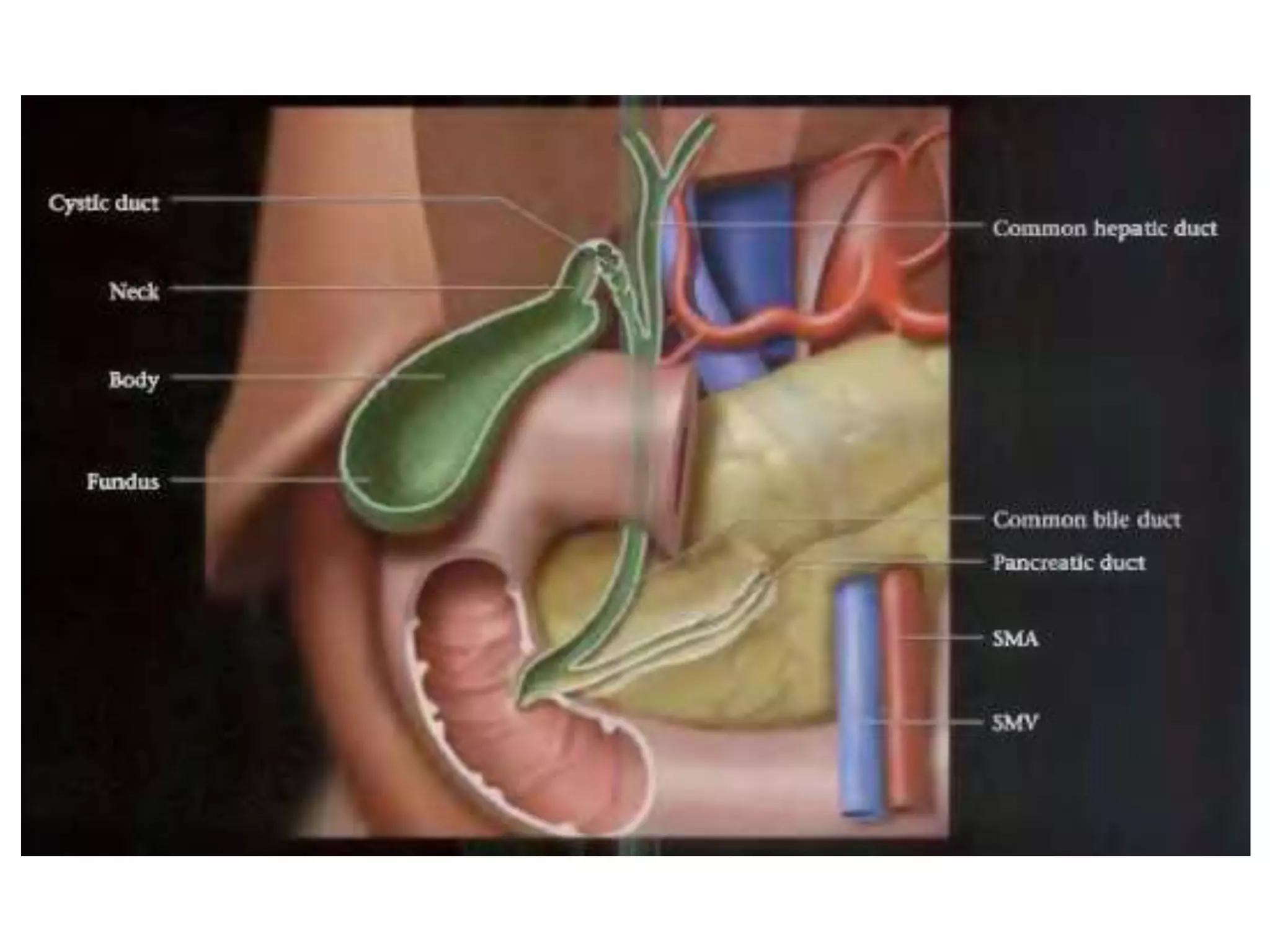


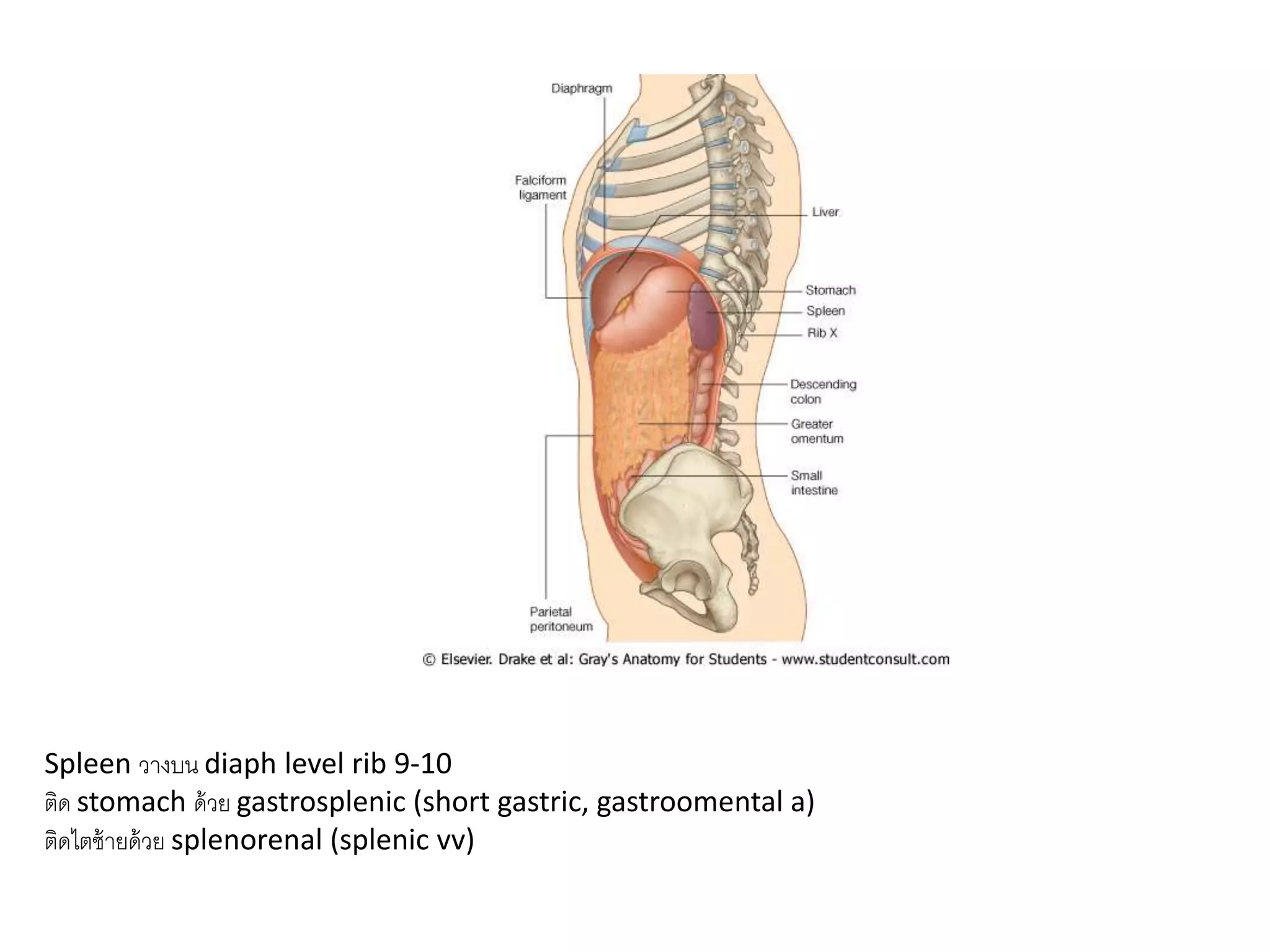

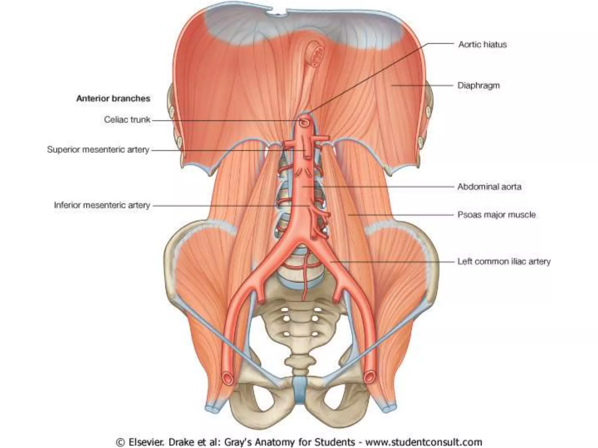
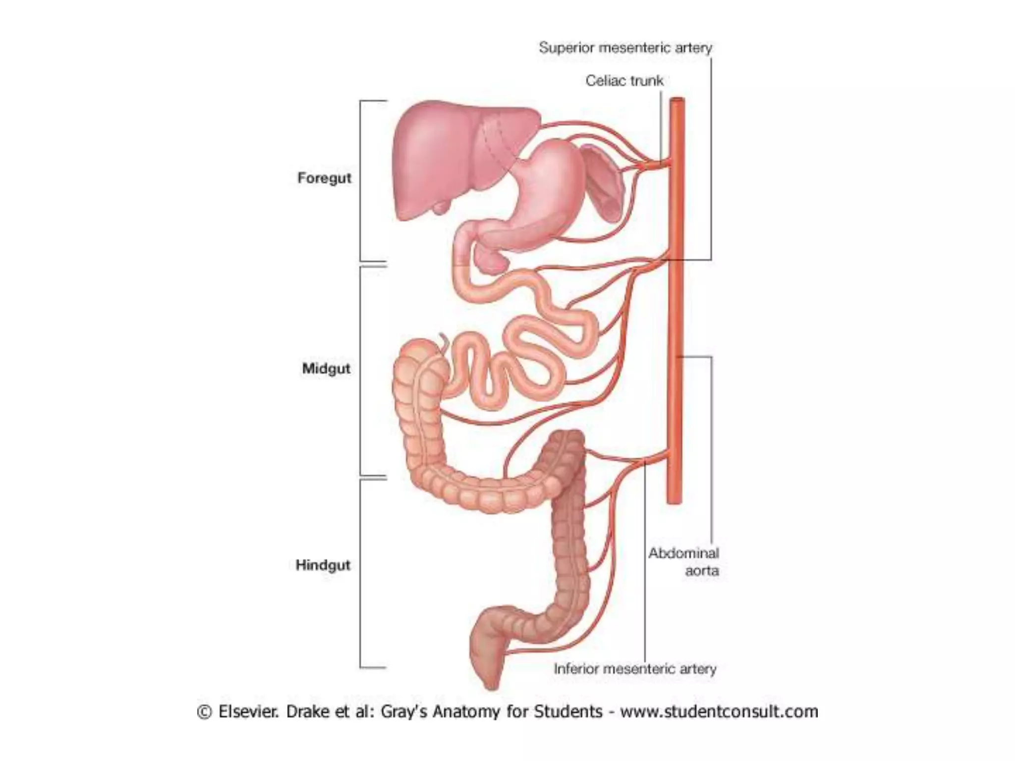
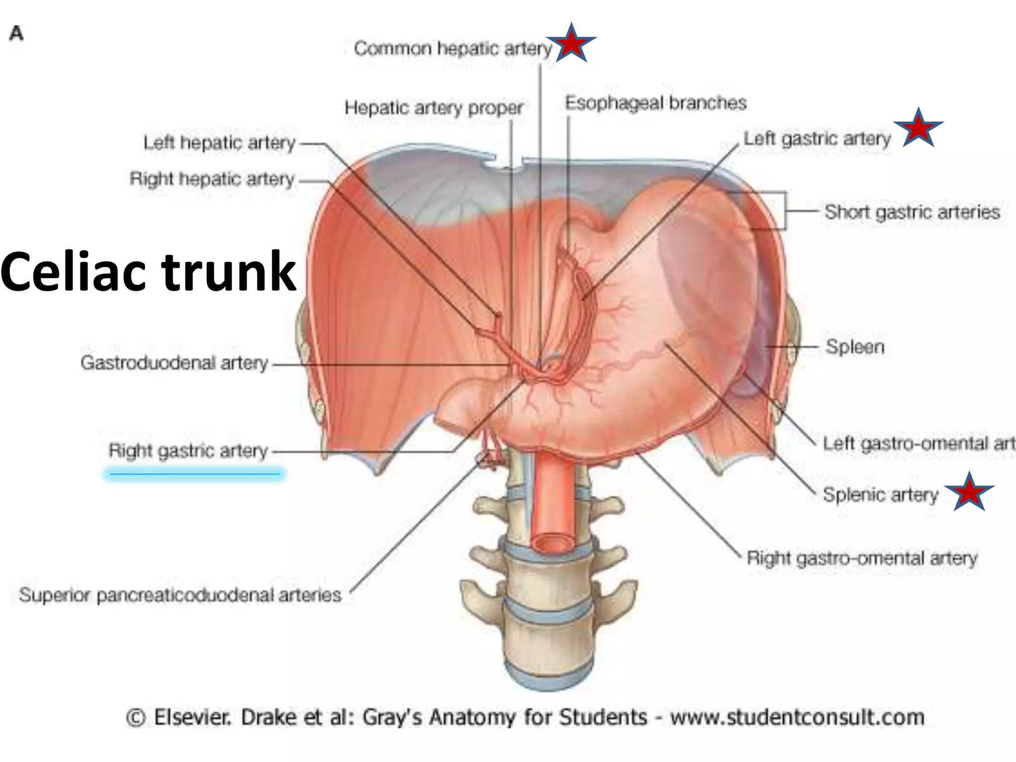
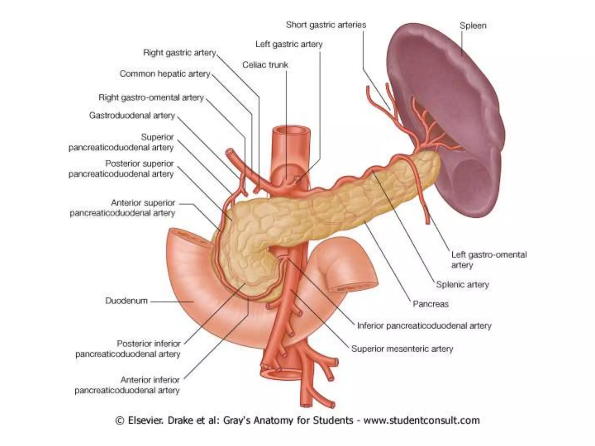
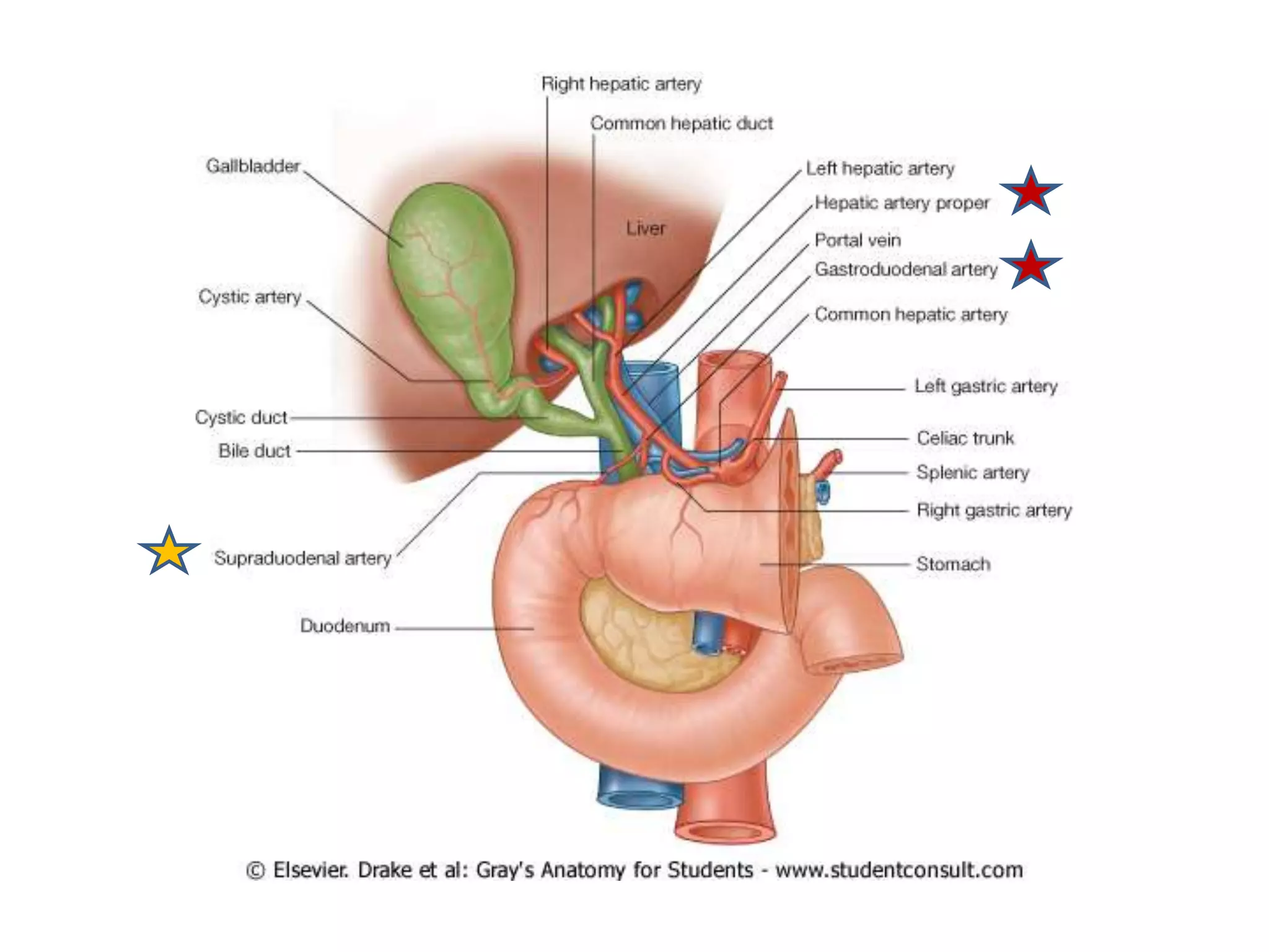
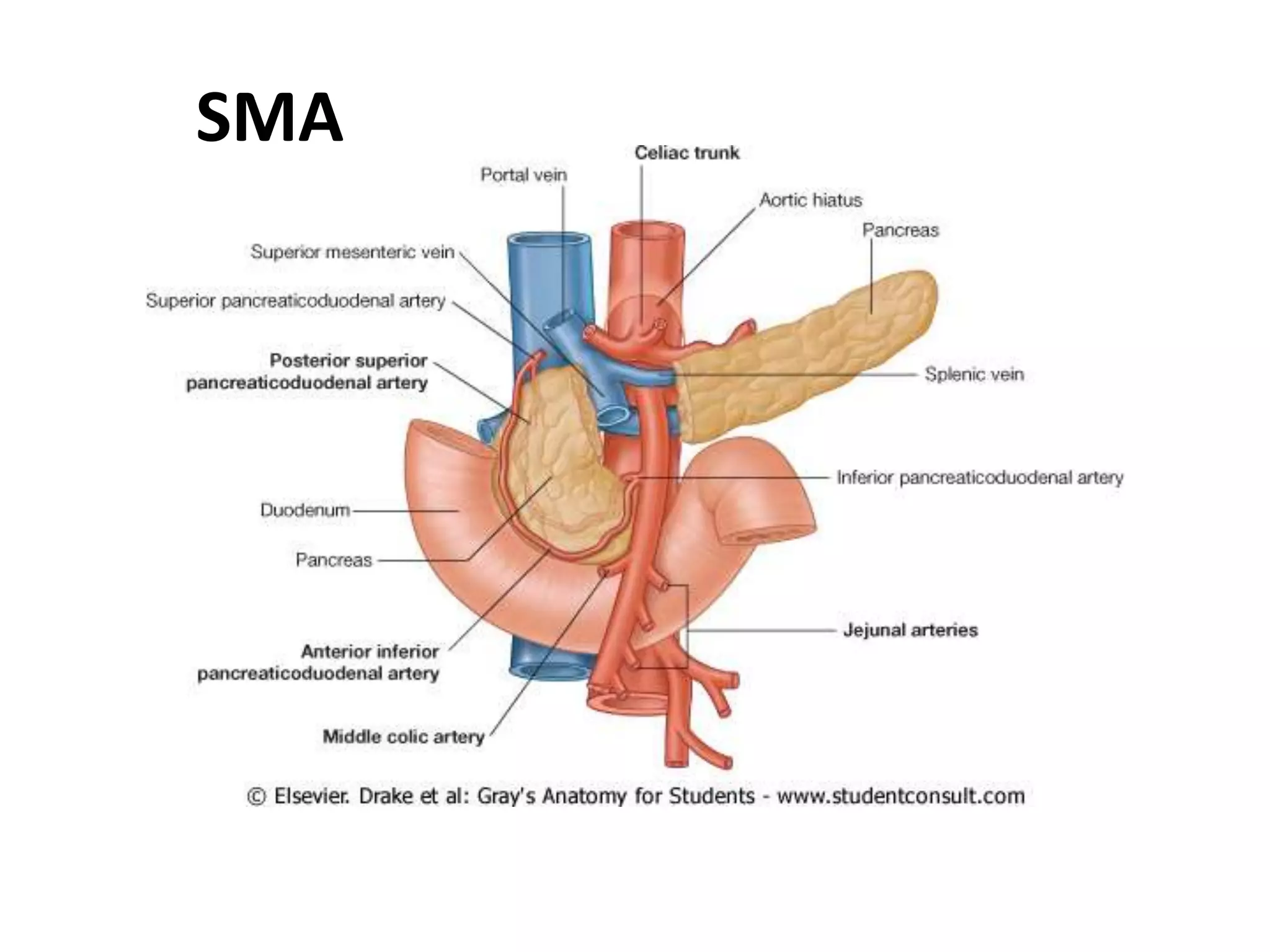
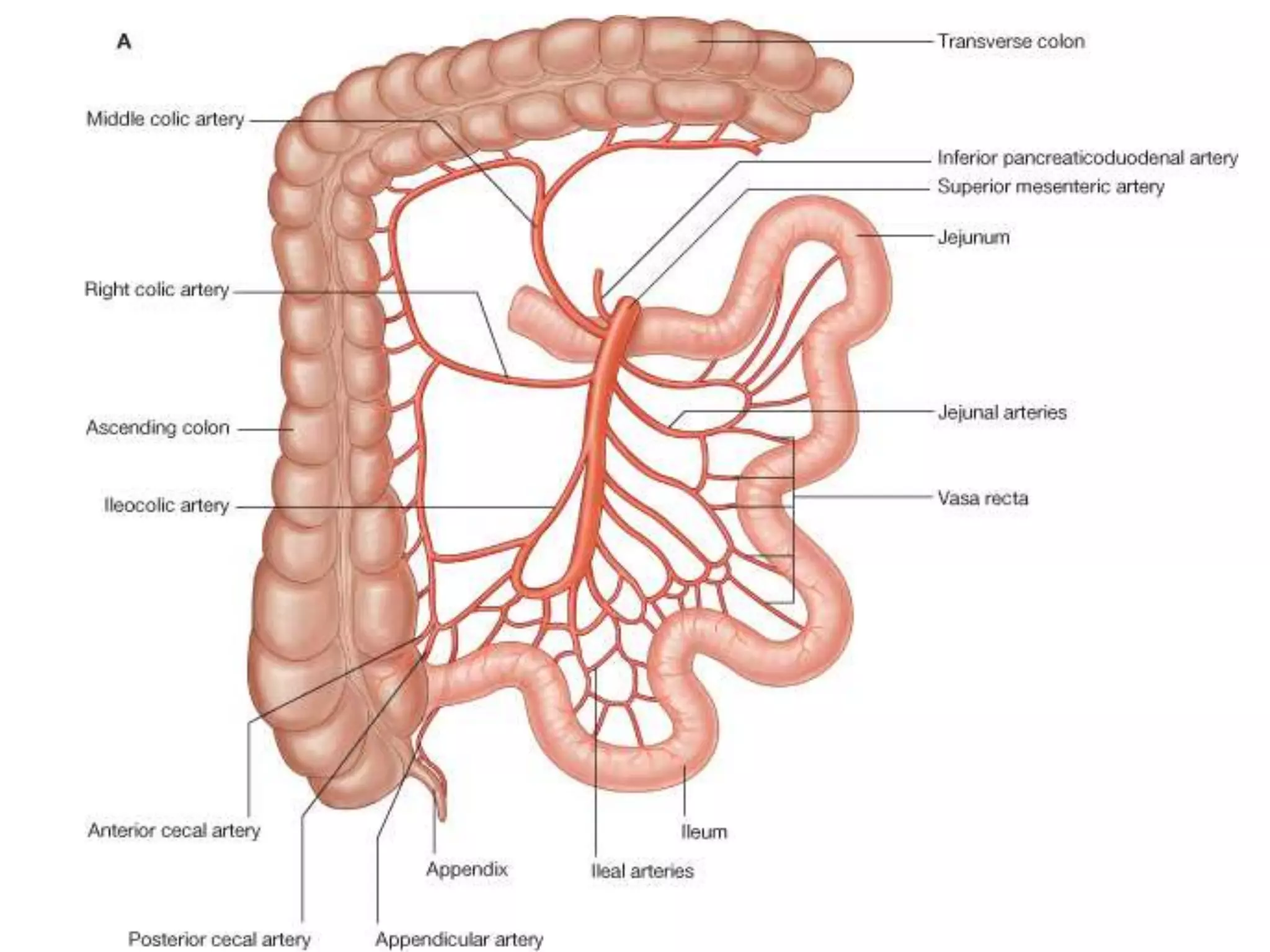
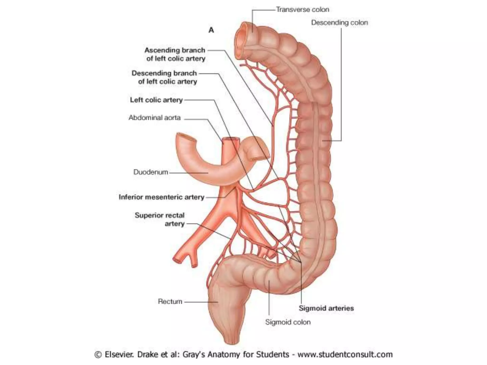
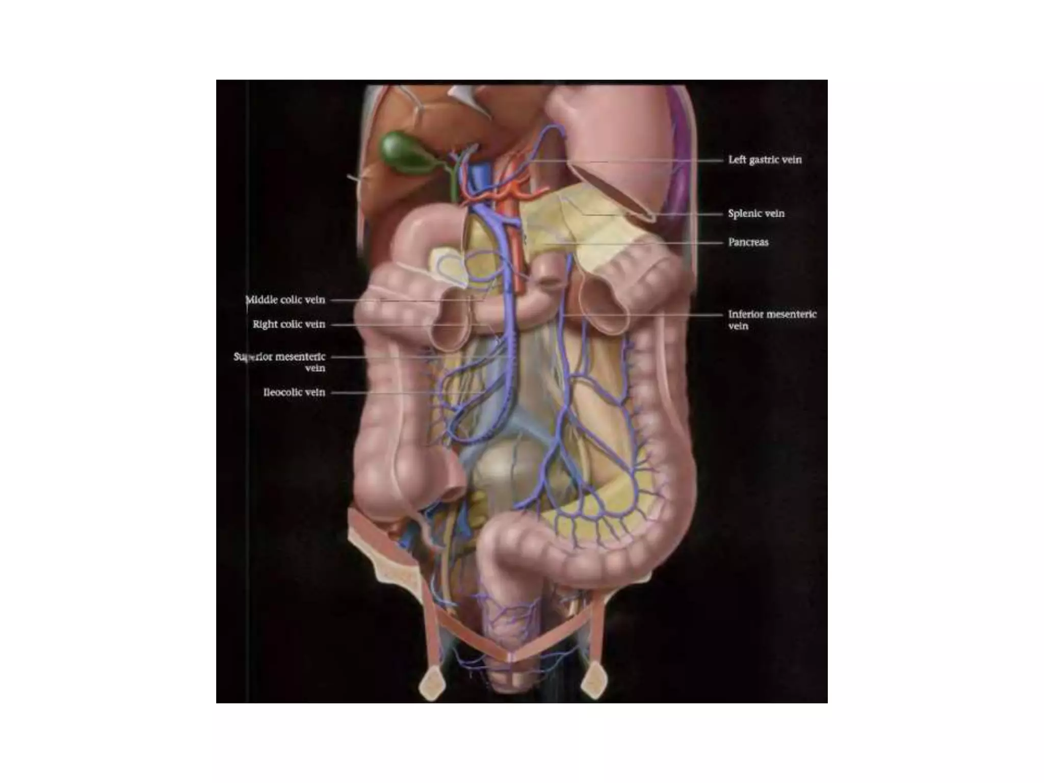
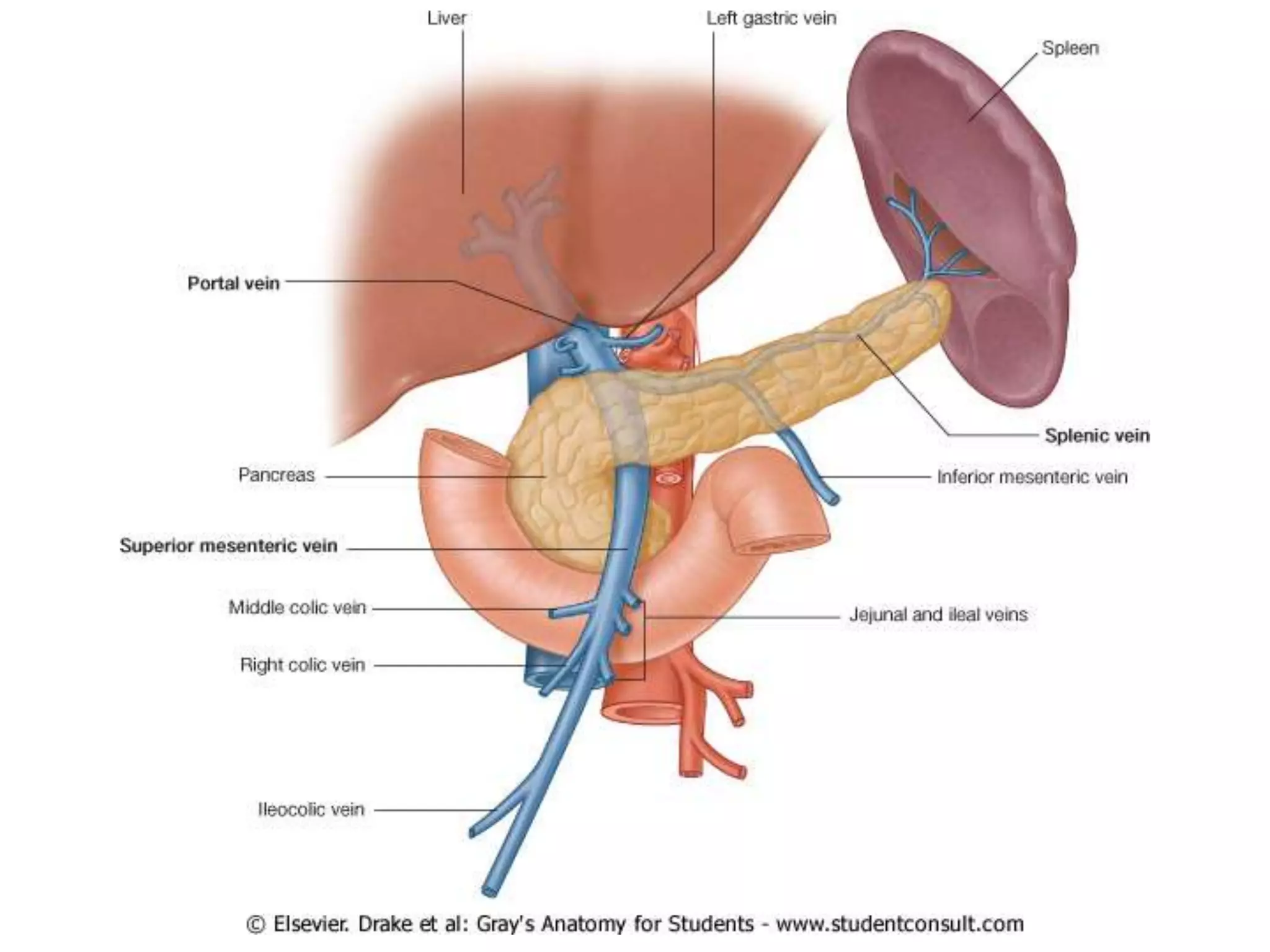
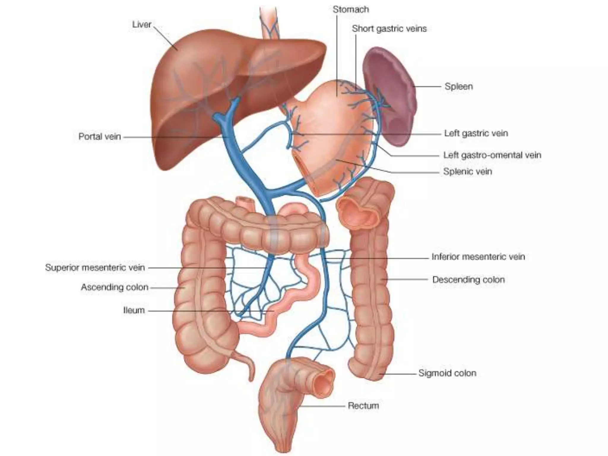


This document provides an overview of the anatomy of the intraperitoneum. It describes the peritoneum and peritoneal spaces, including the parietal and visceral layers. It outlines the greater and lesser sacs and peritoneal ligaments, mesenteries, and omenta. It details the intraperitoneal organs such as the stomach, small intestine, large intestine, liver, gallbladder, pancreas, and spleen. It concludes with notes on vascular structures like the celiac trunk and superior mesenteric artery.


