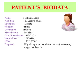
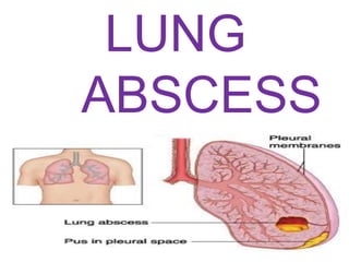
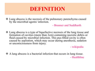
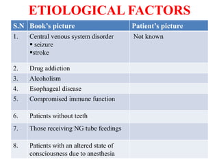
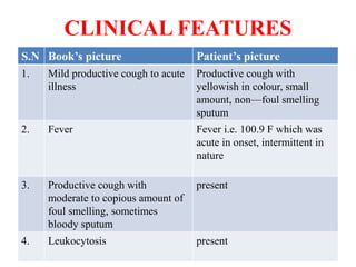
CASE PRESENTATION ON
LUNG ABSCESS
PRESENTED BY:
LAXMI THAPA
BSc NURSING
OBJECTIVES
 To share experience and knowledge to friends and supervisor.
 To get feedback from the friends and supervisor for further
improvement.
 To gain confidence in presenting skills in among the group
PATIENT’S BIODATA
Name : Salina Mahato
Age/ Sex : 20 years/ Female
Education :Literate
Religion :Hindu
Occupation :Student
Marital status :Married
Date of Admission :2017-01-22
Hospital No :16120386
IP No :1701220502
Diagnosis :Right Lung Abscess with operative thoracotomy,
empyema thorasis
Chief Complaints:
 Cough and sputum × 15 Days
 Fever × 3 Days
A. History Collection:
a. Present illness:
Follow up case of lung abscess with right sided empyema thoracic
chest tube insertion with right sided. 15 days earlier presented to
the CTVS opd with sputum since 4days. Cough was productive,
sputum with yellowish in colour small amount, non-foul smelling.
3 days after the onset of cough, the patient developed fever. It was
acute in onset, intermittent in nature, fever was seen in the
evening. Maximum temperature was not recorded. Fever was
associated with chills and rigor. It got relieved after taking
paracetamol medicine. No Aggravating factors were known, no
history of reddish discoloration in sputum, vomiting, loss of
consciousness and chest pain.
b. Past illness:
There was no any history of Hypertension, Tuberculosis, Diabetes
Mellitus, COPD, Asthma at past
c. Present Surgery:
There is history of present surgery i.e. Operative thoracotomy
d. Past Surgery:
There was no any history of past surgery.
e. Family History:
No history of similar illness among family members. There is no any
history of Diabetes Mellitus, Mental illness, Tuberculosis, Hypertension
among family members.
Fig: Family Tree
INDEX
Female
Male
Dead Male
Patient
Personal History
Smoking : Non-smoker
Alcohol: Non-alcoholic
Food habit: Mixed diet , 2-3 times a day
Food allergy : Not known
Drug allergy: Not Known
Bowel and Bladder: Regular
Sleeping pattern: 7-8 hours per day
Hobbies: she likes to watch TV, listen to radio and work
Socio –Economic History
Type of house: Pakka house
No. of rooms: 6 rooms
Kitchen : separate
Type of drainage: open drainage
Type of toilet used: water seal
Sources of water: Hand pump
Type of fuel: wood and gas
Adequate lighting: present
Facilities available in surrounding:Temples, schools available
Monthly income: Rs 28000
Bread winner : Father-in law & Husband
INVESTIGATION
S.N Investigation Patient value Remarks
1 Hemoglobin 10.1mg/dl Anemia
2. Total leucocytes
count
10, 570 cells/ cu mm leukopenia
3. Neutrophils 83% Neutrophilic
leukocytosis
4. lymphocytes 07% lymphocytopenia
5. Platelets count 5,36,000 cells/cu.mm Thrombocytosis
S.N Investigation Patient value Remarks
6. ESR 57 mm/hr Infection
7. Creatinine 0.3 mg/dl Indicate muscular
dystrophy, Liver dz
8. Blood PH 7.30 Metabolic acidosis
9. Pco2 49.1 mm Hg Respiratory acidosis
10. Potassium 2.93 mmol/l Hypercalemia
 Specimen: Fluid
 Bronchial wash cytology:
Smears studies reveal plenty of polymorpho nuclear inflammatory
cells agisnt a degeneratedand preoteinaceous background
Conclusion: Acute inflammatory process
S.N Investigation Patient value Remarks
11. Calcium 0.53 mmol/L Hypocalcemia
MEDICATION
S.N Name of
Drug
Dose Route Frequency Classification
1. Inj. Pantop 40 mg I/V BD Proton pump
inhibitor
2. Inj. Tazopip 4.5gm I/V BD Antibiotic
3. Inj. Metron 500m
g
I/V TDS Antibiotic & Anti-
protozoal
4. Tab. Pcm 500m
g
P/O TDS Anti-pyretics
5. Tab Ifol 1 tab P/O OD Folic Acid
Needs Problems
Physical
Comfort
Elimination
Psychological
Unstable vital sign
ABG analysis
-PH: 7.30
-pco2: 49.1 mm Hg
Shortness of breath
Use of face mask
Pain over the chest tube drainage pipe
on chest
Use of bladder habits i.e.
catherterization
Disturbed sleep pattern
Anxiety.
Development task and
need
• Achieving new and more mature relations with age-mates of both
sexes.
• Achieving a masculine or feminine social role.
• Accepting one’s physic and using one’s body effectively.
• Desiring, accepting, and achieving socially responsible behavior.
• Achieving emotional independence from parents and other adults.
• Preparing for an economic career.
• Preparing for marriage and family life.
• Acquiring a set of values and an ethical system as a guide to
behavior-developing an ideology.
PHYSICAL EXAMINATION
Vital signs :
Blood pressure: 110/70 mm of hg
Heart rate: 88 beat per min
Respiration: 20 breath per min
Temperature: 98.4 degree Fahrenheit
PHYSICAL EXAMINATION
Findings
i. Yellowish with hemoptysis colouration sputum
ii. Chest tube drainage
iii. Dullness, decreased breath sound
iv. Coarse inspiratory crackles.
DISEASE
CONDITI
LUNG
ABSCESS
DEFINITION
 Lung abscess is the necrosis of the pulmonary parenchyma caused
by the microbial agents/ infection.
- Brunner and Suddharth
 Lung abscess is a type of liquefactive necrosis of the lung tissue and
formation of cavities (more than 2cm) containing necrotic debris or
fluid caused by microbial infection. This pus-filled cavity is often
caused by aspiration, which may occur during anesthesia, sedation
or unconsciousness from injury.
- wikipedia
 A lung abscess is a bacterial infection that occurs in lung tissue.
- Healthline
ETIOLOGICAL FACTORS
S.N Book’s picture Patient’s picture
1. Central venous system disorder
 seizure
stroke
Not known
2. Drug addiction
3. Alcoholism
4. Esophageal disease
5. Compromised immune function
6. Patients without teeth
7. Those receiving NG tube feedings
8. Patients with an altered state of
consciousness due to anesthesia
PATHOPHYSIOLOGY
Invasion of infectious agents Defense mechanism of lungs
lose effectiveness and allow organisms to penetrate sterile lower
respiratory tract Inflammatory reaction occur
producing exudates WBC, mostly neutrophils also migrates into
alveoli and fill normally air containing spaces Areas of lung
are not adequately ventilated because of secretion and mucosal
edema partial oclusion of bronchi or alveoli
Decrease in alveolar oxygen tension, alveolar exudates tends to
consolidates so difficult to expectorate ventilation-
perfusion mismatch occurs in affected area of the lung.
CLINICAL FEATURES
S.N Book’s picture Patient’s picture
1. Mild productive cough to acute
illness
Productive cough with
yellowish in colour, small
amount, non—foul smelling
sputum
2. Fever Fever i.e. 100.9 F which was
acute in onset, intermittent in
nature
3. Productive cough with
moderate to copious amount of
foul smelling, sometimes
bloody sputum
present
4. Leukocytosis present
Contd………
S.N Book’s picture Patient’s picture
5. Pleurisy or dull chest pain Absent
6. Dyspnea Present
7. Weakness present
8. Anorexia present
9. Weight loss Absent
DIAGNOSTIC EVALUATION
S.N Book’s picture Patient’s picture
1. History collection Done
2. Physical examination Done
3. Chest X-ray Done
4. Lab test i.e Blood test,
Troponin I , II., HDL, LDL
Lab test i.e Blood test :- CBC, Hb
5. Sputum culture Done
6. Fibetopic bronchoscopy Not done
7. CT-SCAN Done
S.N Book’s picture Patient’s picture
1. Pharmacological therapy:
I/V antimicrobial therapy depend on
results of sputum culture and
sensitivity
Inj. Pantop 40mg BD I/V
2. Clindamycin (cleosin) Inj. Tazopip 4.5gm BD I/V
3. Long term therapy with oral antibiotics
replaces I/V therapy for 4-12 weeks
Inj. Metron 500 mg TDS I/V
4. Adequate drainage of lung abcess
trhrough postural drainage and chest
physiotherapy
Tab Pcm 500mg TDS P/O
5. Insertion of percutaneous chest
catheter for long term drainage of
abcess
Tab Ifol OD P/O
6. A diet in high protein & calories Chest tube drainage
NURSING MANAGMENT
 Administration of antibiotics and iv treatment as prescribed
and monitor for side effects.
 Chest physiotherapy is initiated as prescribed to facilitate
drainage of abscess.
 Educate the patient to perform deep-breathing and coughing
exercise to help expand the lungs.
 Ensure the proper nutritional fluid intake which is high in
protein and calories.
 Administration of oxygen if necessary.
 Mobilization of patient.
 Monitor vital signs.
Contd……
 Monitor intake/output chart.
 Maintain health to prevent the complication.
 Oral care and maintain personal hygiene.
 Encourage lung expansion movements.
 Avoid over exertion and possible exacerbation of symptoms.
 Check for the site of chest-tube insertion and check for
drainage, it’s amount and color.
 Emotional support.
SURGICAL MANAGEMENT
Surgical intervention is rare, but pulmonary resection or
lobectomy is performed if massive hemoptysis (coughing of
blood) occurs or if there is no response to medical
management.
COMPLICATION
 Aspiration pneumoia
 Empyema
 Pneumatocele
 Bronchopleural fistula
 Pleurisy
 Atelectasis
 Shock & respiratory failure
NURSING DIAGNOSIS
1. Ineffective airway clearance r/t broncho constriction,
increased mucus production, ineffective cough.
2. Ineffective breathing pattern r/t mucus, broncho constriction
and airway irritants.
3. Impaired gas exchange r/t ventilation—perfusion inequality.
4. Imbalanced nutrition pattern less than the body requirement
r/t loss of appetite.
5. Activity intolerance r/t fatigue, chest tube drainage and
ineffective breathing pattern.
6. Self-care deficit r/t fatigue secondary to increased work of
breathing and chest tube insertion/
7. Insomnia r/t hospitalization as manifested by sunken eyes.
8. Deficient knowledge about the self-management. Treatment
regimen and preventive measures.
9. Risk for infection r/t prolonged hospitalization.
10. Risk for deficient fluid volume r/t rapid respiratory rate.
HEALTH EDUCATION
1) Personal hygiene
2) Diaphragmatic breathing and coughing exerrcise.
3) Self-care
4) Follow-up
PROGRESS REPORT
Day 1 Day 2
•Patient general condition looks
ill
•Vital sign were stable
•Shortness of breathe
•SpO2 was maintained with
tracheostomy tube of 4L/m
•Kept on liquid diet
•Insertion of foley’s catheter
•Patient general condition looks
satisfactory
•Vital sign were stable
•SpO2 was maintained via
tracheostomy tube of 4L/m
•Kept on liquid diet
•Monitoring of i/o chart
PROGRESS REPORT
Day 3 Day 4
•Patient general condition looks
fair
•Vital sign were stable
•SpO2 was maintained via face
mask
•Tracheostomy was removed
•Chest physio therapy was
provided
•Kept on liquid diet
•Patient general condition looks
fair
•Vital sign were stable
•SpO2 was maintained via face
mask
•Kept on liquid diet
PROGRESS REPORT
Day 5 Day 6
•Patient general condition looks
fair
•Vital sign were stable
•Kept on liquid diet
•Foley’s catheter was removed
•Patient was able to mobilize
•Chest physiotherapy was
provided
•Patient condition was fair and
improved
•Patient was planned to shift
surgery ward
CONCLUSION
A patient named Salina mahato of 20 years female diagnosed
with lung abscess with empyema thorasis was a part of my case
study, she was presented with complaints of cough with
productive yellowish colour sputum and fever for 3 days.
The case study includes detailed information, clinical
features, book picture details on disease condition, etiology,
pathophysiology, management and complications,
The case study on lung abscess with empyema thorasis
allowed me to identify the difference between the diseased and
normal client of same age group. It also provided me an
opportunity to gain knowledge. Experience and enhance the
competency.
REFERENCES
• Brunner and suddharth “ A Textbook of medical surgical
nursing”, 13th edition; page no. 591-592.
• B.T. Basavan thapa “Medical- Surgical Nursing”, 2nd
edition; page no:1062-1066.
• Raj kumar thapa “A companion pocketbook of pharmacy
and pahrmacology” 2nd edition, page no:86-90.
• Ross and wilson “Anatomy and physiology in health and
illness” 11th edition, page no:233-236.
Net sources:
• http://www.wikipedia/com.org/lungabscess.
• https://www.medscape.com/lungabscess.
case presentation on Lung abscess

case presentation on Lung abscess

  • 1.
    CASE PRESENTATION ON LUNGABSCESS PRESENTED BY: LAXMI THAPA BSc NURSING
  • 2.
    OBJECTIVES  To shareexperience and knowledge to friends and supervisor.  To get feedback from the friends and supervisor for further improvement.  To gain confidence in presenting skills in among the group
  • 3.
    PATIENT’S BIODATA Name :Salina Mahato Age/ Sex : 20 years/ Female Education :Literate Religion :Hindu Occupation :Student Marital status :Married Date of Admission :2017-01-22 Hospital No :16120386 IP No :1701220502 Diagnosis :Right Lung Abscess with operative thoracotomy, empyema thorasis
  • 4.
    Chief Complaints:  Coughand sputum × 15 Days  Fever × 3 Days A. History Collection: a. Present illness: Follow up case of lung abscess with right sided empyema thoracic chest tube insertion with right sided. 15 days earlier presented to the CTVS opd with sputum since 4days. Cough was productive, sputum with yellowish in colour small amount, non-foul smelling. 3 days after the onset of cough, the patient developed fever. It was acute in onset, intermittent in nature, fever was seen in the evening. Maximum temperature was not recorded. Fever was associated with chills and rigor. It got relieved after taking paracetamol medicine. No Aggravating factors were known, no history of reddish discoloration in sputum, vomiting, loss of consciousness and chest pain.
  • 5.
    b. Past illness: Therewas no any history of Hypertension, Tuberculosis, Diabetes Mellitus, COPD, Asthma at past c. Present Surgery: There is history of present surgery i.e. Operative thoracotomy d. Past Surgery: There was no any history of past surgery.
  • 6.
    e. Family History: Nohistory of similar illness among family members. There is no any history of Diabetes Mellitus, Mental illness, Tuberculosis, Hypertension among family members. Fig: Family Tree INDEX Female Male Dead Male Patient
  • 7.
    Personal History Smoking :Non-smoker Alcohol: Non-alcoholic Food habit: Mixed diet , 2-3 times a day Food allergy : Not known Drug allergy: Not Known Bowel and Bladder: Regular Sleeping pattern: 7-8 hours per day Hobbies: she likes to watch TV, listen to radio and work
  • 8.
    Socio –Economic History Typeof house: Pakka house No. of rooms: 6 rooms Kitchen : separate Type of drainage: open drainage Type of toilet used: water seal Sources of water: Hand pump Type of fuel: wood and gas Adequate lighting: present Facilities available in surrounding:Temples, schools available Monthly income: Rs 28000 Bread winner : Father-in law & Husband
  • 9.
    INVESTIGATION S.N Investigation Patientvalue Remarks 1 Hemoglobin 10.1mg/dl Anemia 2. Total leucocytes count 10, 570 cells/ cu mm leukopenia 3. Neutrophils 83% Neutrophilic leukocytosis 4. lymphocytes 07% lymphocytopenia 5. Platelets count 5,36,000 cells/cu.mm Thrombocytosis
  • 10.
    S.N Investigation Patientvalue Remarks 6. ESR 57 mm/hr Infection 7. Creatinine 0.3 mg/dl Indicate muscular dystrophy, Liver dz 8. Blood PH 7.30 Metabolic acidosis 9. Pco2 49.1 mm Hg Respiratory acidosis 10. Potassium 2.93 mmol/l Hypercalemia
  • 11.
     Specimen: Fluid Bronchial wash cytology: Smears studies reveal plenty of polymorpho nuclear inflammatory cells agisnt a degeneratedand preoteinaceous background Conclusion: Acute inflammatory process S.N Investigation Patient value Remarks 11. Calcium 0.53 mmol/L Hypocalcemia
  • 12.
    MEDICATION S.N Name of Drug DoseRoute Frequency Classification 1. Inj. Pantop 40 mg I/V BD Proton pump inhibitor 2. Inj. Tazopip 4.5gm I/V BD Antibiotic 3. Inj. Metron 500m g I/V TDS Antibiotic & Anti- protozoal 4. Tab. Pcm 500m g P/O TDS Anti-pyretics 5. Tab Ifol 1 tab P/O OD Folic Acid
  • 13.
    Needs Problems Physical Comfort Elimination Psychological Unstable vitalsign ABG analysis -PH: 7.30 -pco2: 49.1 mm Hg Shortness of breath Use of face mask Pain over the chest tube drainage pipe on chest Use of bladder habits i.e. catherterization Disturbed sleep pattern Anxiety.
  • 14.
    Development task and need •Achieving new and more mature relations with age-mates of both sexes. • Achieving a masculine or feminine social role. • Accepting one’s physic and using one’s body effectively. • Desiring, accepting, and achieving socially responsible behavior. • Achieving emotional independence from parents and other adults. • Preparing for an economic career. • Preparing for marriage and family life. • Acquiring a set of values and an ethical system as a guide to behavior-developing an ideology.
  • 15.
    PHYSICAL EXAMINATION Vital signs: Blood pressure: 110/70 mm of hg Heart rate: 88 beat per min Respiration: 20 breath per min Temperature: 98.4 degree Fahrenheit
  • 16.
    PHYSICAL EXAMINATION Findings i. Yellowishwith hemoptysis colouration sputum ii. Chest tube drainage iii. Dullness, decreased breath sound iv. Coarse inspiratory crackles.
  • 17.
  • 18.
  • 19.
    DEFINITION  Lung abscessis the necrosis of the pulmonary parenchyma caused by the microbial agents/ infection. - Brunner and Suddharth  Lung abscess is a type of liquefactive necrosis of the lung tissue and formation of cavities (more than 2cm) containing necrotic debris or fluid caused by microbial infection. This pus-filled cavity is often caused by aspiration, which may occur during anesthesia, sedation or unconsciousness from injury. - wikipedia  A lung abscess is a bacterial infection that occurs in lung tissue. - Healthline
  • 20.
    ETIOLOGICAL FACTORS S.N Book’spicture Patient’s picture 1. Central venous system disorder  seizure stroke Not known 2. Drug addiction 3. Alcoholism 4. Esophageal disease 5. Compromised immune function 6. Patients without teeth 7. Those receiving NG tube feedings 8. Patients with an altered state of consciousness due to anesthesia
  • 21.
    PATHOPHYSIOLOGY Invasion of infectiousagents Defense mechanism of lungs lose effectiveness and allow organisms to penetrate sterile lower respiratory tract Inflammatory reaction occur producing exudates WBC, mostly neutrophils also migrates into alveoli and fill normally air containing spaces Areas of lung are not adequately ventilated because of secretion and mucosal edema partial oclusion of bronchi or alveoli Decrease in alveolar oxygen tension, alveolar exudates tends to consolidates so difficult to expectorate ventilation- perfusion mismatch occurs in affected area of the lung.
  • 22.
    CLINICAL FEATURES S.N Book’spicture Patient’s picture 1. Mild productive cough to acute illness Productive cough with yellowish in colour, small amount, non—foul smelling sputum 2. Fever Fever i.e. 100.9 F which was acute in onset, intermittent in nature 3. Productive cough with moderate to copious amount of foul smelling, sometimes bloody sputum present 4. Leukocytosis present
  • 23.
    Contd……… S.N Book’s picturePatient’s picture 5. Pleurisy or dull chest pain Absent 6. Dyspnea Present 7. Weakness present 8. Anorexia present 9. Weight loss Absent
  • 24.
    DIAGNOSTIC EVALUATION S.N Book’spicture Patient’s picture 1. History collection Done 2. Physical examination Done 3. Chest X-ray Done 4. Lab test i.e Blood test, Troponin I , II., HDL, LDL Lab test i.e Blood test :- CBC, Hb 5. Sputum culture Done 6. Fibetopic bronchoscopy Not done 7. CT-SCAN Done
  • 26.
    S.N Book’s picturePatient’s picture 1. Pharmacological therapy: I/V antimicrobial therapy depend on results of sputum culture and sensitivity Inj. Pantop 40mg BD I/V 2. Clindamycin (cleosin) Inj. Tazopip 4.5gm BD I/V 3. Long term therapy with oral antibiotics replaces I/V therapy for 4-12 weeks Inj. Metron 500 mg TDS I/V 4. Adequate drainage of lung abcess trhrough postural drainage and chest physiotherapy Tab Pcm 500mg TDS P/O 5. Insertion of percutaneous chest catheter for long term drainage of abcess Tab Ifol OD P/O 6. A diet in high protein & calories Chest tube drainage
  • 27.
    NURSING MANAGMENT  Administrationof antibiotics and iv treatment as prescribed and monitor for side effects.  Chest physiotherapy is initiated as prescribed to facilitate drainage of abscess.  Educate the patient to perform deep-breathing and coughing exercise to help expand the lungs.  Ensure the proper nutritional fluid intake which is high in protein and calories.  Administration of oxygen if necessary.  Mobilization of patient.  Monitor vital signs.
  • 28.
    Contd……  Monitor intake/outputchart.  Maintain health to prevent the complication.  Oral care and maintain personal hygiene.  Encourage lung expansion movements.  Avoid over exertion and possible exacerbation of symptoms.  Check for the site of chest-tube insertion and check for drainage, it’s amount and color.  Emotional support.
  • 29.
    SURGICAL MANAGEMENT Surgical interventionis rare, but pulmonary resection or lobectomy is performed if massive hemoptysis (coughing of blood) occurs or if there is no response to medical management.
  • 30.
    COMPLICATION  Aspiration pneumoia Empyema  Pneumatocele  Bronchopleural fistula  Pleurisy  Atelectasis  Shock & respiratory failure
  • 31.
    NURSING DIAGNOSIS 1. Ineffectiveairway clearance r/t broncho constriction, increased mucus production, ineffective cough. 2. Ineffective breathing pattern r/t mucus, broncho constriction and airway irritants. 3. Impaired gas exchange r/t ventilation—perfusion inequality. 4. Imbalanced nutrition pattern less than the body requirement r/t loss of appetite. 5. Activity intolerance r/t fatigue, chest tube drainage and ineffective breathing pattern. 6. Self-care deficit r/t fatigue secondary to increased work of breathing and chest tube insertion/
  • 32.
    7. Insomnia r/thospitalization as manifested by sunken eyes. 8. Deficient knowledge about the self-management. Treatment regimen and preventive measures. 9. Risk for infection r/t prolonged hospitalization. 10. Risk for deficient fluid volume r/t rapid respiratory rate.
  • 33.
    HEALTH EDUCATION 1) Personalhygiene 2) Diaphragmatic breathing and coughing exerrcise. 3) Self-care 4) Follow-up
  • 34.
    PROGRESS REPORT Day 1Day 2 •Patient general condition looks ill •Vital sign were stable •Shortness of breathe •SpO2 was maintained with tracheostomy tube of 4L/m •Kept on liquid diet •Insertion of foley’s catheter •Patient general condition looks satisfactory •Vital sign were stable •SpO2 was maintained via tracheostomy tube of 4L/m •Kept on liquid diet •Monitoring of i/o chart
  • 35.
    PROGRESS REPORT Day 3Day 4 •Patient general condition looks fair •Vital sign were stable •SpO2 was maintained via face mask •Tracheostomy was removed •Chest physio therapy was provided •Kept on liquid diet •Patient general condition looks fair •Vital sign were stable •SpO2 was maintained via face mask •Kept on liquid diet
  • 36.
    PROGRESS REPORT Day 5Day 6 •Patient general condition looks fair •Vital sign were stable •Kept on liquid diet •Foley’s catheter was removed •Patient was able to mobilize •Chest physiotherapy was provided •Patient condition was fair and improved •Patient was planned to shift surgery ward
  • 37.
    CONCLUSION A patient namedSalina mahato of 20 years female diagnosed with lung abscess with empyema thorasis was a part of my case study, she was presented with complaints of cough with productive yellowish colour sputum and fever for 3 days. The case study includes detailed information, clinical features, book picture details on disease condition, etiology, pathophysiology, management and complications, The case study on lung abscess with empyema thorasis allowed me to identify the difference between the diseased and normal client of same age group. It also provided me an opportunity to gain knowledge. Experience and enhance the competency.
  • 38.
    REFERENCES • Brunner andsuddharth “ A Textbook of medical surgical nursing”, 13th edition; page no. 591-592. • B.T. Basavan thapa “Medical- Surgical Nursing”, 2nd edition; page no:1062-1066. • Raj kumar thapa “A companion pocketbook of pharmacy and pahrmacology” 2nd edition, page no:86-90. • Ross and wilson “Anatomy and physiology in health and illness” 11th edition, page no:233-236. Net sources: • http://www.wikipedia/com.org/lungabscess. • https://www.medscape.com/lungabscess.