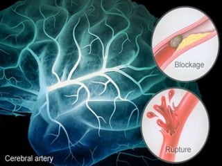
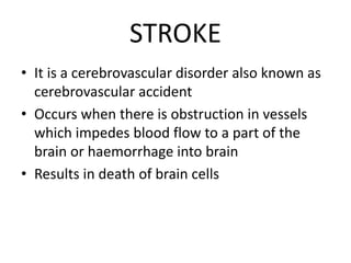
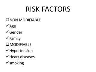
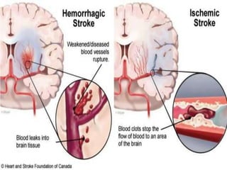
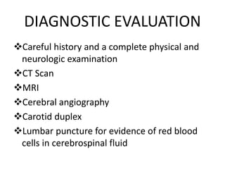
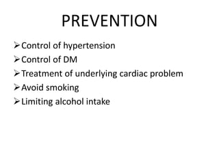
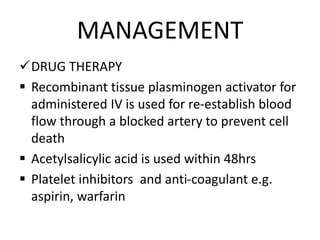
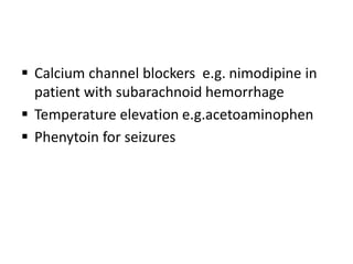
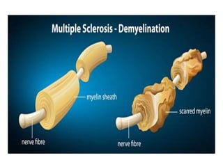
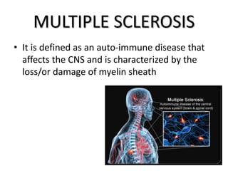
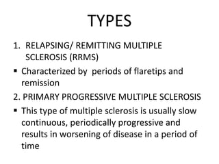
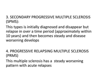
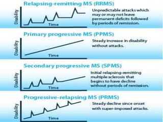
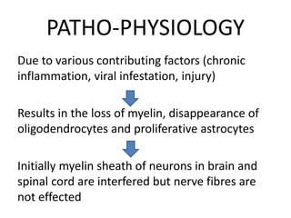
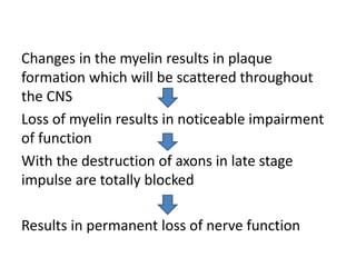
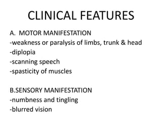
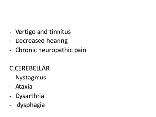
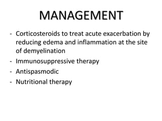
STROKE
AND
MULTIPLE SCLEROSIS
LAXMI THAPA
BSC 3RD YEAR
COMS-TH
STROKE
• It is a cerebrovascular disorder also known as
cerebrovascular accident
• Occurs when there is obstruction in vessels
which impedes blood flow to a part of the
brain or haemorrhage into brain
• Results in death of brain cells
RISK FACTORS
NON MODIFIABLE
Age
Gender
Family
MODIFIABLE
Hypertension
Heart diseases
smoking
Obesity
Sleep apnea
Metabolic syndrome
Lack of physical exercises
TYPES
1. ISCHEMIC STROKE
An ischemic stroke results from inadequate
blood flow to the brain from partial or complete
occlusion of an atery
It divides into thrombotic and embolic
a) THROMBOTIC STROKE
It occurs from injury to a vessels wall and
formation of blood clot
B) EMBOLIC STROKE
It occurs when an embolus lodge in and
occludes a cerebral artery
2. HEMORRHAGIC STROKE
It is caused by bleeding into the brain tissue, the
ventricles or subarachnoid space
-primary intracerebral haemorrhage
-secondary intracerebral haemorrhage
PATHO-PHYSIOLOGY
Due to various etiological factors persisting for a
long time
There is disruption of the cerebral blood flow due
to obstruction of a blood vessels
Due to the obstruction of blood vessels, initially
there is abnormal infiltration of lipid in the intimal
layer of the artery
This fatty streaks further develops into the plague
This formation of the palgue may rupture which
may travel and occlude the vessels thus limiting the
blood flow causing cerebal ischemia
The ischemic cascade begins when cerebral blood
flow falls to less than 25ml/ 100 g/min
Due to the cerebral hypoxia, neurons cannot
longer maintain aerobic respiration
The mitochondria then switch to anaerobic
respiration, which generates large amount of
lactic acid
Initiation of anaerobic respiration
Neurons incapable of producing sufficient
quantities of adenosine triphosphate (ATP) to
fuel the depolarization processes of brain tissue
Maintenance of electrolyte balances begin to
fall and cell function ceases
Manifest features like syncope, hemiperesis,
weakness of the limb
CLINICAL FEATURES
ISCHEMIC STROKE
GENERAL
 Numbness or weakness of face
 Visual disturbance
 Sudden severe headache
 Confusion
MOTOR LOSS
 Hemiplegia
 Hemiparesis
 Flaccid paralysis
COMMUNICATION LOSS
 Dysarthria
 Dysphasia
 Apraxia
PERCEPTUAL DISTURBANCES
 Headache
 Pain and rigidity
 Visual disturbances
 Tinnitus, dizziness and hemiparesis
DIAGNOSTIC EVALUATION
Careful history and a complete physical and
neurologic examination
CT Scan
MRI
Cerebral angiography
Carotid duplex
Lumbar puncture for evidence of red blood
cells in cerebrospinal fluid
PREVENTION
Control of hypertension
Control of DM
Treatment of underlying cardiac problem
Avoid smoking
Limiting alcohol intake
MANAGEMENT
DRUG THERAPY
 Recombinant tissue plasminogen activator for
administered IV is used for re-establish blood
flow through a blocked artery to prevent cell
death
 Acetylsalicylic acid is used within 48hrs
 Platelet inhibitors and anti-coagulant e.g.
aspirin, warfarin
 Calcium channel blockers e.g. nimodipine in
patient with subarachnoid hemorrhage
 Temperature elevation e.g.acetoaminophen
 Phenytoin for seizures
SURGICAL TREATMENT
Evacuation of aneurysm induced hematomas
or cerebral hematomas larger than 3cm
Aneurysm involves clipping wrapping or
coiling the aneurysm to prevent right bleeding
Merei Retriver removes blood clots in patient
who are experiencing ischemic stroke
MULTIPLE SCLEROSIS
• It is defined as an auto-immune disease that
affects the CNS and is characterized by the
loss/or damage of myelin sheath
TYPES
1. RELAPSING/ REMITTING MULTIPLE
SCLEROSIS (RRMS)
 Characterized by periods of flaretips and
remission
2. PRIMARY PROGRESSIVE MULTIPLE SCLEROSIS
 This type of multiple sclerosis is usually slow
continuous, periodically progressive and
results in worsening of disease in a period of
time
3. SECONDARY PROGRESSIVE MULTIPLE SCLEROSIS
(SPMS):
This types is initially diagnosed and disappear but
relapse in over a time period (approximately within
10 years) and then becomes steady and disease
worsening develops
4. PROGRESSIVE RELAPSING MULTIPLE SCLEROSIS
(PRMS)
This multiple sclerosis has a steady worsening
pattern with acute relapses
ETIOLOGY
Idiopathic
Aging Neuro degeneration
Possible precipitating factors:
 Infection
 Physical injury
 Emotional stress
 Excessive fatigue
 Pregnancy
 Poor state of health
PATHO-PHYSIOLOGY
Due to various contributing factors (chronic
inflammation, viral infestation, injury)
Results in the loss of myelin, disappearance of
oligodendrocytes and proliferative astrocytes
Initially myelin sheath of neurons in brain and
spinal cord are interfered but nerve fibres are
not effected
Changes in the myelin results in plaque
formation which will be scattered throughout
the CNS
Loss of myelin results in noticeable impairment
of function
With the destruction of axons in late stage
impulse are totally blocked
Results in permanent loss of nerve function
CLINICAL FEATURES
A. MOTOR MANIFESTATION
-weakness or paralysis of limbs, trunk & head
-diplopia
-scanning speech
-spasticity of muscles
B.SENSORY MANIFESTATION
-numbness and tingling
-blurred vision
- Vertigo and tinnitus
- Decreased hearing
- Chronic neuropathic pain
C.CEREBELLAR
- Nystagmus
- Ataxia
- Dysarthria
- dysphagia
D. EMOTIONAL
- Anger
- Depression
- Euphoria
DIAGNOSTIC EVALUATION
A. History collection
B. MRI
C. Evoked potential tests (EVPs)
D. Cerebrospinal fluid analysis
MANAGEMENT
- Corticosteroids to treat acute exacerbation by
reducing edema and inflammation at the site
of demyelination
- Immunosuppressive therapy
- Antispasmodic
- Nutritional therapy
stroke and multiple sclerosis

stroke and multiple sclerosis

  • 1.
  • 3.
    STROKE • It isa cerebrovascular disorder also known as cerebrovascular accident • Occurs when there is obstruction in vessels which impedes blood flow to a part of the brain or haemorrhage into brain • Results in death of brain cells
  • 4.
  • 5.
  • 7.
    TYPES 1. ISCHEMIC STROKE Anischemic stroke results from inadequate blood flow to the brain from partial or complete occlusion of an atery It divides into thrombotic and embolic
  • 8.
    a) THROMBOTIC STROKE Itoccurs from injury to a vessels wall and formation of blood clot B) EMBOLIC STROKE It occurs when an embolus lodge in and occludes a cerebral artery
  • 9.
    2. HEMORRHAGIC STROKE Itis caused by bleeding into the brain tissue, the ventricles or subarachnoid space -primary intracerebral haemorrhage -secondary intracerebral haemorrhage
  • 10.
    PATHO-PHYSIOLOGY Due to variousetiological factors persisting for a long time There is disruption of the cerebral blood flow due to obstruction of a blood vessels Due to the obstruction of blood vessels, initially there is abnormal infiltration of lipid in the intimal layer of the artery
  • 11.
    This fatty streaksfurther develops into the plague This formation of the palgue may rupture which may travel and occlude the vessels thus limiting the blood flow causing cerebal ischemia The ischemic cascade begins when cerebral blood flow falls to less than 25ml/ 100 g/min
  • 12.
    Due to thecerebral hypoxia, neurons cannot longer maintain aerobic respiration The mitochondria then switch to anaerobic respiration, which generates large amount of lactic acid Initiation of anaerobic respiration
  • 13.
    Neurons incapable ofproducing sufficient quantities of adenosine triphosphate (ATP) to fuel the depolarization processes of brain tissue Maintenance of electrolyte balances begin to fall and cell function ceases Manifest features like syncope, hemiperesis, weakness of the limb
  • 14.
    CLINICAL FEATURES ISCHEMIC STROKE GENERAL Numbness or weakness of face  Visual disturbance  Sudden severe headache  Confusion MOTOR LOSS  Hemiplegia  Hemiparesis
  • 15.
     Flaccid paralysis COMMUNICATIONLOSS  Dysarthria  Dysphasia  Apraxia PERCEPTUAL DISTURBANCES  Headache  Pain and rigidity  Visual disturbances  Tinnitus, dizziness and hemiparesis
  • 16.
    DIAGNOSTIC EVALUATION Careful historyand a complete physical and neurologic examination CT Scan MRI Cerebral angiography Carotid duplex Lumbar puncture for evidence of red blood cells in cerebrospinal fluid
  • 17.
    PREVENTION Control of hypertension Controlof DM Treatment of underlying cardiac problem Avoid smoking Limiting alcohol intake
  • 18.
    MANAGEMENT DRUG THERAPY  Recombinanttissue plasminogen activator for administered IV is used for re-establish blood flow through a blocked artery to prevent cell death  Acetylsalicylic acid is used within 48hrs  Platelet inhibitors and anti-coagulant e.g. aspirin, warfarin
  • 19.
     Calcium channelblockers e.g. nimodipine in patient with subarachnoid hemorrhage  Temperature elevation e.g.acetoaminophen  Phenytoin for seizures
  • 20.
    SURGICAL TREATMENT Evacuation ofaneurysm induced hematomas or cerebral hematomas larger than 3cm Aneurysm involves clipping wrapping or coiling the aneurysm to prevent right bleeding Merei Retriver removes blood clots in patient who are experiencing ischemic stroke
  • 22.
    MULTIPLE SCLEROSIS • Itis defined as an auto-immune disease that affects the CNS and is characterized by the loss/or damage of myelin sheath
  • 23.
    TYPES 1. RELAPSING/ REMITTINGMULTIPLE SCLEROSIS (RRMS)  Characterized by periods of flaretips and remission 2. PRIMARY PROGRESSIVE MULTIPLE SCLEROSIS  This type of multiple sclerosis is usually slow continuous, periodically progressive and results in worsening of disease in a period of time
  • 24.
    3. SECONDARY PROGRESSIVEMULTIPLE SCLEROSIS (SPMS): This types is initially diagnosed and disappear but relapse in over a time period (approximately within 10 years) and then becomes steady and disease worsening develops 4. PROGRESSIVE RELAPSING MULTIPLE SCLEROSIS (PRMS) This multiple sclerosis has a steady worsening pattern with acute relapses
  • 26.
    ETIOLOGY Idiopathic Aging Neuro degeneration Possibleprecipitating factors:  Infection  Physical injury  Emotional stress  Excessive fatigue  Pregnancy  Poor state of health
  • 27.
    PATHO-PHYSIOLOGY Due to variouscontributing factors (chronic inflammation, viral infestation, injury) Results in the loss of myelin, disappearance of oligodendrocytes and proliferative astrocytes Initially myelin sheath of neurons in brain and spinal cord are interfered but nerve fibres are not effected
  • 28.
    Changes in themyelin results in plaque formation which will be scattered throughout the CNS Loss of myelin results in noticeable impairment of function With the destruction of axons in late stage impulse are totally blocked Results in permanent loss of nerve function
  • 29.
    CLINICAL FEATURES A. MOTORMANIFESTATION -weakness or paralysis of limbs, trunk & head -diplopia -scanning speech -spasticity of muscles B.SENSORY MANIFESTATION -numbness and tingling -blurred vision
  • 30.
    - Vertigo andtinnitus - Decreased hearing - Chronic neuropathic pain C.CEREBELLAR - Nystagmus - Ataxia - Dysarthria - dysphagia
  • 31.
    D. EMOTIONAL - Anger -Depression - Euphoria
  • 32.
    DIAGNOSTIC EVALUATION A. Historycollection B. MRI C. Evoked potential tests (EVPs) D. Cerebrospinal fluid analysis
  • 33.
    MANAGEMENT - Corticosteroids totreat acute exacerbation by reducing edema and inflammation at the site of demyelination - Immunosuppressive therapy - Antispasmodic - Nutritional therapy